Introduction
Arsenic (Z = 33) is a metalloid element exhibiting a remarkable mineralogy, with one of the highest diversity index, D, among chemical elements (D = 10.3 – Christy, Reference Christy2015). In agreement with Majzlan et al. (Reference Majzlan, Drahota and Filippi2014), about 58% of all known As-bearing minerals are arsenates. Considering that As is one of the most worrying among the potentially toxic elements (PTE), the mineralogical investigation of arsenate-bearing assemblages plays an important role in understanding the fate of As in the environment, following the alteration of primary As minerals (e.g. As-bearing pyrite, arsenopyrite, and several other arsenic sulfides and sulfosalts). Possible sources of As include members of the tetrahedrite group, whose alteration processes have been investigated recently by several authors (e.g. Borčinová Radková et al., Reference Borčinová, Jamieson, Lalinská-Voleková, Majzlan, Števko and Chovan2017; Keim et al., Reference Keim, Staude, Marquardt, Bachmann, Opitz and Markl2018).
An interesting locality for the investigation of supergene phases related to the alteration of tetrahedrite-group minerals is Grosses Chalttal, where a formerly exploited small Cu-(U) deposit is hosted in a coarse fanglomeratic facies of the Permian Verrucano Formation belonging to the Helvetic nappes of eastern Switzerland. Primary mineralogy is represented by tennantite-(Cu), associated with pyrite, bornite, chalcopyrite, galena, uraninite and brannerite in a quartz ± baryte gangue. Bächtiger (Reference Bächtiger1963) also cited molybdenite, wittichenite and stromeyerite as minor primary minerals. Spionkopite and yarrowite are secondary phases intimately associated with tennantite-(Cu) (Malcherek et al., Reference Malcherek, Mihailova, Schlüter, Roth and Meisser2024). At the end of the 1990s, Meisser (Reference Meisser1999) described the oxidized supergene phases occurring at Grosses Chalttal, identifying several arsenates and sulfates along with rare carbonates. The recent study of some unknown arsenates lead to the description of two new Pb-Cu arsenates, heimite, ideally PbCu2(AsO4)(OH)3·2H2O, and its dehydrated counterpart metaheimite, PbCu2(AsO4)(OH)3 (Malcherek et al., Reference Malcherek, Mihailova, Schlüter, Roth and Meisser2024, Reference Malcherek, Mihailova, Schlüter, Roth and Meisser2025).
The systematic screening using energy-dispersive spectroscopy of old material from the dumps of the Grosses Chalttal mine allowed the discovery of an additional new Pb-Cu arsenate, identified in a sample collected in 2003. The full characterization of this species lead to its proposal as a new mineral. The mineral, its name, zanelliite, and its symbol, Znl, have been approved by the Commission on New Minerals, Nomenclature and Classification of the International Mineralogical Association under voting number IMA 2024-061. The name honours Dr. Remo Zanelli (b. 1977), a mineral collector, mathematics and geography teacher, and former specialist in pedogenic clay minerals. His extensive exploration of the (economically) modest ore deposits of the Glarus Alps and his keen eye, both in the field and under the microscope, have made him an expert on the secondary minerals of the region. He is also an excellent photographer of these minerals. Repeatedly, including for this new species, he has liberally placed unique specimens at the disposal of science. The type material of zanelliite is represented by two cotype specimens. One specimen (oxidized ore sample and polished sections used for EDS, WDS and optics) is kept in the Muséum cantonal des sciences naturelles (Naturéum), Département de géologie, Université de Lausanne, Switzerland, under catalogue number MGL 087285. The other specimen (oxidized ore sample and crystals used for X-ray diffraction – both single-crystal and powder techniques – and Raman spectroscopy) is deposited in the mineralogical collection of the Museo di Storia Naturale, Università di Pisa, Via Roma 79, Calci (Pisa, Italy), under catalogue number 20077.
In this paper, the description of zanelliite is reported, along with a discussion of its crystal chemistry.
Occurrence and physical properties
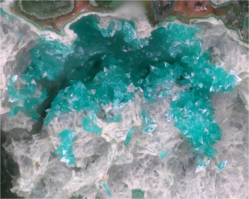
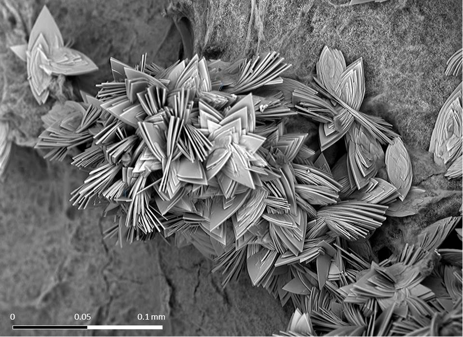
Zanelliite was found in the dumps of the Grosses Chalttal deposit, Mürtschenalp district, Glarus, Switzerland (47°04’10.12”N, 9°11’24.01”E). This mineral occurs as star-like aggregates of oval-shaped, tabular crystals, flattened on {100}, up to 0.05 mm in size (Figs 1, 2). Colour is turquoise-green and streak is pale green. Lustre is vitreous. Zanelliite is transparent and it does not fluoresce under both short- or long-wavelength UV radiation. The mineral is brittle, with a perfect {100} cleavage and a Mohs hardness of 3.5. Zanelliite reacts with Clerici solution, sinking in diiodomethane; its measured density is higher than 3.32 g cm–3. Calculated density, based on the empirical formula and unit-cell parameters determined using single-crystal X-ray diffraction, is 4.425 g cm–3.
Zanelliite, star-like aggregates of turquoise-green platy crystals on parnauite. Grosses Chalttal deposit, Mürtschenalp district, Glarus, Switzerland. Field of view: 0.9 mm. Photo R. Zanelli.

Back-scattered electron image of zanelliite, showing the crystal morphology. Grosses Chalttal deposit, Mürtschenalp district, Glarus, Switzerland. Photo P. Roth.

Zanelliite reacts immediately with immersion index liquids with n > 1.74 and for this reason the refractive index could not be measured. It is biaxial, with a weak dispersion and a weak pleochroism. On the basis of the Gladstone-Dale relationship, the mean refractive index should be 1.796 (Mandarino, Reference Mandarino1979, Reference Mandarino1981). Despite the high refractive index, an attempt was made to measure the 2V angle using Wright’s (Reference Wright1905) method of isogyre curvature, yielding a value of 2V > 65°.
In type material, zanelliite is associated with brochantite, chrysocolla, heimite and parnauite. Its genesis is probably related to the oxidation of the primary Cu ores. Due to the discovery of the type material as a small sample in the mine dumps, a more detailed contextualization of the occurrence of zanelliite in the ore body is not currently possible.
Experimental
Chemical and spectroscopic analysis
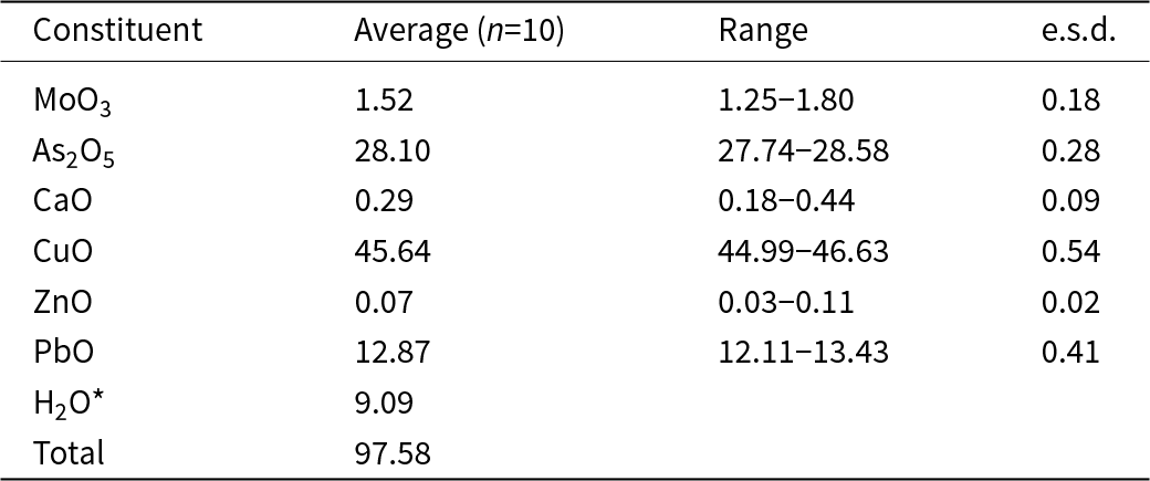
Quantitative chemical analysis of zanelliite were carried out using a JEOL JXA-8530F HyperProbe electron microprobe (WDS mode, accelerating voltage 15 kV, beam current 15 nA, beam size 5 μm) on polished crystal surfaces in epoxy mounts of 13.4 mm in diameter (Institute of Earth Sciences, University of Lausanne, Switzerland). Counting times were 30 s on peak and 15 s on the background on either side of the peak. Reference materials (element, emission line) were: adamite (AsKα, ZnKα), covellite (CuKα), anglesite (PbMα, SKα), wollastonite (SiKα, CaKα), pure Mo (MoLα). Matrix correction procedures using the φ(ρz) model of Pouchou and Pichoir (Reference Pouchou, Pichoir and Armstrong1985) were applied to the data. Results of chemical analysis are given in Table 1. Low analytical totals are probably due to sample porosity and to the thinness of the polished crystals. Direct H2O measurements were not possible owing to the small amount of available material and it was inferred based on the structural study (see below).
Electron microprobe data (in wt.%) for zanelliite

Table 1 Long description
Electron microprobe results summarize oxide constituents for zanelliite, reporting average values (in wt%) from 10 spot analyses plus ranges and estimated standard deviations where available. CuO is the largest component at an average of 45.64, ranging from 44.99 to 46.63, with an estimated standard deviation of 0.54. As2O5 averages 28.10 with a range of 27.74 to 28.58 and an estimated standard deviation of 0.28. PbO averages 12.87, ranging from 12.11 to 13.43, with an estimated standard deviation of 0.41. Minor constituents include MoO3 at 1.52 (range 1.25 to 1.80), CaO at 0.29 (range 0.18 to 0.44), and ZnO at 0.07 (range 0.03 to 0.11), each with small estimated standard deviations. Water is listed as a calculated value of 9.09 . The reported total averages 97.58 wt%, indicating the summed measured and calculated components fall slightly below 100 wt%, which may reflect analytical and calculation limits.
n = number of spot analyses; *Calculated in agreement with structural data; e.s.d. = estimated standard deviation.
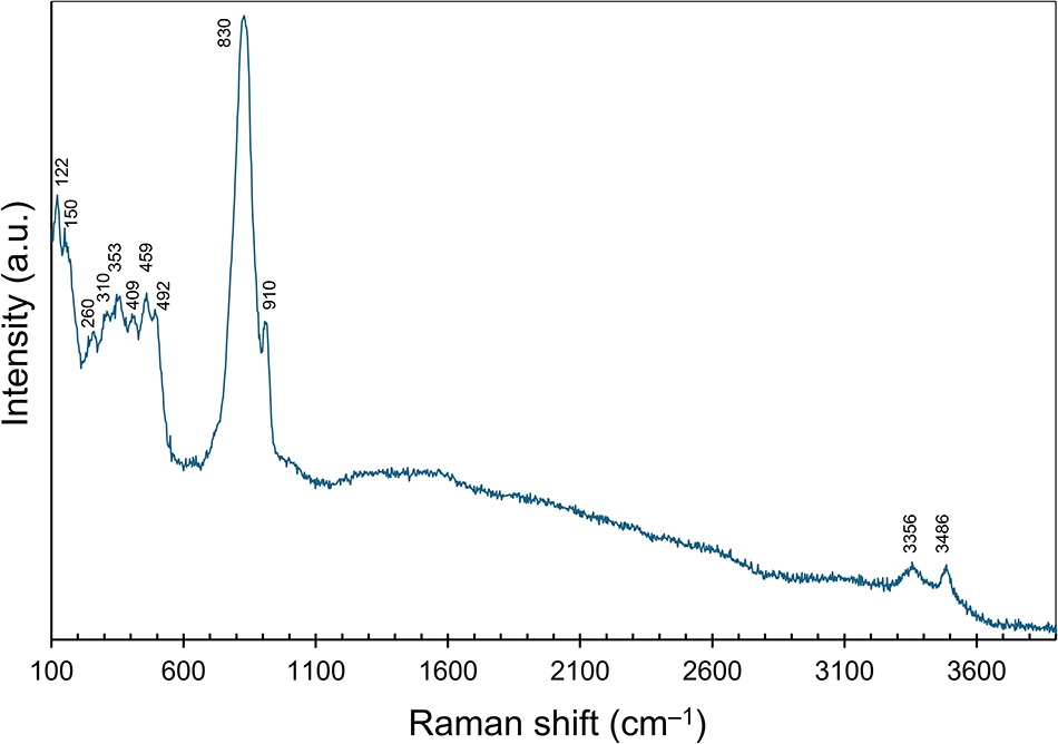
Micro-Raman spectra of zanelliite were collected on an unpolished grain in the range 100–4000 cm–1 in nearly back-scattered geometry with a Jobin-Yvon Horiba XploRA Plus spectrometer, equipped with a motorized x-y stage and an Olympus BX41 microscope with a 50× objective (Dipartimento di Scienze della Terra, University of Pisa, Italy). The 532 nm line of a solid-state laser was used. Laser power was 6.25 mW. The minimum lateral and depth resolution was set to a few μm. The system was calibrated using the 520.6 cm–1 Raman band of silicon before each experimental session. Spectra were collected through multiple acquisitions (10) with single counting times of 30 s. Back-scattered radiation was analysed with a 1200 grooves mm–1 grating monochromator. Possible thermal damage of the measured points was excluded by visual inspection of the excited surface after measurement, by observing the possible decay of spectral features in the start of the excitation and by checking for thermal downshift of Raman lines. The Raman spectrum of zanelliite is shown in Fig. 3.
Raman spectrum of zanelliite in the range 100–4000 cm–1.

X-ray crystallography
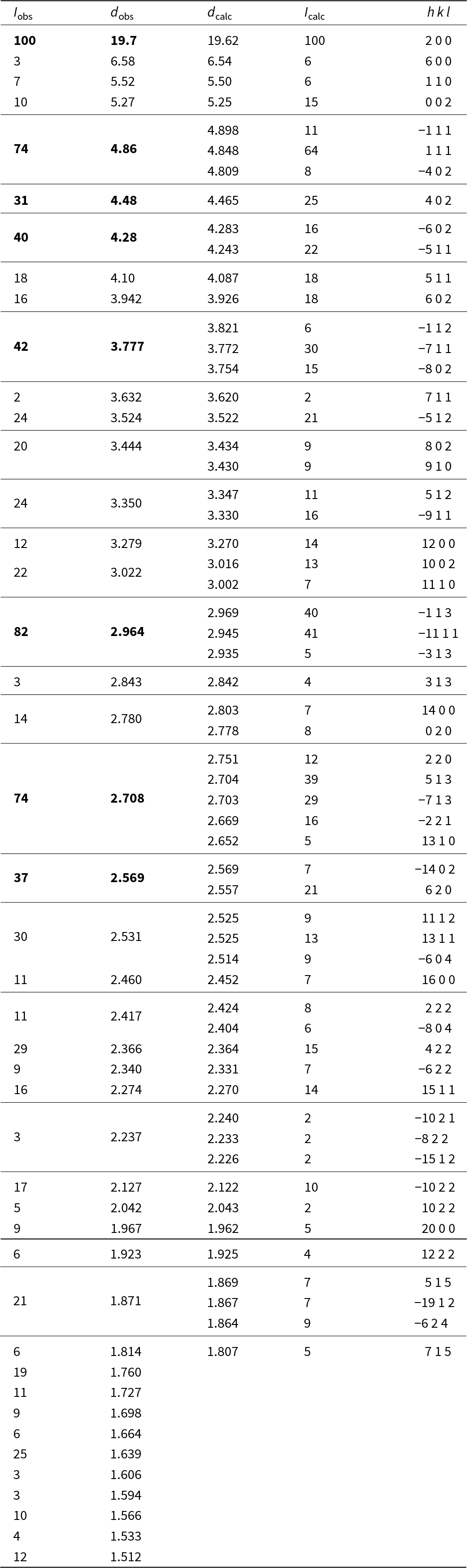
Powder X-ray diffraction data of zanelliite were collected using a Bruker D8 Venture single-crystal X-ray diffractometer equipped with a Photon III CCD area detector (Centro per l’Integrazione della Strumentazione scientifica dell’Università di Pisa, Università di Pisa, Italy) and microfocus CuKα radiation, simulating a Gandolfi-like geometry. Observed X-ray diffraction lines are reported in Table 2, along with the calculated pattern based on the structural model discussed below.
Powder X-ray diffraction data (d in Å) for zanelliite

Table 2 Long description
Powder X-ray diffraction results for zanelliite list observed peak intensity, observed d spacing in angstroms, calculated d spacing, calculated intensity, and the indexed h k l reflection. The most intense observed peak occurs at about 19.7 Å (h k l 2 0 0), with other strong observed peaks near 4.86 Å (minus 1 1 1), 2.96 Å (minus 1 1 3), and 2.71 Å (2 2 0), plus a notable peak near 4.28 Å (minus 6 0 2). Across most listed reflections, observed and calculated d spacings are very close, typically differing only slightly, supporting the indexing and structural model used for the calculations. Calculated intensities are provided for many reflections, including some that are weak or not clearly observed, while calculated reflections below about 1.80 Å are not included.
Notes: I calc and d calc were obtained using PowderCell 2.4 (Kraus and Nolze, Reference Kraus and Nolze1996) on the basis of the structural model of zanelliite given in Table 4. Only calculated reflections with I calc > 5 are reported (if not observed); d calc < 1.80 Å are not reported. The eight strongest observed reflections are shown in bold.
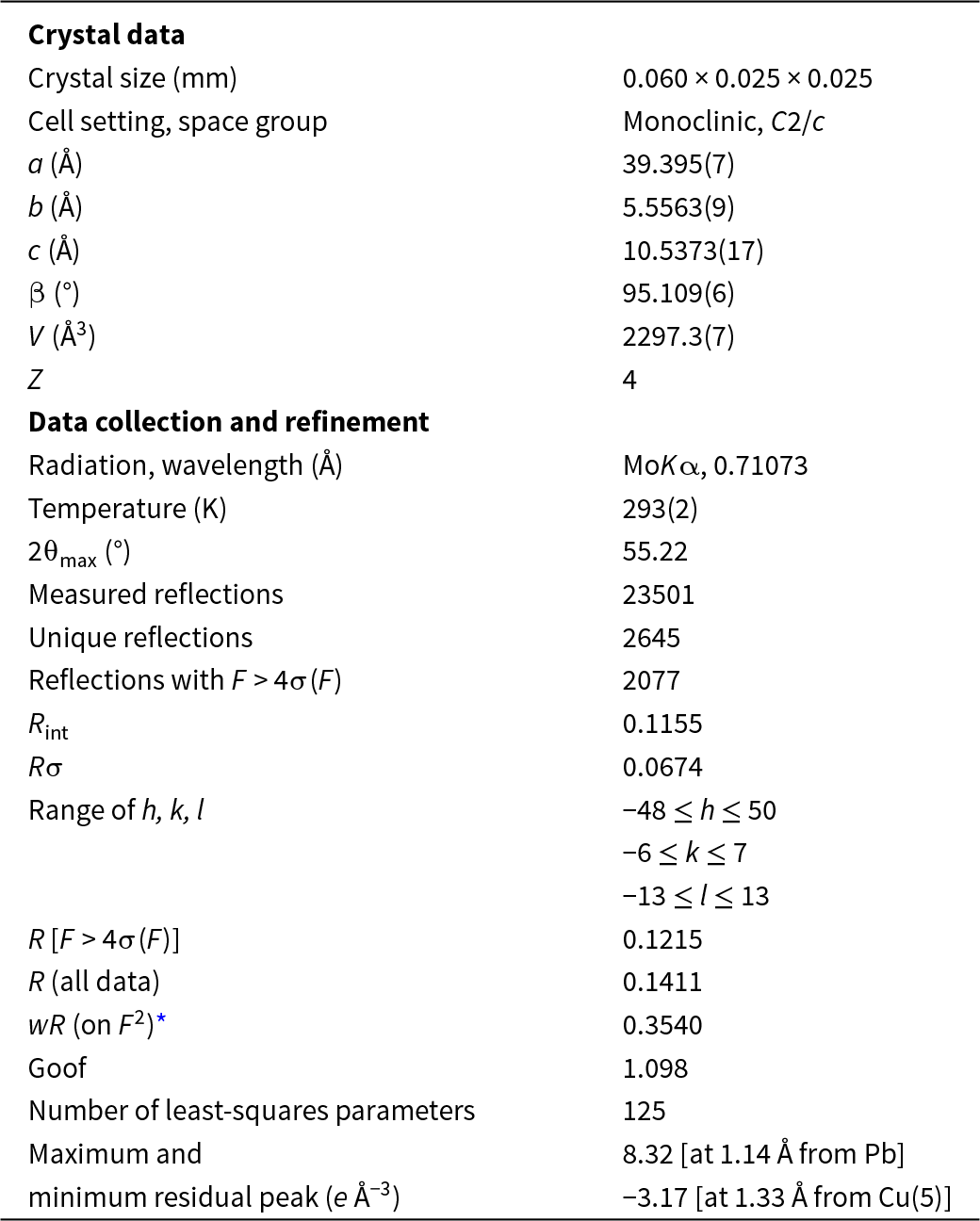
Single-crystal X-ray diffraction intensity data were collected on the same instrument using microfocus MoKα radiation. The detector-to-crystal distance was set at 38 mm. Data were collected using ω and φ scan modes, in 0.5° slices, with an exposure time of 20 s per frame. A total of 1682 frames was collected. The frames were integrated with the Bruker SAINT software package using a narrow-frame algorithm. A total of 23,501 reflections were measured up to 2θmax = 55.22°, resulting in 2645 unique reflections. Data were corrected for Lorentz-polarization, absorption and background using the Apex4 software package (Bruker AXS, Reference Bruker2022). Unit-cell parameters, refined on the basis of the XYZ centroid of 3185 reflections above 20σ(I) with 7.407° < 2θ < 54.47°, are a = 39.395(7), b = 5.5563(9), c = 10.5373(17) Å, β = 95.109(6)° and V = 2297.3(7) Å3. The statistical tests on the distribution of |E| values (|E 2 – 1| = 0.845) may suggest (but not unequivocally) the presence of a centre of symmetry. According to the systematic absences, and the uncertainty about the occurrence of a centre of symmetry, the crystal structure of zanelliite was solved in three different space groups using Shelxtl (Sheldrick, Reference Sheldrick2015a), i.e. C2/c, C2, and Cc. The main structural features are very similar in these three models. However, the C2 and Cc structural models gave racemic twin ratios close to 0.5, suggesting the presence of a centre of symmetry. For this reason, the crystal structure of zanelliite was refined, using Shelxl-2018 (Sheldrick, Reference Sheldrick2015b) in the space group C2/c. Neutral scattering curves, taken from the International Tables for Crystallography (Wilson, Reference Wilson1992), were used. Eight independent cation sites and 14 anion positions were located. The occurrence of a two-fold axis along [001] was suggested by Platon (Spek, Reference Spek2003) but the refined ratio between the two twinned domains is close to 0, i.e. 0.0044(16). After several cycles of anisotropic refinement for cation positions only, the conventional R 1 factor converged to 0.1215 for 2077 unique reflections with F > 4σ(F) and 125 refined parameters. An anisotropic modelling for anion sites resulted in some of them being negatively defined. Consequently, an isotropic model for anion positions was preferred.
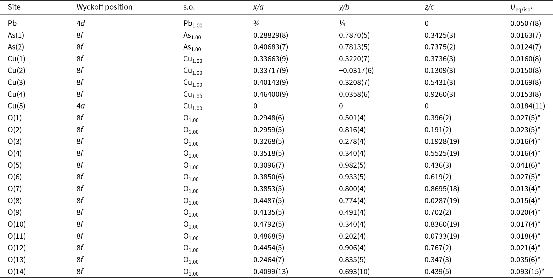
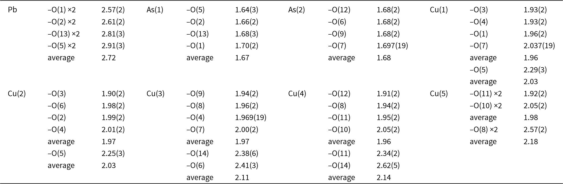
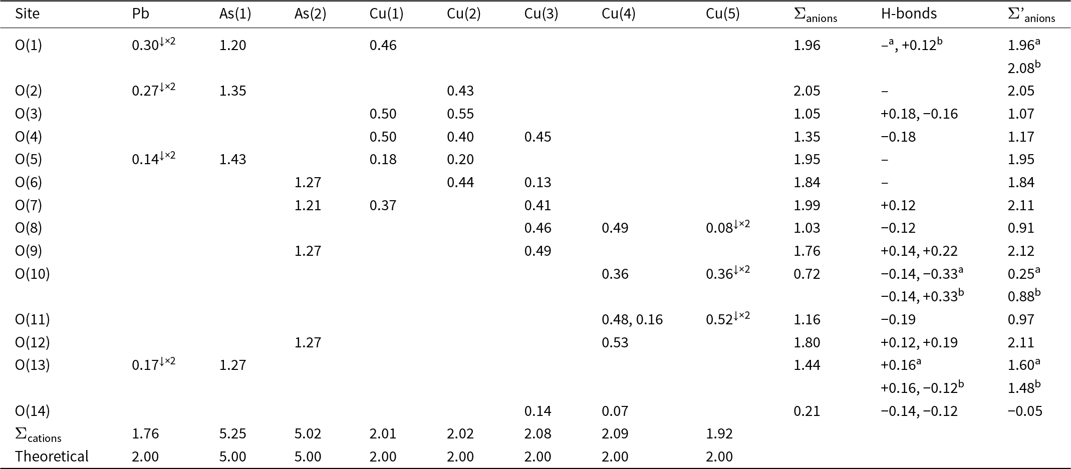
Details of the data collection and crystal structure refinement are given in Table 3. Atom coordinates and isotropic or equivalent isotropic displacement parameters are reported in Table 4, whereas Table 5 gives selected bond distances. The bond-valence calculation, shown in Table 6, was performed using the bond parameters of Gagné and Hawthorne (Reference Gagné and Hawthorne2015). The crystallographic information file has been deposited with the Principal Editor of Mineralogical Magazine and is available as Supplementary material (see below).
Crystal and experimental data for zanelliite

Table 3 Long description
Crystal and diffraction refinement metrics for zanelliite are summarized, including crystal size, unit-cell geometry, data collection limits, and refinement quality indicators. The crystal measured 0.060 by 0.025 by 0.025 millimeters and was refined in the monoclinic space group C2/c. Unit-cell parameters were a 39.395 angstroms, b 5.5563 angstroms, c 10.5373 angstroms, beta 95.109 degrees, volume 2297.3 cubic angstroms, with Z equal to 4. Data were collected using molybdenum K alpha radiation at 293 kelvin to a maximum two-theta of 55.22 degrees, yielding 23,501 measured reflections and 2,645 unique reflections. Of these, 2,077 reflections met the criterion of structure-factor amplitude exceeding four times its uncertainty; merging indicators were Rint 0.1155 and Rsigma 0.0674. Index ranges were h from minus 48 to 50, k from minus 6 to 7, and l from minus 13 to 13. Refinement agreement factors were R 0.1215 for the stronger reflections and 0.1411 for all data, with weighted R on squared structure factors 0.3540 and goodness of fit 1.098 using 125 parameters. Residual electron-density extrema were 8.32 electrons per cubic angstrom near lead and minus 3.17 electrons per cubic angstrom near copper site 5, suggesting remaining local misfit around heavy-atom positions.
* w = 1/[σ2(F o2)+0.1562P 2+1231.6315P]
Sites, Wyckoff positions, site occupancy (s.o.), fractional atom coordinates and equivalent isotropic or isotropic (*) displacement parameters (in Å2) for zanelliite

Table 4 Long description
Atomic site list for zanelliite giving Wyckoff positions, site occupancies, fractional coordinates along a, b, and c, and equivalent isotropic or isotropic-star displacement parameters. All listed sites have full occupancy (1.00) for Pb, two As sites, five Cu sites, and fourteen O sites. Pb is at Wyckoff 4d with coordinates (0.75, 0.25, 0) and the largest displacement parameter, 0.0507(8). As(1) and As(2) are both at 8f ; their displacement parameters are 0.0163(7) and 0.0124(7). Cu(1) through Cu(4) are at 8f and displacement parameters around 0.015 to 0.017, while Cu(5) is at 4a at the origin with equivalent isotropic displacement parameter 0.0184(11). Oxygen sites are all 8f with isotropic displacement parameters ranging from 0.013(4) for O(7) up to 0.093(15) for O(14), indicating notably higher positional uncertainty or motion for O(14). Values in parentheses indicate reported uncertainties in the final digits, so small differences between similar sites may not be significant.
Selected bond distances (in Å) in zanelliite

Table 5 Long description
Bond distances in angstroms are listed for Pb, two As sites, and five Cu sites to specific oxygen atoms, with some contacts repeated twice and with site averages. Pb shows the longest Pb to O distances: about 2.57 to 2.91 Å, with an average near 2.72 Å. As sites have the shortest bonds: As(1) to O ranges about 1.64 to 1.70 Å with an average near 1.67 Å, and As(2) to O is tightly grouped around 1.68 to 1.697 Å with an average near 1.68 Å. Cu(1) has four shorter Cu to O distances near 1.93 to 2.04 Å (average about 1.96 Å) plus a longer contact to O(5) at about 2.29 Å, giving a higher overall average near 2.03 Å. Cu(2), Cu(3), and Cu(4) each show four short bonds around 1.90 to 2.05 Å (short-bond averages near 1.96 to 1.97 Å) and one or two longer contacts around 2.25 to 2.62 Å, raising their overall averages to about 2.03, 2.11, and 2.14 Å respectively. Cu(5) includes two short pairs near 1.92 and 2.05 Å (average about 1.98 Å) and a much longer pair reported as 2..57 Å,.
Weighted bond valences (in vu) for zanelliite

Table 6 Long description
Bond-valence contributions in valence units are listed for each oxygen site O1 to O14 from Pb, two As sites, and five Cu sites, along with each oxygen’s anion total, hydrogen-bond adjustments, and a hydrogen-bond-corrected anion total. Most corrected anion totals cluster near about 2.0, including O2 at 2.05, O5 at 1.95, O6 at 1.84, O7 at 2.11, O9 at 2.12, and O12 at 2.11. Several sites show notable deviations: O8 corrects downward to 0.91, O11 corrects to 0.97, and O14 is strongly underbonded with a corrected total of minus 0.05 after negative hydrogen-bond adjustments. Two oxygen sites have alternative hydrogen-bond configurations that change the corrected totals: O1 is 1.96 in option a and 2.08 in option b, and O10 is 0.25 in option a versus 0.88 in option b. Cation bond-valence sums at the bottom are close to theoretical expectations: Pb 1.76 versus 2.00, As1 5.25 versus 5.00, As2 5.02 versus 5.00, and Cu sites range from 1.92 to 2.09 versus 2.00. Down-arrow entries indicate bonds counted twice, and corrected anion totals depend on the chosen hydrogen-bond configuration and the method used to estimate hydrogen-bond strengths.
Notes: Superscript a and b refer to two different H-bond configurations. Σ’anions reports bond-valence sums corrected for H-bonds whose values were calculated according to Ferraris and Ivaldi (Reference Ferraris and Ivaldi1988). Right superscripts indicate the number of bonds involving cations.
Results and discussion
Chemical composition of zanelliite
The empirical formula of zanelliite was calculated on the basis of 28 O atoms per formula unit (apfu), in agreement with the results of the structural study (see below). It can be written as (Pb0.90Ca0.08)Σ0.98(Cu9.00Zn0.01)Σ9.01(As3.84Mo0.17)Σ4.01O15.17(OH)9.83(H2O)3. The ideal formula is PbCu9As4O15(OH)10(H2O)3 or PbCu9[AsO3.5(OH)0.5]2(AsO4)2(OH)9(H2O)3 (Z = 4). It corresponds to (in wt.%) As2O5 29.79, CuO 46.40, PbO 14.47, H2O 9.34, for a total of 100.00 wt.%.
Micro-Raman spectrum of zanelliite
The Raman spectrum of zanelliite is in Fig. 3. The main bands in the region between 100 and 1200 cm–1 are (in cm–1): 122, 150, 260, 310, 353, 409, 459, 492, 830 and 910. The very strong Raman band at 830 cm–1 is probably due to the ν1 symmetric stretching vibration of AsO4 groups (e.g. Frost and Weier, Reference Frost and Weier2004; Reddy et al., Reference Reddy, Frost and Martens2005; Kloprogge and Wood, Reference Kloprogge and Wood2017). The weaker band at 910 cm–1 may be due to OH-stretching deformation of OH groups bonded to Cu atoms (e.g. Martens et al., Reference Martens, Frost and Williams2003; Reddy et al., Reference Reddy, Frost and Martens2005). Bending modes of AsO4 groups can be associated with Raman bands at 353, 409, 459, and 492 cm–1, whereas the band at 310 cm–1 may be interpreted either as an AsO4 bending mode (e.g. Martens et al., Reference Martens, Frost and Williams2003; Frost and Weier, Reference Frost and Weier2004) or as M–φ (M = Cu, Pb; φ = OH, H2O) stretching vibrations (e.g. Frost et al., Reference Frost, López, de Oliveira Gonçalves, Scholz and Xi2014; Kloprogge and Wood, Reference Kloprogge and Wood2017). Bands below 300 cm–1 are related to M–φ vibrations and lattice modes.
The occurrence of H2O and OH groups is confirmed by the presence of Raman bands at 3356 and 3486 cm–1, which are related to O–H stretching modes.
Crystal structure of zanelliite
General features
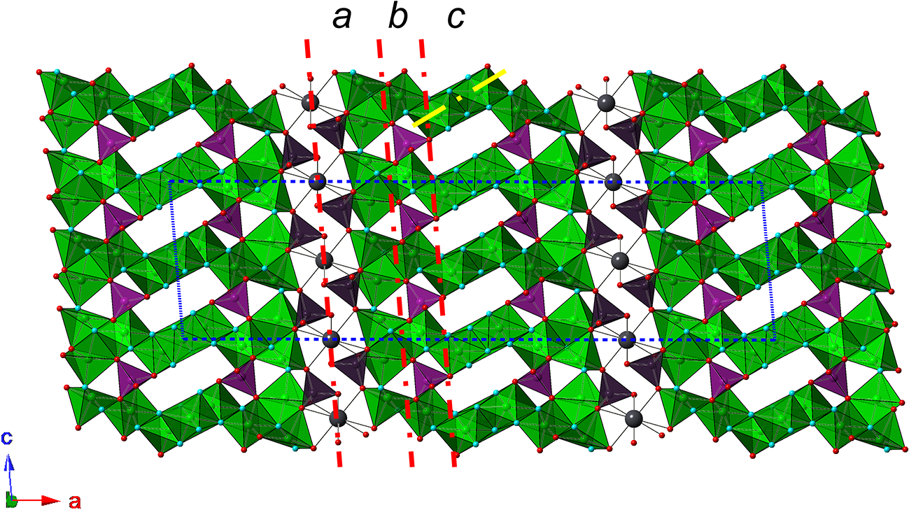
The crystal structure of zanelliite can be described as layered. Figure 4 shows the different modules that can be identified.
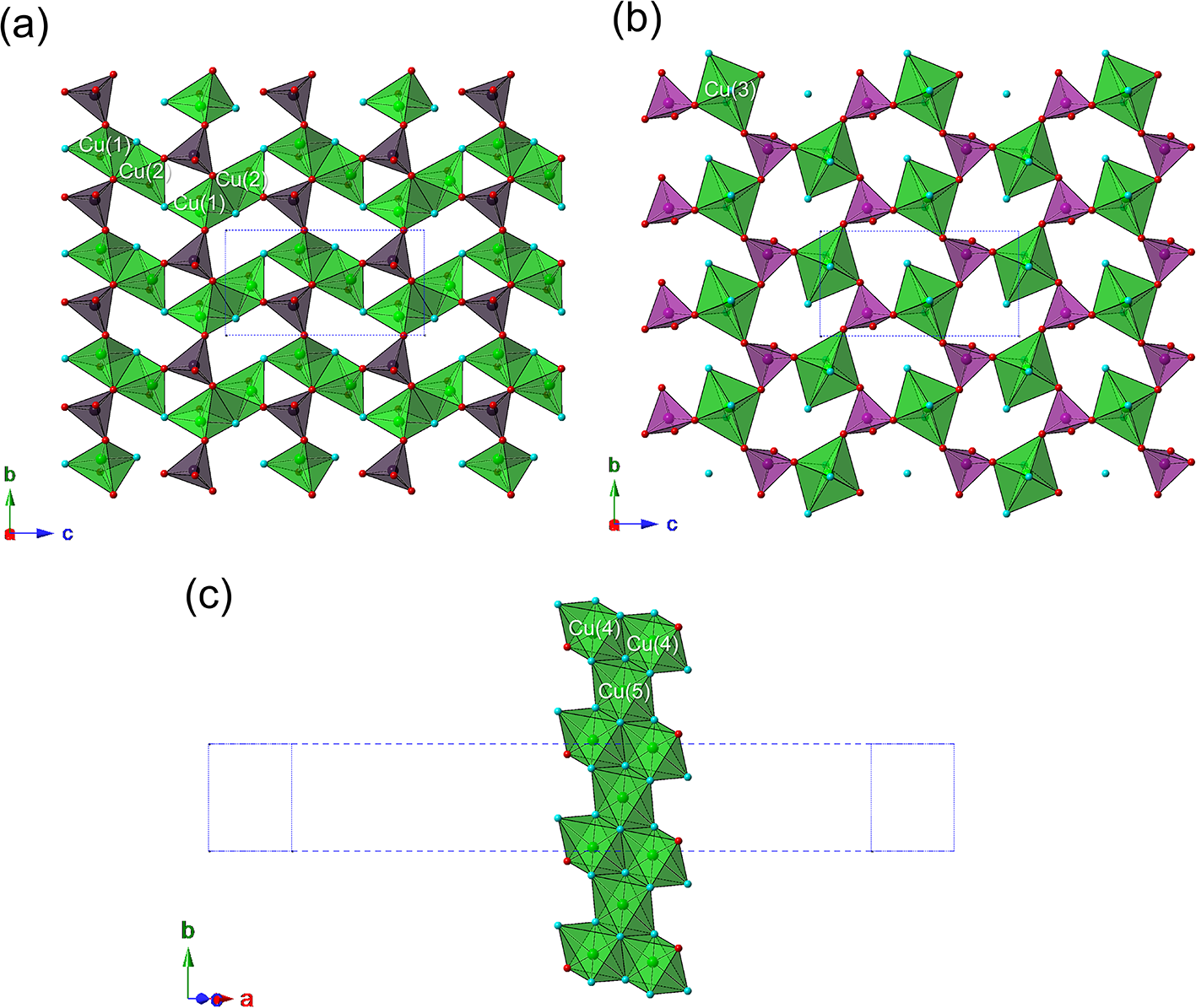
Five-fold coordinated Cu atoms at the Cu(1) and Cu(2) sites form dimers through edge-sharing. These dimers are connected along the c-axis giving rise to zig-zag chains. Each chain is decorated, on one side, by As(1) tetrahedra through the sharing of three O atoms of the As(1)O4 groups, that connect the chains along b forming a heteropolyhedral Cu–As layer (Fig. 5a). Two of these Cu–As layers face each other along a, with Pb atoms sandwiched between them. On the other side of the zig-zag chains formed by the Cu(1) and Cu(2) sites, there is another Cu–As layer formed by Cu(3) and As(2) sites (Fig. 5b). The former is bonded through edge-sharing to one Cu(1)–Cu(2) dimer, whereas the As(2)O4 groups share two O atoms with adjacent dimers. In this way, a complex Pb–Cu–As heteropolyhedral layer can be identified.
Crystal structure of zanelliite as seen down b. Symbols: green polyhedra = Cu-centred polyhedra; dark and light violet = As(1) and As(2) tetrahedra. Spheres: dark grey = Pb atoms; green = Cu atoms, red = O sites; light blue = OH and H2O groups. The red lines indicate the two layers a and b, whereas the yellow line shows the  $\{ \bar 201\} $ ribbons c. Dashed blue lines indicate the unit cell. Crystal structures were drawn using CrystalMaker (Palmer, Reference Palmer2014).

Figure 4 Long description
The crystal structure of zanelliite is depicted with various polyhedra and atoms. Green polyhedra represent copper-centered polyhedra, while dark and light violet tetrahedra indicate As(1) and As(2). Dark grey spheres denote lead atoms, green spheres represent copper atoms, red spheres indicate oxygen sites and light blue spheres show (OH) groups. Red lines highlight two layers and a yellow line marks the ribbons. Dashed blue lines outline the unit cell. The structure is arranged in a layered pattern, with the axes labeled as a, b and c at the top. A coordinate system is shown at the bottom left, indicating the directions of the a, b and c axes.
The different modules shown in Fig. 4. (a) The Cu(1)–Cu(2) zig-zag chains and As(1)O4 groups; (b) the layer formed by Cu(3) and As(2)O4 groups; and (c) the  $\{ \bar 201\} $ Cu(4)–Cu(5) ribbons. Same symbols as in Fig. 4.

Figure 5 Long description
The image consists of three parts. In the first part, labeled (a), Cu(1) and Cu(2) atoms form zig-zag chains with As(1)O4 groups. These chains are arranged in a repeating pattern, with the Cu atoms connected through edge-sharing, forming dimers. The As(1) tetrahedra are attached to the chains, sharing three oxygen atoms. In the second part, labeled (b), a layer is formed by Cu(3) atoms and As(2)O4 groups. The Cu(3) atoms are bonded through edge-sharing and the As(2)O4x groups share two oxygen atoms with adjacent dimers. The third part, labeled (c), shows Cu(4) and Cu(5) forming ribbons. These ribbons are depicted in a linear arrangement, with the Cu atoms connected in a sequence. Each part is oriented with axes labeled a, b and c, indicating the spatial arrangement of the structures.
Along a, these heteropolyhedral layers alternate, in a one-to-one ratio, with  $\{ \bar 201\} $ ribbons formed by Cu(4) and Cu(5) sites (Fig. 5c).
Cation sites
Arsenic is hosted at the As(1) and As(2) sites and shows a tetrahedral coordination, with average <As–O> bond distances of 1.67 and 1.68 Å, respectively, comparable with the characteristic bond length of 1.688 Å for the As–O bond reported by Hawthorne and Gagné (Reference Hawthorne and Gagné2024). The bond-valence sums at As(1) and As(2) are 5.25 and 5.02 valence units (vu), respectively, in agreement with As5+. The position of minor Mo (assumed as Mo6+) was not identified, owing to the low amount of Mo (up to 0.08 atoms per site, taking into account site multiplicity) and the relatively low quality of the crystal structure refinement. However, it is likely that Mo6+ could enter the structure of zanelliite through the heterovalent substitution As5+ + (OH)– = Mo6+ + O2–.
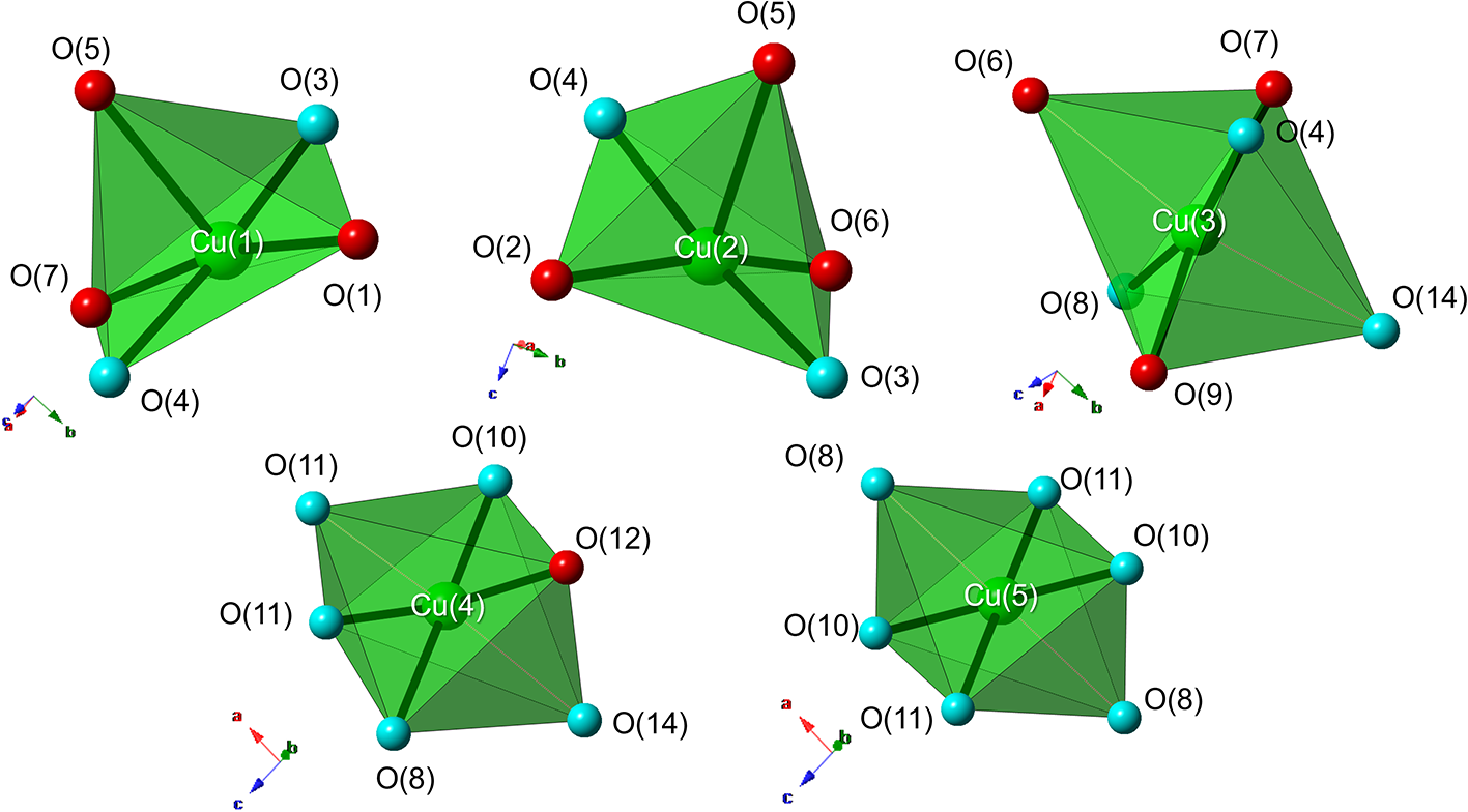
Copper atoms are hosted at five independent sites showing distorted (4+1) or (4+2) coordination (Fig. 6). Copper atoms at the Cu(1) and Cu(2) sites show a square pyramidal (4+1) coordination, with four <Cu–φ> (φ = O, OH, H2O) bond distances ranging between 1.90 and 2.04 Å, and a fifth one at ca. 2.25–2.30 Å. Bond-valence sums at these positions are 2.01 and 2.02 vu, respectively, in agreement with Cu2+. Copper atoms at the Cu(3), Cu(4) and Cu(5) sites have a distorted (4+2) octahedral coordination, with four <Cu–φ> bond distances in the range 1.91–2.05 Å, and two distinctly longer distances (up to ca. 2.6 Å). These Cu atoms also display bond-valence sums, ranging from 1.92 to 2.09 vu, consistent with the presence of divalent copper. The geometry of the Cu coordination polyhedra is in accordance with that observed in other Cu2+ compounds (e.g. the Cu-arsenate-carbonate-sulfate mineral claraite – Biagioni and Orlandi, Reference Biagioni and Orlandi2017) and with the occurrence of the Jahn-Teller effect (Burns and Hawthorne, Reference Burns and Hawthorne1996). Minor Zn, detected through electron microprobe analysis, could not be located.
Coordination environment of Cu atoms. Same symbols as in Fig. 4. Thick bonds represent Cu–φ distances shorter than 2.3 Å, whereas thin dotted lines are distances longer than this value.

Figure 6 Long description
Coordination environment of copper atoms at five independent sites, each represented by a green polyhedron. The copper atoms are labeled as Cu(1), Cu(2), Cu(3), Cu(4) and Cu(5), positioned at the center of each polyhedron. Surrounding each copper atom are oxygen atoms labeled O(1) through O(14), depicted as spheres in different colors. The polyhedra illustrate distorted coordination geometries, with thick bonds indicating shorter Cu–O distances and thin dotted lines representing longer distances. Each polyhedron is accompanied by a small coordinate axis diagram, indicating orientation. The arrangement highlights the spatial relationship and coordination between copper and oxygen atoms, emphasizing the structural complexity of the copper sites.
Lead atoms occur at the Pb site and show eight-fold coordination, with an average <Pb–O> bond distance of 2.72 Å. The bond-valence sum is 1.76 vu, in agreement with the presence of Pb2+. Minor Ca most likely replaces Pb at this site.
Anion sites and possible H-bonds
Fourteen independent anion sites have been located in the crystal structure of zanelliite. Bond-valence sums (Table 6) allow the identification of different groups of anions: (i) O(1), O(2), O(5), O(6), O(7), O(9) and O(12) have bond-valence sums in the range 1.76–2.05 vu, probably corresponding to O2– anions; (ii) O(3), O(8) and O(11) have bond-valence sums of ∼1 vu, ranging between 1.03 and 1.16 vu, probably corresponding to (OH)– groups; (iii) O(4) and O(13) have a low bond-valence sums intermediate between those of an O2– and (OH)– group, i.e., 1.35 and 1.44 vu, respectively; (iv) O(10) has a bond-valence sum lower than that of an (OH)– group but significantly higher than that of an H2O group; and finally, (v) O(14) has a bond-valence sum of 0.14 vu, corresponding to an H2O group.
The examination of O···O distances shorter than 3 Å and O···O···O angles makes it possible to propose the H-bond network in zanelliite.
The atoms hosted at O(1), O(2), O(5) and O(7) have bond-valence sums very close to 2 vu and are therefore O2– anions, whereas O(3) and O(4) are (OH)– groups. Indeed, the anion at O(3) acts as an acceptor of H-bond from O(4) [O(4)···O(3) = 2.81 Å] and as a donor to O(13) [O(3)···O(13) = 2.89 Å]. Anions at O(6), O(9) and O(12) are slightly underbonded and may act as H-bond acceptors. In particular, the anions at O(9) and O(12) are acceptors of two H-bonds. The former is an acceptor from O(10) [O(10)···O(9) = 2.96 Å] and O(14) [O(14)···O(9) = 2.71 Å], whereas the latter is an acceptor from O(8) [O(8)···O(12) = 3.09 Å] and O(11) [O(11)···O(12) = 2.79 Å]. The anion at O(14), strongly underbonded (0.14 vu), is the oxygen atom of an H2O group. This O atom is at H-bond distance from three other O atoms, i.e. O(6) (at 2.58 Å), O(7) (at 3.05 Å), and O(9) (at 2.71 Å). However, different bond angles can be observed, i.e. O(6)···O(14)···O(7) = 69°, O(7)···O(14)···O(9) = 100°, and O(9)···O(14)···O(6) = 158°. According to Ferraris and Franchini-Angela (Reference Ferraris and Franchini-Angela1972), the A1···W···A2 angle (where A1 and A2 are the acceptors and W is the H2O group) has an average value of 108°, ranging between ca. 80° and 140° (see their figure 2d). Consequently, the O(6)···O(14)···O(7) and O(9)···O(14)···O(6) angles are respectively too small and too large. For this reason, the O(7)···O(14)···O(9) is here considered as the H-bond involving O(14), although O(7) does not require H-bonds whereas O(6) is slightly underbonded.
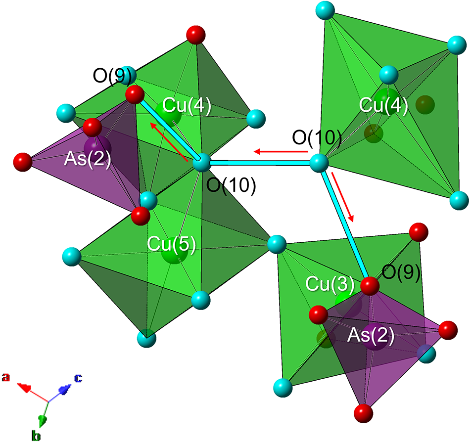
A more important question regards the nature of both O(10) and O(13). The former has a bond-valence sum of 0.72 vu, being shared between Cu(4) and Cu(5). The examination of its local environment suggests that it should act as a donor of an H-bond to O(9), as discussed above. Moreover, it forms an O(10)···O(10) distance of 2.55 Å, with an O(9)···O(10)···O(10) angle of 105°. Thus, in one case O(10) is donor to both O(9) and O(10), whereas in the other case O(10) acts as an acceptor from O(10) and donor to O(9) (Fig. 7). The corresponding bond-valence sums are thus 0.25 and 0.88 vu, respectively, indicating the statistical nature of O(10), which is occupied by H2O and (OH)– groups. The anion at O(13) is bonded to As(1) and Pb and has a bond-valence sum of 1.44 vu. It acts as an acceptor of an H-bond from O(3), and it is at an H-bond distance from O(2) [O(13)···O(2) = 3.16 Å] and O(1) [O(13)···O(1) = 3.05 Å]. Thus this position could be occupied by an O atom (underbonded, with a bond-valence sum of 1.60 vu) or by an (OH)– group that could be donor to O(1) [slightly underbonded with respect to O(2) that is slightly overbonded], and has a bond-valence sum of 1.48 vu overbonded with respect to the other (OH)– groups. This discrepancy could be due to the average nature of this position. It is worth noting that the mixed nature of O(10) [(OH)0.5(H2O)0.5] could be related to the mixed nature of O(13), which could be occupied by [O0.5(OH)0.5], thus corresponding to the substitution mechanism O(10)H2O + O(13)O2– = O(10)(OH)– + O(13)(OH)–.
The local environment around O(10) in zanelliite. Same symbols as in Fig. 4. The red arrows indicate the H-bonds, with the direction from the donor to the acceptor. Thick light blue lines represent O···O distances.

Figure 7 Long description
The structure displays interconnected polyhedra representing copper and arsenic atoms. Copper atoms are labeled Cu(4), Cu(5) and Cu(3), while arsenic atoms are labeled As(2). Oxygen atoms are labeled O(9) and O(10), positioned at vertices of the polyhedra. Red arrows indicate hydrogen bonds, showing direction from donor to acceptor, specifically from O(10) to O(9) and between O(10) atoms. Thick light blue lines represent O to O distances, connecting O(10) to other O atoms. The polyhedra are colored differently to distinguish between copper and arsenic, with green for copper and purple for arsenic. A small coordinate system in the bottom left corner indicates axes labeled a, b and c.
The bond-valence sums of anions corrected for H-bonds according to the relation of Ferraris and Ivaldi (Reference Ferraris and Ivaldi1988) are also reported in Table 6.
Structural formula of zanelliite
Taking into account both the modular description given above, where two different types of layers have been identified, and the results of the bond-valence calculations, the structural formula of zanelliite can be written as the sum of the chemistry of the Cu–Pb–As heteropolyhedral layers and that of the Cu layer, and therefore as [PbCu6As4O14(OH)6(H2O)] + [Cu3O(OH)4(H2O)2] = PbCu9As4O15(OH)10(H2O)3 (Z = 4). If one wants to highlight the two different As sites, a better description of the crystal-chemical formula of zanelliite could be written as PbCu9[AsO3.5(OH)0.5]2(AsO4)2(OH)9(H2O)3 (Z = 4).
Shortcomings of the crystal structure refinement
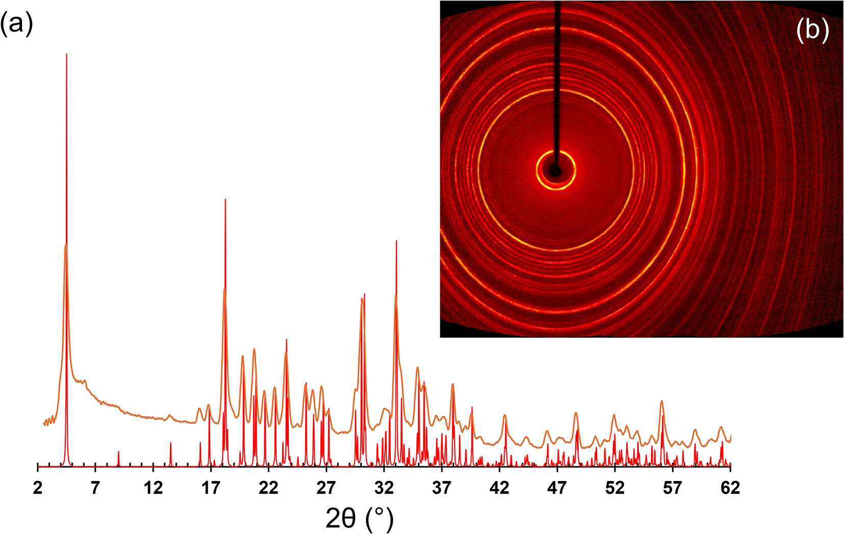
As discussed in the experimental section, the crystal structure of zanelliite was refined to a relatively high R 1 factor, with anions refined isotropically. Indeed, the diffraction quality of the available crystals was not ideal, owing to the nature of the studied material and the difficulty in isolating a single crystal suitable for intensity data collection (see Fig. 2). For this reason, the high residuals around Pb atoms (ca. 8 e Å–3) are due to the crystal quality, with the intensity data affected by the occurrence of other misorientated individuals within the studied grain. However, despite this limitation, the crystal structure of zanelliite is physically reasonable and is supported by the discussed bond distances and bond-valence sums. Moreover, the fit between the observed and calculated X-ray powder diffraction patterns is satisfactory, further supporting the reliability of the structural model (Fig. 8).
Powder X-ray diffraction pattern of zanelliite. (a) Comparison between observed (orange) and calculated (red) patterns; (b) the observed X-ray diffraction pattern.

A second finding of zanelliite
After the first identification of zanelliite at the Grosses Chalttal deposit, a second discovery of this Pb-Cu arsenate was made at the Hochmättli deposit, Mürtschenalp district, Glarus, Switzerland (47°03’12.25”N, 9°10’29.62”E), located 2.1 km SSW from the type locality. At the Hochmättli deposit, zanelliite was first analysed in 1994 using powder X-ray diffraction and energy-dispersive spectroscopy qualitative chemical analysis (Meisser, Reference Meisser1999). It is associated with bayldonite, plumboagardite, chrysocolla, baryte and quartz (Bächtiger, Reference Bächtiger1963; Meisser, Reference Meisser1999; Roth and Zanelli, Reference Roth and Zanelli2022; Roth et al., Reference Roth, Zanelli and Meisser2025). Material from this second finding is represented by an oxidized ore sample and a polished section and it was used for the collection of X-ray diffraction, Raman and chemical data. It is deposited in the Muséum cantonal des sciences naturelles (Naturéum), Départment de géologie, Université de Lausanne, Bâtiment Anthropole, Switzerland, with catalogue number MGL 087286. The average of ten spot analyses collected using the same electron microprobe used for the chemical characterization of the type material gave (in wt.%): MoO3 1.45, As2O5 28.88, CaO 0.75, CuO 47.21, PbO 11.31, H2Ocalc 9.36, total 98.96. On the basis of 28 O apfu, the chemical formula of this second finding of zanelliite corresponds to (Pb0.77Ca0.20)Σ0.97Cu9.05(As3.83Mo0.15)Σ3.98O15.15(OH)9.85(H2O)3. The only detectable difference between the Grosses Chalttal and Hochmättli zanelliite is related to the slightly higher Ca content of the latter, whereas no significant differences were found for the other elements.
Zanelliite and currently known Pb–Cu arsenates
Zanelliite is a new addition to the series of Pb–Cu arsenates which currently consists of six other mineral species (Table 7). Notably, all of them have been reported from the Grosses Chalttal deposit. Zanelliite has the highest Cu/(Cu+Pb) atomic ratio (0.90), close to that of end-member plumboagardite (0.86), and a (Cu+Pb)/As atomic ratio of 2.50, which is smaller than that of heimite and metaheimite (3.00) and larger than those of plumboagardite (2.33), bayldonite (2.00), duftite (2.00), and thometzekite (1.50). Moreover, like heimite, plumboagardite and thometzekite, zanelliite contains both (OH) and (H2O) groups.
Lead-Cu natural arsenates

Table 7 Long description
Seven lead copper arsenate minerals are listed with chemical composition, unit-cell parameters a, b, and c in angstroms, beta angle in degrees, space group, and literature reference. Zanelliite stands out with a very large a axis of 39.40 angstroms, while its b and c axes are 5.56 and 10.54 angstroms. Most other minerals have a axes between about 5.83 and 13.77 angstroms, with plumboagardite showing equal a and b values of 13.77 angstroms. Beta angles are reported for bayldonite at 106.0 degrees, thometzekite at 117.0 degrees, and zanelliite at 95.1 degrees; heimite and metaheimite are listed at 90.0 degrees. Space groups include C2/c for bayldonite and zanelliite, P21/n for heimite and metaheimite, C2/m for thometzekite, P212121 for duftite, and P63/m for plumboagardite.
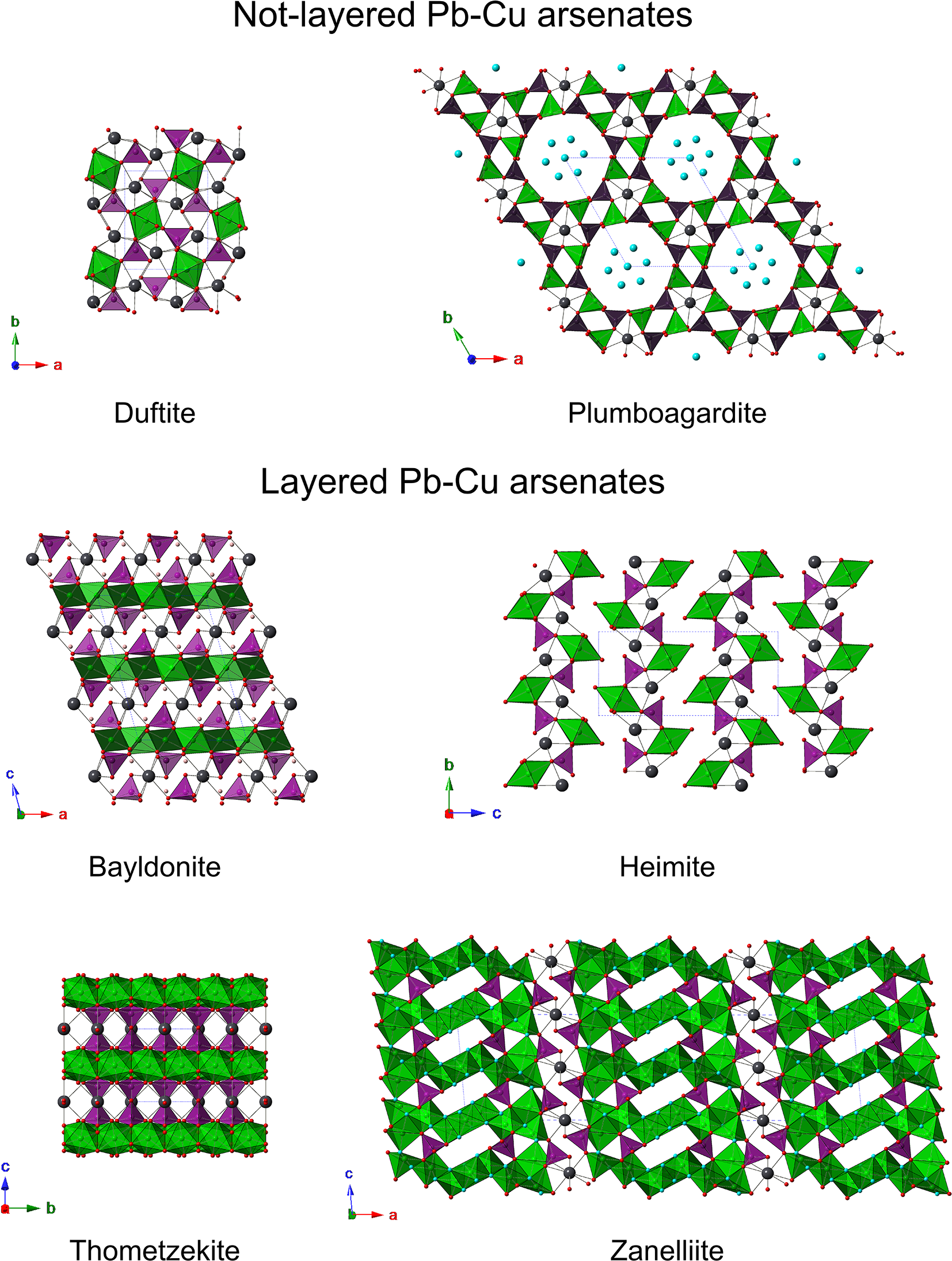
Lead-Cu arsenates display different kinds of crystal structures (Fig. 9) which can be divided into layered and non-layered types. The latter kind of structure is shown by duftite and plumboagardite. Duftite is a member of the adelite group within the descloizite supergroup. Its crystal structure is formed by columns of edge-sharing Cu octahedra running along c; every column is connected to four adjacent octahedral columns through corner-sharing with AsO4 groups (Kharisun et al., Reference Kharisun, Taylor, Bevan and Pring1998). Plumboagardite belongs to the mixite group and its crystal structure can be described as a honeycomb-like framework formed by chains of Cu-polyhedra linked through AsO4 groups. Two kinds of channels occur: the first one is delimited by a six-membered ring and hosts Pb atoms; the second one is a twelve-membered ring and hosts H2O groups (e.g. Hess, Reference Hess1983).
Crystal structures of currently known Pb-Cu arsenates. Cu-centred polyhedra are shown in green, As-centred tetrahedra are light violet. Dark grey and red circles represent Pb and O atoms, respectively.

Figure 9 Long description
Crystal structures of lead-copper arsenates are depicted, divided into non-layered and layered types. The non-layered section includes duftite and plumboagardite. Duftite is shown with a compact arrangement of polyhedra, while plumboagardite displays a honeycomb-like framework with large circular channels. The layered section features bayldonite, heimite, thometzekite and zanelliite. Bayldonite and heimite exhibit a structured arrangement of polyhedra forming layers. Thometzekite is characterized by rows of polyhedra with circular voids and zanelliite shows a repeating pattern of polyhedra forming distinct layers. Each structure is accompanied by axes labeled a, b and c, indicating orientation.
Bayldonite, heimite and thometzekite show a layered crystal structure. Bayldonite is characterized by the alternation of Cu sheets and Pb-As sheets, stacked along c (Ghose and Wang, Reference Ghose and Wang1979). Thometzekite is a member of the tsumcorite group, a series of monoclinic and triclinic species with general formula M(1)M(2)2(XO4)2Φ2. In the crystal structure of these species, M(2) octahedra form [010] chains through edge-sharing which are bonded by XO4 tetrahedra forming {001} layers. Successive layers are connected by M(1) cations exhibiting a [6+2] coordination (e.g. Krause et al., Reference Krause, Belendorff, Bernhardt, McCammon, Effenberger and Mikenda1998). In thometzekite, M(1) = Pb, M(2) = Cu, X = As, and Φ = H2O (Schmetzer et al., Reference Schmetzer, Nuber and Medenbach1985). In heimite, Malcherek et al. (Reference Malcherek, Mihailova, Schlüter, Roth and Meisser2024) described the occurrence of chains of edge-sharing Cu octahedra laterally connected to AsO4 and Pb atoms, giving rise to layers stacked along [001]. The crystal structure of metaheimite has not yet been published; however, it is hypothesized that it is analogous to the crystal structure of the more hydrated phase.
Zanelliite belongs to the second group, i.e., layered Pb-Cu arsenates. However, its crystal structure is novel, with no known analogues among currently described mineral species.
Conclusions
Zanelliite is a further addition to the small number of species belonging to the PbO–CuO–As2O5–H2O system. It displays a previously undescribed crystal structure, highlighting the necessity to investigate natural occurrences in order to discover novel and unexpected structural arrangements. This finding aligns with the conclusions of earlier studies that indicated the predictive capabilities of synthetic compounds is often overestimated. This is evidenced by the existence of several new mineral species that have never been synthesized or were not synthesized at the time of their discovery (e.g. Grew et al., Reference Grew, Hystad, Hazen, Krivovichev and Gorelova2017; Biagioni, Reference Biagioni2025). Malcherek et al. (Reference Malcherek, Mihailova, Schlüter, Roth and Meisser2024) hypothesized that heimite could be a metastable phase; a similar hypothesis may also apply to zanelliite. These species could have a very short geological life and may be a vestige of glacial–interglacial cycles that have affected the Alps, thereby underscoring the intricate interconnection between the evolution of lithosphere, atmosphere, hydrosphere and biosphere in the so-called Earth’s critical zone. The presence of euchroite in this mineral association at Grosses Challtal, with its proven metastability (Majzlan et al., Reference Majzlan, Števko, Dachs, Benisek, Plášil and Sejkora2017), and Mindel-Riss interglacial 238U–234U–230Th age for metazeunerite from Höchmattli (the second occurrence of zanelliite), further supports this hypothesis (Meisser, Reference Meisser1999; Meisser, Reference Meisser2012, Roth and Zanelli, Reference Roth and Zanelli2022; Roth et al., Reference Roth, Zanelli and Meisser2025).
Grosses Chalttal, as well as similar localities in the Mürtschenalp district, can thus be considered an important reference site for the study of the physical-chemical phenomena affecting the alteration of primary ores and the genesis and crystal chemistry of Pb-Cu arsenates. Further investigation of the mineralogy of these localities may reveal additional species and the examination of the textural relationship among different Pb-Cu arsenates and other supergene minerals may provide insights into the fate of As in the Earth’s critical zone.
Supplementary material
The supplementary material for this article can be found at https://doi.org/10.1180/mgm.2025.10164.
Acknowledgements
The Centro per l’Integrazione della Strumentazione scientifica dell’Università di Pisa (C.I.S.U.P.) is gratefully acknowledged for providing the access to the single-crystal X-ray diffraction laboratory. The characterization and preservation of zanelliite have benefited from funding by the Swiss Academy of Sciences (SCNAT) through the SwissCollNet project no. SCN201-VD to NM.
Competing interests
The authors declare none.