





Introduction
Medical education is presently acknowledged as one of the most challenging disciplines. The traditional medical educational experience has been improved through the incorporation of artificial intelligence and three-dimensional technologies such as augmented reality, virtual reality, and three-dimensional printing. Reference Tokuç and Varol1–Reference Mir, Mir and Raina4 Compared with augmented/virtual reality technologies, three-dimensional-printed medical educational models could offer better accessibility and user-friendliness for both trainers and trainees. Reference Urlings, de Jong, Maal and Henssen2
Since its introduction in the 1980s, three-dimensional printing has been gradually adopted in health care and medical education. Reference Schubert, van Langeveld and Donoso5 Three-dimensional-printed models are more cost-effective than are commercial models. Reference Asif, Lee, Caputo, Biglino and Shearn6–Reference Costello, Olivieri and Su9 Moreover, trainers can easily design, create, and customise these models according to the target students. Studies have revealed the superior efficacy of three-dimensional-printed models in medical education, particularly for enhancing learning skills and knowledge among medical students. Undergraduates also expressed high satisfaction with these technologies, and the outcomes consistently favoured the three-dimensional group. Reference Ardila, Gonzalez-Arroyave and Zuluaga-Gomez10 Many medical fields have embraced the use of simulation in education and training, using three-dimensional-printed anatomical models. These models have been applied in various training courses, including neurosurgery, otolaryngology, cardiac surgery, and adult cardiology. Reference Anwar, Singh and Miller11–Reference Ziv, Wolpe, Small and Glick15
Cardiac catheterisation is one of the most frequently performed cardiac procedures. Reference Mozaffarian, Benjamin and Go16 Despite improvements in equipment and techniques, beginners find it challenging to learn this procedure. Reference Manda and Baradhi17–Reference Bagai, O’Brien and Al Lawati19 Medical education in cardiac catheterisation traditionally follows the apprenticeship model. In this model, trainees observe procedures, gradually assume responsibilities, and practise under supervision until they can perform interventions independently. Reference Hopfner, Jakob and Tengler20 The apprenticeship model has enabled the effective transfer of skills and knowledge from one generation to the next; however, several authors argue that the apprenticeship model is no longer acceptable to the medical profession or the well-informed public. Simulator training has been demonstrated to be effective in medicine, and the demand for three-dimensional-printed models in medical education continues to grow. Reference Ziv, Wolpe, Small and Glick15,Reference Lau and Sun21
The technology of three-dimensional printing has been widely integrated into medical education, especially in simulation-based clinical training. Reference Lichtenberger, Tatum and Gada22,Reference Gallagher, Smith and Sheppard23 The primary advantage of three-dimensional printing is its cost-effective reproduction of detailed anatomical structures, offering students tangible models that bridge the gap between theoretical knowledge and clinical practice. Reference Ye, Dun and Jiang24
This study developed a cost-effective three-dimensional-printed heart model for medical education. The objectives of this study were to demonstrate the effectiveness of this model in teaching basic cardiac catheterisation and to improve the learning experience for medical students.
Methods
Study design
This open-label, single-site, randomised controlled trial was conducted at Fu Jen Catholic University in Northern Taiwan from October 2023 to April 2024. We conducted the trial in accordance with good clinical practice guidelines and the Declaration of Helsinki. The study was approved by the Institutional Review Board of Fu Jen Catholic University Hospital, New Taipei City, Taiwan (FJUH110157) and was registered on ClinicalTrials.gov (NCT05760365, 27/2/2023). The study results were reported in accordance with the CONSORT statement. Reference Schulz, Altman and Moher25
Production of three-dimensional-printed heart model
We reasoned that a life-size human heart model might not best meet the educational requirements of medical students and junior doctors. Consequently, we developed a three-dimensional-printed heart model that was twice the size of a life-size model and that enhanced educational outcomes by improving teaching and demonstration methods. This larger model featured a hollow design, facilitating the practice of catheter insertion for both trainers and trainees. Additionally, we ensured that the model was watertight, allowing for realistic simulation of a blood-filled cardiac vessel, thereby augmenting the learning experience (Figure 1).
3D-printed heart model. ( a ) Three-dimensional model displayed using the 3ds max image processing software (Autodesk); ( b ) 3D-printed heart model; ( c ) ready for simulant blood injection with syringe; and ( d ) filled with simulant blood. 3D: three-dimensional.

A three-dimensional heart model was specifically created by our three-dimensional medical design team for this project under the guidance of CYL. The three-dimensional heart model was designed using Autodesk 3ds Max 2017 image processing software (Autodesk, San Rafael, CA, USA) and printed using a stereolithography three-dimensional printer (Jupiter, Elegoo, Shenzhen, China). The model was constructed using a transparent, durable, water-resistant resin. The printer deposited 0.05-mm-thick layers of photopolymer for both the model and the support material, which were immediately cured in ultraviolet light. The support material was removed using manual cutting. The entire three-dimensional printing process consumed approximately 72 hours, with an additional hour devoted to postprocessing tasks.
Participants
This study enrolled undergraduate medical students and postgraduate junior doctors (including postgraduate year 1, year 2, and resident doctors) from Fu Jen Catholic University. The participants were aged between 20 and 50 years. Individuals who refused to sign the informed consent form were excluded.
Randomisation
Individuals who met the inclusion criteria underwent block randomisation. All participants were randomly assigned to either the three-dimensional model group or the control group. Random assignment was performed through block randomisation using the website http://www.randomization.com. Written informed consent was obtained from all participants before including them in the study.
Medical education
All participants underwent a 20-minute, slide-based learning session. The three-dimensional model group received this slide-based presentation in conjunction with a 20-minute hands-on practice session using a three-dimensional heart model (Figure 2). This practical session included an initial demonstration of cardiac catheterisation by the trainer, followed by the trainees practicing the procedure themselves under the guidance and feedback of the trainer. The control group received only the slide-based presentation without any hands-on component.
Photograph of the 3D-printed heart model with a cardiac catheter.

Student evaluation
A written examination consisting of 5 questions was administered to the participants before and after the experiment to evaluate their theoretical knowledge of the anatomy of the coronary artery and percutaneous coronary intervention (Supplementary Table S1). Additionally, the participants evaluated their attitude towards the training course through a questionnaire consisting of 5 questions that were answered using a 5-point Likert scale; higher scores indicated higher levels of agreement (Supplementary Table S2).
Statistical analysis
Continuous data are presented as means ± standard deviations or medians with interquartile ranges depending on the distribution of the data (Shapiro–Wilk test). Categorical variables were compared using Fisher’s exact test. Because the sample size was small, Wilcoxon’s signed-rank test was performed to analyse continuous variables. Statistical analyses were performed using SPSS (version 25.0 for Windows; IBM, Chicago, IL, USA). Statistical significance was indicated by P < 0.05.
Results
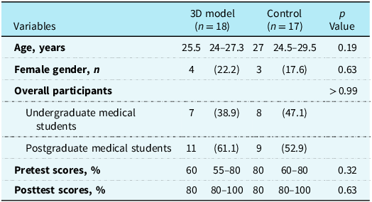
Between October 2023 and April 2024, 39 individuals were screened for participation in this study, of whom 4 declined to participate (Fig. S1). The remaining 35 individuals were randomly assigned to either the three-dimensional model group (n = 18) or the control group (n = 17). The demographic characteristics of the 2 groups were similar (P > 0.05 for all; Table 1). The participants’ median age was 25.5 years (interquartile range: 24 to 27.3 years) in the three-dimensional model group and 27 years (interquartile range: 24.5 to 29.5 years) in the control group. In the three-dimensional model group, 7 participants (38.9%) were undergraduate medical students, and 11 (61.1%) were postgraduate medical students. In the control group, 8 participants (47.1%) were undergraduate medical students, and 9 (52.9%) were postgraduate medical students.
Participant characteristics and written examination scores

Table 1. Long description
A table comparing participant characteristics and examination scores between two groups. The table has 10 rows and 4 columns. The columns are labeled Variables, 3D model (n = 18), Control (n = 17), and p Value. The rows include Age, Female gender, Overall participants, Undergraduate medical students, Postgraduate medical students, Pretest scores, and Posttest scores. Each row provides specific data for the 3D model and Control groups, along with p values indicating statistical significance. For example, the median age is 25.5 years for the 3D model group and 27 years for the Control group. The table also shows the distribution of undergraduate and postgraduate medical students in each group, as well as pretest and posttest scores in percentages.
Data are presented as medians, interquartile ranges, or numbers (%).
3D = three-dimensional.
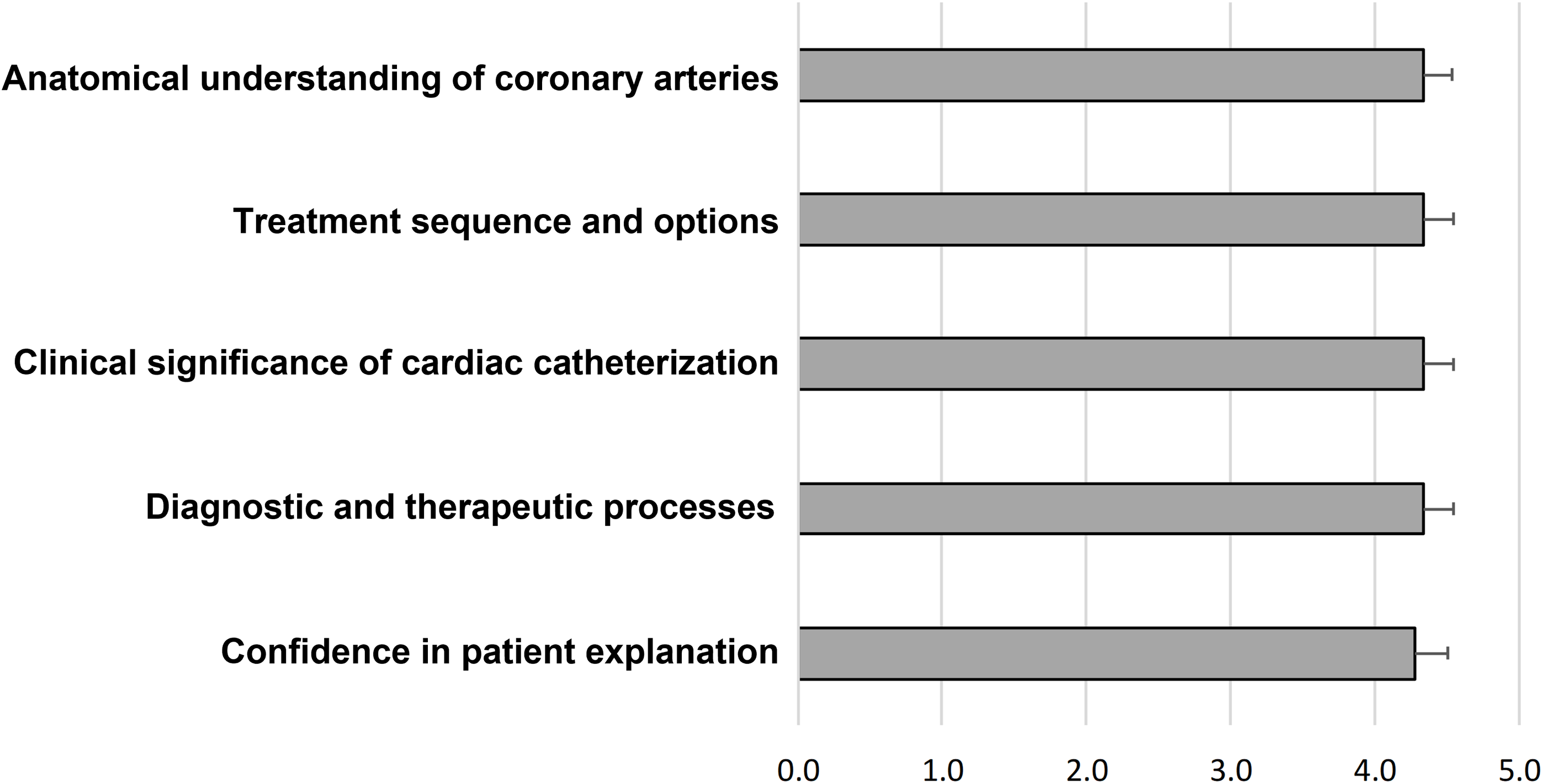
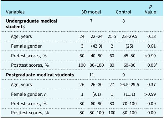
The pre-examination and postexamination scores were similar. A subgroup analysis was performed to assess the pre-examination and postexamination scores by level of education (undergraduate versus postgraduate). The trends in the pre-examination and postexamination scores of the 20 postgraduate medical students were consistent with the trend in the scores of the whole group; however, among the 15 undergraduate medical students, the postexamination scores were significantly higher in the three-dimensional model group than in the control group (100% [interquartile range: 80% to 100%] versus 80% [interquartile range: 60% to 80%], P = 0.03; Table 2). All participants in the three-dimensional model group responded to the attitude questionnaire and generally provided positive feedback (Figure 3). This study successfully fabricated an economical three-dimensional-printed heart model costing US$200 per model.
Results of the attitude questionnaire assessed using a 5-point Likert scale (1 = strongly disagree, 5 = strongly agree). Bars indicate mean scores, and error bars represent the standard error of the mean.

Participant characteristics and written examination scores for undergraduate and postgraduate medical students

Table 2. Long description
A table comparing characteristics and examination scores of undergraduate and postgraduate medical students. The table has 12 rows and 4 columns. The columns are labeled Variables, 3D model, Control, and P Value. The rows are grouped into undergraduate medical students and postgraduate medical students. For undergraduate medical students, the variables include the number of students, age, female gender, pretest scores, and posttest scores. For postgraduate medical students, the variables include the number of students, age, female gender, pretest scores, and posttest scores. The table provides specific values for each variable under the 3D model and Control columns, along with corresponding P Values.
Data are presented as medians, interquartile ranges, or numbers (%).
* P < 0.05.
3D = three-dimensional.
Discussion
This study enrolled 35 participants, none of whom dropped out. The results suggested the potential educational value and cost-efficiency of three-dimensional-printed heart models as teaching aids, with the educational benefit appearing more evident among undergraduate medical students. Given the small sample size and single-centre design, this study should be considered exploratory in nature. These findings suggest that three-dimensional-printed models have the potential to substantially enhance traditional slide-based learning in undergraduate medical education. In contrast, the educational impact among postgraduate year 1 and year 2 residents was less pronounced, and the applicability of these findings to postgraduate training therefore remains uncertain. These results differ from those of Wang et al., Reference Wang, Liu and Luo26 who reported that three-dimensional-printed cardiac models were not superior to traditional teaching models for undergraduate medical education on cardiac diseases. However, the three-dimensional-printed model in Wang et al. was simple and lacked interactivity, being designed solely for anatomical education rather than for demonstrating procedures such as cardiac catheterisation.
Historically, cardiology training for undergraduate medical students and junior doctors has primarily relied on traditional methods, such as theoretical lectures, and has not incorporated practical components. Reference McMahon, Tretter and Redington27 The conventional pedagogical strategy for complex medical procedures, such as cardiac catheterisation, has predominantly followed the apprenticeship model, which emphasises observation and hands-on practice, adhering to the traditional “see one, do one, teach one” doctrine. Reference Hopfner, Jakob and Tengler20 Training in this framework occurs directly within the catheterisation laboratory, which might not provide the most conducive learning environment, given that training opportunities often occur spontaneously. This can potentially increase stress among trainees and adversely affect the learning process. Despite its historical success in transferring practical knowledge, the apprenticeship model faces significant issues, including extended procedure times, increased costs, reduced operational efficiency, and heightened stress among trainees, which can prevent students from effectively achieving educational outcomes. Reference Babineau, Becker and Gibbons28,Reference Wetzel, Kneebone and Woloshynowych29
The process of learning cardiac procedures can vary in complexity, ranging from simple to highly intricate. Therefore, the proportional adjustment of costs for introductory training models is essential. Simulation-based clinical training is particularly effective for teaching technical skills. High-fidelity mannequin simulators are excellent tools used in the demonstration of procedural skills, including cardiac catheterisation. These simulators can replicate physical findings such as oxygen saturation, respond physiologically to medications, and undergo various procedures. However, these advanced mannequins can cost more than US$250,000. Reference Srinivasan, Hwang, West and Yellowlees30
Nowadays, three-dimensional printing technology has become a common component of clinical and medical education in cardiology. Reference Lindquist, Gosnell and Khan31,Reference Chetan, Valverde and Yoo32 Medical education is a lifetime learning process, spanning from undergraduate to postgraduate to specialty training and beyond.
Using three-dimensional cardiac models in cardiology education enhances trainees’ understanding of complex anatomy, provides high-fidelity simulation experiences, and increases exposure to rare pathologies, thereby reducing the steepness of the learning curve. Additionally, three-dimensional printing improves engagement, accelerates comprehension of anatomical defects, and exposes students to a wide range of malformations, even within the same condition. Reference Anwar, Singh and Miller11,Reference Valverde33 Studies on the teaching of CHD have demonstrated that compared with traditional imaging methods, three-dimensional-printed models significantly enhanced the anatomical knowledge of medical students, paediatric cardiology fellows, and consultants. Reference Costello, Olivieri and Su9,Reference Valverde, Gomez and Byrne34 Participants reported that three-dimensional models improved their understanding of CHD, increased their interest in anatomy, enhanced their grasp of imaging techniques, and were beneficial for patient communication. The participants advocated for the inclusion of three-dimensional models in medical workshops and for their use as substitutes for cadaveric specimens in teaching. Reference Valverde, Gomez and Byrne34
The feasibility of using three-dimensional-printed heart models to plan endovascular stenting for transverse arch hypoplasia has been demonstrated. Reference Valverde, Gomez and Coserria35 Participants found these models advantageous for planning interventions, enhancing communication, and reducing catheterisation complications. Additionally, research involving individuals with varying levels of cardiac catheterisation experience indicated that practice with three-dimensional models led to decreased fluoroscopy times. Most participants reported that the three-dimensional models were effective for learning the process of catheter intervention, familiarising themselves with catheterisation devices, and improving patient safety through increased practice opportunities. Reference Brunner, Thierij and Jakob36 Although data supporting training in interventional cardiology are limited, the Society for Cardiovascular Angiography and Intervention’s Simulation Committee anticipates that simulation will become a more crucial component of training and certification maintenance over time. Reference Green, Klein and Pancholy37
Three-dimensional models have been employed in the training of students in medical, nursing, respiratory therapy, and resident programmes. Reference Biglino, Capelli and Koniordou38–Reference Yang, Chen, Liu, Liu and Chao41 Three-dimensional models have been demonstrated to improve spatial awareness and the ability to identify anatomical structures. Despite these positive findings, several studies have reported no significant long-term benefits of three-dimensional models. Reference McKinley, Stuart, Ailawadi and Brunswick42–Reference Ochoa, Segal, Garcia and Fischer44 Considering the different stages of learning and the varying levels of difficulty associated with various techniques, we must effectively integrate three-dimensional-printed models into medical education curricula.
This study has several limitations. First, the sample size was small, and the participants were recruited from a single institution, restricting the generalizability of the findings. In addition, a formal power calculation was not performed, which may further limit the statistical robustness of the results. Second, immediate testing may not be the most practical method for evaluating the learning process. In the short term, focusing on enhancing students’ motivation and appreciation is more advantageous. However, this approach does not allow assessment of long-term knowledge retention or objectively measured procedural skills, including catheterisation proficiency. Overall test performance or pass rates would be more appropriate metrics for long-term evaluation. Additionally, enrolling all medical students without a calculated sample size may introduce bias.
Further evaluations are necessary to determine the effectiveness of three-dimensional-printed models in advanced educational settings. Larger multicentre studies with follow-up assessments and objective procedural-skill assessments are warranted to evaluate knowledge retention and skill transfer, particularly in postgraduate medical education, to provide a more comprehensive understanding of the educational benefits and limitations of these models. Although this study lacks sufficient statistical power, its primary objective was to encourage faculty and school administrators to adopt these tools to enhance students’ educational experiences.
Conclusions
In conclusion, this study demonstrated the cost-effectiveness and feasibility of using three-dimensional-printed models as learning tools in medical education on cardiac catheterisation. To achieve effective teaching, the integration of three-dimensional printing models in medical education curricula should be adjusted in accordance with students’ learning stages and the difficulty of the techniques being taught.
Supplementary material
The supplementary material for this article can be found at https://doi.org/10.1017/S1047951126124846
Data availability statement
The datasets used and analysed in this study are available from the corresponding author upon reasonable request.
Acknowledgements
This manuscript was edited by Wallace Academic Editing.
Author contributions
KYC, LYH, and CYL conceived and designed the study. KYC, CYC, and CYL supervised and assisted with the postprocessing of three-dimensional-printed models. LYH, KLH, and CYL conducted experiments and collected data. LYH and CYC contributed to data analysis. KYC and CYL wrote the paper. All authors revised the manuscript for important intellectual content and approved the final version.
Financial support
This study was funded by Fu Jen Catholic University Hospital (PL-202108032-V). The funder was not involved in the study design, data collection, analysis, interpretation, report writing, or the decision to submit the article for publication.
Competing interests
The authors declare no competing interests.
Ethical standard
This study was reviewed and approved by the Institutional Review Board of Fu Jen Catholic University Hospital (FJUH110157). Written informed consent was obtained from all participants before their inclusion in the study.
Consent for publication
Not applicable.
Trial registration
This study was registered with ClinicalTrials.gov (Registration ID NCT05760365, registered on 27/2/2023).




 Open access

