Introduction
Primary irradiation is frequently used in head and neck cancer patients. Post-treatment dysphagia and aspiration are frequently seen, and are directly related both to disruption of the neuromuscular swallowing mechanism and to stricture formation of the pharynx or oesophagus. Although the patient's nutritional status can easily be restored by tube feeding, swallowing problems generally have a considerable impact on quality of life, and may lead to social isolation.Reference de Boer, Pruyn, van den Borne, Knegt, Ryckman and Verwoerd1
Strictures are reported in 3.4 per cent of patients receiving primary irradiation,Reference Laurell, Kraepelien, Mavroidis, Lind, Fernberg and Beckman2 and in 18–26 per cent after chemoradiation.Reference Lawson, Otto, Grist and Johnstone3, Reference Guadagnolo, Haddad, Posner, Weeks, Wirth and Norris4 They are usually found in the hypopharynx or cervical oesophagus, but can also occur at more cephalid, pharyngeal levels. Depending on the stricture site and severity, the patient may suffer dyspnoea, dysphagia or life-threatening aspiration.
Endoscopic bougienage or balloon dilatation is usually a successful treatment strategy (repeated if necessary).Reference Piotet, Escher and Monnier5
We report a patient in whom stricture formation had advanced to complete stenosis of the upper oesophageal sphincter; thus, other treatment options were required.
Case report
A 75-year-old man was treated elsewhere with primary irradiation for a supraglottic laryngeal carcinoma, staged as tumour 2 node 0 metastasis 0. During radiotherapy, he encountered progressive difficulty swallowing solid food, for which a percutaneous endoscopic gastrostomy was performed. He subsequently became almost completely dependent upon his gastrostomy for nutritional intake, but initially retained the ability to swallow thin liquids. However, over several months he gradually developed complete obstruction, even for liquids. This was confirmed by rigid oesophagoscopy, which found complete stenosis of the cervical oesophagus at the level of the upper oesophageal sphincter. The patient had to spit out his saliva to prevent aspiration.
It was suggested that the patient undergo resection of the stenosed segment with reconstruction by means of a gastric pull-up procedure. However, the patient requested a second opinion, and was referred to our dysphagia clinic.
At the time of presentation to our clinic, it had been almost one year since the patient had completed radiotherapy. No efforts had been made to dilate his oesophageal stenosis, because the risk of creation of a false route was considered too high.
The patient's medical history included severe gastroesophageal reflux with recurrent gastric bleeding from ulcers, prior to diagnosis of his laryngeal carcinoma. He had still experienced occasional reflux episodes prior to and during radiotherapy, despite taking proton pump inhibitors twice daily.
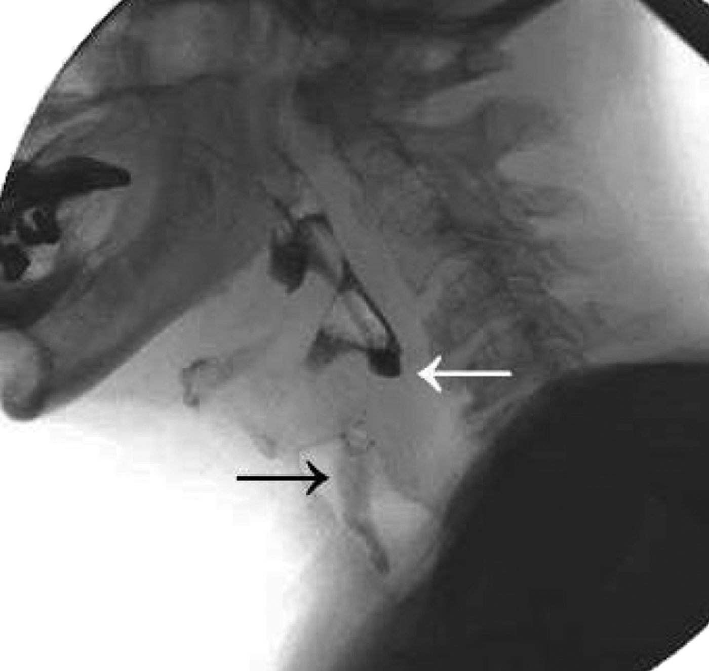
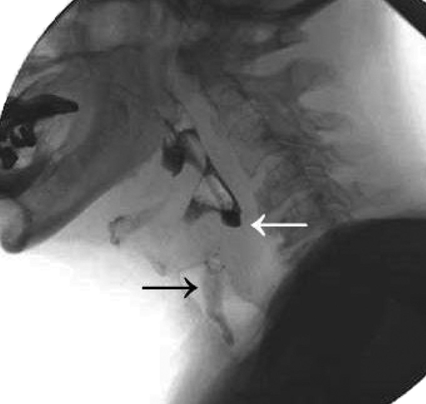
On flexible videolaryngoscopy in our dysphagia clinic, stasis of saliva was seen in both pyriform sinuses. Both vocal folds were mobile. There were no signs of locoregional recurrence of malignancy. Videofluoroscopy showed a total blockage of passage, with severe aspiration. Laryngeal elevation was late and impaired (Figure 1).
Pre-operative lateral videofluoroscopy demonstrating stenosis in the post-cricoid area (white arrow) and severe aspiration (black arrow).
Rigid oesophagoscopy demonstrated multiple adhesions in the hypopharynx and complete stenosis in the post-cricoid area. The stenosis was considered to be induced by a combination of radiotherapy and gastroesophageal reflux.
Blind dissection of the upper oesophageal sphincter stenosis from the proximal side (by laser or other means) was considered too dangerous, because of the risk of creating a false route into the mediastinum. In addition, it proved impossible to introduce a flexible endoscope from the stomach into the oesophagus; the endoscope just curled up in the stomach.
Therefore, a rigid bronchoscope was introduced through the gastrostomy opening. Using a 60° telescope in a retrograde fashion, the oesophageal lumen was identified. The bronchoscope was introduced through the lower oesophageal sphincter into the distal oesophagus, and a Savary dilatation wire (Wilson Cook Medical, Winston Salem, North Carolina, USA) was introduced through the bronchoscope until the stenosis was reached (Figure 2). Simultaneous endoscopy from the proximal end allowed palpation of the tip of the dilatation wire, which could be seen pushing against the stenosis from below. Using CO2 laser, a small perforation was created from the proximal side, through which the dilatation wire could be passed from below. A new dilatation wire was then attached to the first dilatation wire and passed in an anterograde fashion through the stenosis. Dilatation with Savary bougies could now be performed, in the normal anterograde fashion, gradually achieving a lumen of 15 mm (Figure 3) diameter. Following this modified anterograde–retrograde rendezvous procedure,Reference van Twisk, Brummer and Manni6 a nasogastric feeding tube was introduced.
Introduction of the bronchoscope through the percutaneous endoscopic gastrostomy opening, with subsequent introduction of a Savary dilatation wire (arrow). The flexible tube attached to the bronchoscope had been used for insufflation purposes.

The tip of the Savary dilatator over-wire (arrow) exiting the percutaneous endoscopic gastrostomy opening, during anterograde dilatation.

Post-operative antibiotics were administered and recovery was unremarkable.
One month post-operatively, endoscopy of the hypopharynx and oesophagus was repeated. Using a small, rigid oesophagoscope, it was possible to pass through the stenosis from above. Dilatation was performed again to 15 mm, and the nasogastric feeding tube was replaced. Again, recovery was unremarkable. After one day, oral intake with water was commenced.
Two months later, this procedure was repeated once more. At the end of this procedure, only a small thread was left behind, to facilitate the next dilatation; no nasogastric feeding tube was placed.
At this stage, thick liquid oral intake was possible without significant aspiration. Subsequently, the patient was able to return to a normal diet provided he chewed carefully and took liquids during his meal (Figure 4).
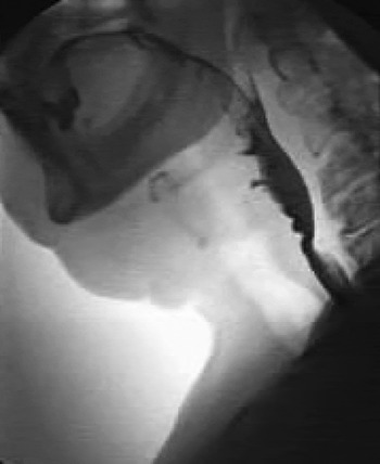
Post-operative lateral videofluoroscopy demonstrating bolus passage into the oesophagus, with moderate residual stenosis and absence of aspiration.

After more than six months, safe and secure swallowing activity was still present. It was therefore decided to remove the patient's percutaneous endoscopic gastrostomy, which had not been used since the second dilatation procedure.
Thereafter, dilatation of the upper oesophageal sphincter was performed every six months in our out-patient clinic.
At five years' follow up, no dysphagic complaints were reported, and the patient had a normal oral intake of all food consistencies.
Discussion
The cervical oesophagus and hypopharynx are known to be fragile and radio-sensitive, and late radiation injury is a well known sequel of head, neck and oesophageal cancer treatment. Development of strictures is related to mucositis, progressive obliterative endarteritis, ischaemia and slowly progressive fibrosis.Reference Nguyen, Sallah, Karlsson and Antoine7, Reference Silvain, Barrioz, Besson, Babin, Fontanel, Daban, Matuchansky and Beauchant8 Risk factors for stricture formation include high dose radiotherapy (>60Gy),Reference Laurell, Kraepelien, Mavroidis, Lind, Fernberg and Beckman2 twice-daily fractioning,Reference Lee, Akst, Adelstein, Saxton, Wood and Strome9 concomitant chemotherapy,Reference Laurell, Kraepelien, Mavroidis, Lind, Fernberg and Beckman2 limited oral intake during treatmentReference Lawson, Otto, Grist and Johnstone3 and poor nutritional status.Reference Laurell, Kraepelien, Mavroidis, Lind, Fernberg and Beckman2 Some authors recommend that patients experiencing treatment-induced dysphagia should be managed with a nasogastric feeding tube rather than a percutaneous gastrostomy, because the former also has a stenting function and a consequently lower frequency of re-dilatation.Reference Mekhail, Adelstein, Rybicki, Larto, Saxton and Lavertu10
Following radiotherapy, differing times of stricture onset have been reported. Eisbruch et al. Reference Eisbruch, Lyden, Bradford, Dawson, Haxer and Miller11 found that most strictures developed one to three months after chemoradiation for head and neck cancer, and remained stable thereafter. Other authors have reported later onset of strictures, with median values varying from six to 26 months post-radiotherapy.Reference Laurell, Kraepelien, Mavroidis, Lind, Fernberg and Beckman2, Reference Silvain, Barrioz, Besson, Babin, Fontanel, Daban, Matuchansky and Beauchant8
Strictures are often not detected early on, mainly due to the fact that other toxicity side effects (e.g. xerostomia, odynophagia and suppressed cough reflex) can mask the patient's reduced bolus passage capacity. A stricture typically presents with increasing effort required to swallow a food bolus, together with weight loss, and may be associated with increased risk of (silent) aspiration. Only those patients with symptomatic dysphagia are formally investigated for stricture evaluation; therefore, the overall incidence of strictures may be underestimated.
Stricture severity may vary from a fibrous ring at the oesophageal inlet to deep fibrosis with subtotal or total obliteration of the lumen (the latter frequently observed following hypopharyngeal cancer treatment).Reference Lee, Akst, Adelstein, Saxton, Wood and Strome9
Post-radiation hypopharyngeal and oesophageal strictures are commonly managed with bougie dilatation, provided there is still some degree of lumen present. Laurell and colleaguesReference Laurell, Kraepelien, Mavroidis, Lind, Fernberg and Beckman2 have reported a 78 per cent success rate with anterograde dilatation of hypopharyngeal strictures occurring secondary to radiotherapy for head and neck malignancies. Patients with moderate to severe strictures required one to eight dilatations. One patient with a complete stricture of the oesophageal inlet died as a consequence of oesophageal perforation with mediastinitis.
• Strictures of the hypopharynx and oesophagus are recognised sequelae of chemoradiation
• Anterograde dilatation of a complete stenosis carries a high risk of perforation
• In cases of complete obstruction of the oesophageal inlet, an anterograde–retrograde dilatation approach is advisable
• The main advantage of this technique is that the stenosis can be punctured with a guide-wire moving away from the mediastinum, avoiding creation of a false passage in this direction and thereby reducing the risk of mediastinitis
• Stenosis puncture can be monitored endoscopically from above, further reducing the risk of perforation
The anterograde–retrograde rendezvous technique was first described by Van Twisk et al. Reference van Twisk, Brummer and Manni6 in 1998. Several other small series were later reported.Reference Petro, Wein and Minocha12–Reference Oxford and Ducic14 The advantage of this technique is that a stenosis can be punctured with a dilatation guide-wire moving away from the mediastinum, avoiding the creation of a false route in this direction and thus reducing the risk of mediastinitis. In addition, stenosis puncture can be monitored endoscopically from above, further reducing the risk of perforation. Transillumination is sometimes used from both sides to determine the direction of puncture. If the stenosis extends over a longer distance, anterograde dissection with a blunt instrument or CO2 laser can be performed, progressing towards an illuminated post-stenotic lumen, or, as in our case, towards a palpable probe. Most authors have described the use of flexible endoscopes in the anterograde–retrograde rendezvous technique; however, in our patient it proved impossible to enter the oesophagus from below, because the flexible endoscope curled up in the stomach without advancing into the oesophagus. Therefore, a rigid bronchoscope proved very useful as a means of entering the oesophagus from below.
The majority of patients treated with the anterograde–retrograde rendezvous technique respond well to subsequent serial dilatations, and most are able to discontinue gastrostomy tube usage.Reference van Twisk, Brummer and Manni6, Reference Petro, Wein and Minocha12–Reference Oxford and Ducic14
In the presented case, it seemed likely that gastroesophageal reflux had contributed to the patient's oesophagitis and consequently to the development of severe stenosis. However, we were unable to find any published evidence to substantiate a relationship between gastroesophageal reflux and post-irradiation stenosis in the upper oesophageal sphincter.
Conclusion
In patients with complete stenosis, the anterograde–retrograde rendezvous dilatation technique offers an alternative to high risk blind dilatation of the upper oesophagus and hypopharynx. The risk of perforation during the anterograde–retrograde technique is reduced by adequate endoscopic visualisation and by performing dilatation away from rather than towards the mediastinum.