I. INTRODUCTION
Dapoxetine ((S)-N,N-Dimethyl-3-(naphthalen-1-yloxy)-1-phenylpropan-1-amine, C21H23NO, DAP, see Figure 1) is a potent selective serotonin reuptake inhibitor (SSRI) used for the treatment of premature ejaculation (PE), the most prevalent sexual dysfunction in men (Russo et al., Reference Russo, Capogrosso, Ventimiglia, La Croce, Boeri, Montorsi and Salonia2016). Its hydrochloride form (C21H24ClNO, DAPHCl), marketed under the name Priligy™, has been approved for the treatment of PE in over 50 countries worldwide (Russo et al., Reference Russo, Capogrosso, Ventimiglia, La Croce, Boeri, Montorsi and Salonia2016). It is the only pharmacological agent specially developed for the treatment of men with PE (Hoy and Scott, Reference Hoy and Scott2010). All other medical treatments used are based on antidepressant SSRIs (such as fluoxetine, paroxetine, and sertraline) with off-label indications. The presence of the naphthyl moiety in dapoxetine seems to be the reason for its efficiency and its improved pharmacokinetic characteristics. Its rapid absorption and elimination in the body, which results in minimal accumulation, and its dose-proportional pharmacokinetics render dapoxetine unaffected by multiple dosing. This pharmacokinetic profile makes it effective for the “on-demand” oral treatment of PE (McCarty and Dinsmore, Reference McCarty and Dinsmore2012).
Molecular diagram of Dapoxetine hydrochloride (DAPHCl).

To our knowledge, three studies associated with the characterization of dapoxetine have been published in the open literature (Attia et al., Reference Attia, Souaya and Soliman2015; Darcsi, Reference Darcsi2017; Selvakumar, Reference Selvakumar2018). The first report deals with the thermal characterization of dapoxetine and vardenafil hydrochlorides using conventional thermal analysis techniques (TGA, DTA, and DSC) complemented with semi-empirical molecular orbital calculations. In this study, the chemical reactions of DAPHCl in every step of its thermal decomposition and their thermodynamic parameters (ΔH, ΔS, and ΔG) are reported (Attia et al., Reference Attia, Souaya and Soliman2015). The other two references are a Doctoral dissertation and a Master's degree thesis. In the Doctoral dissertation (Darcsi, Reference Darcsi2017), the synthesis of dapoxetine and cyclodextrin complexes, phase solubility studies, and their characterization using mass spectrometry, NMR, circular dichroism, UV spectroscopy, and capillary electrophoresis are presented. In his Master's degree thesis, Selvakumar (Reference Selvakumar2018) prepared nanoparticles of DAPHCl with the purpose of enhancing its solubility and bioavailability. The resulting materials were subjected to stability and solubility studies and were characterized by using UV spectroscopy, FT-IR spectroscopy, scanning electron microscopy (SEM), and X-ray powder diffraction (XRPD). It must be noted that although the powder pattern recorded shows relatively well-defined peaks, no structural information was extracted from it.
A search in the Cambridge Structural Database (CSD) version 5.42 (Groom et al., Reference Groom, Bruno, Lightfoot and Ward2016) did not lead to any report of the crystal structure of dapoxetine or of any closely related phases. Also, a search in the ICDD PDF-4/Organics database (Gates-Rector and Blanton, Reference Gates-Rector and Blanton2019) produced no results.
After searching for “dapoxetine” in the Google Patents site (https://patents.google.com), many entries are encountered. Some of them refer to the preparation of various crystal forms of anhydrous and hydrated DAPHCl phases. In two patents CN103130661B (Ren et al., Reference Ren, Ren, Qi, Le, Hong, Cao and Chen2011) and WO2013075669A1 (Ren et al., Reference Ren, Ren, Chen, Qi, Yue, Hong and Cao2013), information about the unit-cell parameters of a DAPHCl phase is provided. In both patents, the reported phase crystallizes in an orthorhombic unit cell with a = 6.314(2) Å, b = 10.658(2) Å, c = 28.150(6) Å, V = 1894.3(7) Å3, and Z = 4 as determined by a single crystal X-ray diffraction study. In these patents, a powder diffracti`on pattern and a plot of the molecular structure for the reported phase called Form B are shown. However, the atomic positions are not reported. These authors also report a powder pattern of a phase called Form A.
Within a project of the Grant-in-Aid program supported by the International Centre for Diffraction Data (ICDD) intended to register high-quality X-ray powder diffraction data of pharmaceutical materials of interest with none or limited structural information in the literature, several compounds of pharmaceutical and general chemical interest have been studied in our laboratory (Dugarte-Dugarte et al., Reference Dugarte-Dugarte, van de Streek, Díaz de Delgado, Rafalska-Lasocha and Delgado2021; Toro et al., Reference Toro, Dugarte-Dugarte, van de Streek, Henao, Delgado and Díaz de Delgado2022). Since there is no structural information on Dapoxetine hydrochloride, it was decided to record its powder pattern and undertake the structure determination of this compound from powder diffraction data. The structural model obtained was further validated by dispersion-corrected DFT calculations. The material under study was also characterized by FT-IR.
II. EXPERIMENTAL METHODS
A small portion of the sample, provided by Tecnoquímicas (Cali, Colombia), was ground and mounted on a zero-background holder. The X-ray powder diffraction data were registered at room temperature with a BRUKER D8 ADVANCE diffractometer working in the Bragg-Brentano geometry. This instrument is equipped with a CuKα source, working at 40 kV and 30 mA, and a LynxEye detector. The pattern was recorded from 5.00 to 60.00° in steps of 0.01526° (2θ) at 1 s/step. The standard instrument settings (Ni filter of 0.02 mm, a primary and secondary Soller slits of 2.5°, divergence slit of 0.2 mm, and scatter screen height of 3 mm) were used. ATR-FTIR spectra were recorded in a Bruker Alpha spectrophotometer with an ATR eco ZnSe accessory, from 4000 to 500 cm−1, in 24 scans collected in 30 s, with a resolution of 4 cm−1.
III. COMPUTATIONAL STUDIES
A. DFT-D calculations
The crystal structure obtained from XRPD was energy-minimized with the program GRACE (Neumann et al., Reference Neumann2002), which calls VASP (Kresse and Furthmüller, Reference Kresse and Furthmüller1996) for single-point DFT calculations with the PBE functional (Perdew et al., Reference Perdew, Burke and Ernzerhof1996) to which a dispersion correction (Neumann and Perrin, Reference Neumann and Perrin2005) has been added. The method has been extensively validated against about 600 crystal structures and the upper limit for the root-mean-square Cartesian displacement between the structure from the Rietveld refinement, if correct, and the energy-minimized structure was established to be approximately 0.35 Å (van de Streek and Neumann, Reference van de Streek and Neumann2014). Details of the calculations can be found elsewhere (Neumann and Perrin, Reference Neumann and Perrin2005).
B. Hirshfeld surface analysis
The software CrystalExplorer21 (Spackman et al., Reference Spackman, Turner, McKinnon, Wolff, Grimwood, Jayatilaka and Spackman2021) was used to produce “Fingerprint plots” of the intermolecular interactions present in the structure determined in this work. The map of the parameter d norm onto the Hirshfeld surface (HS) (Spackman and Jayatilaka, Reference Spackman and Jayatilaka2009) was calculated. This parameter is useful for visualizing the atoms involved in intermolecular contacts and the strengths of such contacts.
IV. RESULTS AND DISCUSSION
A. Preliminary study
The ATR-IR spectrum (Supplementary Figure S1) shows the expected absorptions of the functional groups in the title compound. Among the most relevant absorptions are the Csp2–H stretch which appears in the range of 3050–3007 cm−1, the Csp3–H stretches at 2931–2887 cm−1, the stretch of C = C(aromatic) at 1574 cm−1, and the stretch of the C–O–C group which appears at 1270 cm−1. The C3N–H+ stretch is observed in the 2540–2445 cm−1 range.
B. Structure determination
The powder pattern recorded has been submitted to the ICDD through the GiA program to be incorporated in the Powder Diffraction File. The indexing of the pattern with DICVOL14 (Louër and Boultif, Reference Louër and Boultif2014) as implemented in the PreDICT graphical user interface (Blanton et al., Reference Blanton, Papoular and Louër2019) using the first 25 peaks produced an orthorhombic unit cell.
The analysis of all the 60 diffraction maxima registered, performed with NBS*AIDS86 (Mighell et al., Reference Mighell, Hubbard and Stalick1981), using the unit cell obtained by DICVOL14 (Louër and Boultif, Reference Louër and Boultif2014) led to the following unit-cell parameters: a = 6.325(1) Å, b = 10.678(2) Å, c = 28.200(3) Å, V = 1904.5(4) Å3. The de Wolff (de Wolff, Reference de Wolff1968) and Smith–Snyder (Smith and Snyder, Reference Smith and Snyder1979) figures of merit obtained were M 20 = 59.4 and F 30 = 85.4 (0.0050, 70), respectively. The unit cell with the highest figures of merit, obtained with CONOGRAPH (Esmaeili et al., Reference Esmaeili, Kamiyama and Oishi-Tomiyasu2017) using the first 30 peaks, was similar to the cell obtained by DICVOL14. The figures of merit obtained with CONOGRAPH were M 30 = 27.732,  $M_{30}^{\rm Wu}$ = 28.021,
 $M_{30}^{\rm Rev}$ = 2.695,
 $M_{30}^{\rm Sym}$ = 74.747, NN = 68, and number of solutions = 3 (Oishi-Tomiyasu, Reference Oishi-Tomiyasu2013). It must be noted that the cell parameters are similar to the values reported in patents CN103130661B (Ren et al., Reference Ren, Ren, Qi, Le, Hong, Cao and Chen2011) and WO2013075669A1 (Ren et al., Reference Ren, Ren, Chen, Qi, Yue, Hong and Cao2013), indicating that the material under study corresponds to the so-called Form B reported in the patents. A reduced cell search in the CSD (Groom et al., Reference Groom, Bruno, Lightfoot and Ward2016) combined with a chemical elements search having only C, H, N, O, and Cl yielded no hits.
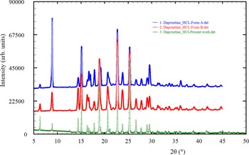
The superposition of the powder patterns reported in the patents (Ren et al., Reference Ren, Ren, Qi, Le, Hong, Cao and Chen2011, Reference Ren, Ren, Chen, Qi, Yue, Hong and Cao2013) for Forms A and B digitized using the on-line JADE® Pattern Digitizer (ICDD, 2022) and the pattern recorded in the present work is shown in Figure 2. The patterns of Form A and B look strikingly similar. Based on the similarity of the unit-cell parameters obtained in the present study with the parameters of the phase reported as Form B and their powder patterns, it can be concluded that they correspond to the same phase. The pattern reported by Selvakumar (Reference Selvakumar2018) was also digitized. Even though the plot reported is somewhat blurry, rendering a low-quality digitized pattern, its comparison with the pattern recorded in the present study shows that it also corresponds to the same phase. This comparison is included in Supplementary Figure S2.
Superposition of the reported powder patterns of Form A and Form B with the powder pattern recorded in the present study.

The fit of the recorded pattern was carried out with the Pawley algorithm by modeling the background, sample displacement errors, absorption, surface roughness, cell parameters, and peak shape parameters (including anisotropic broadening) using TOPAS-Academic (Coelho, Reference Coelho2016). A 12-term Chebyshev polynomial was used to model the background. The intermediate Gaussian–Lorentzian function was employed with a correction for axial divergence as proposed by the program. The Pawley refinement produced a good fitting of all the diffraction maxima recorded with residuals R p = 0.0339, R wp = 0.0481, and GoF = 1.836, confirming the correctness of the unit cell and the single-phase nature of the material. The analysis of the reflection conditions implemented in the CONOGRAPH software suggested P212121 as the possible space group. This space group was also suggested by the Bayesian extinction symbol algorithm in DASH 3.4.9 (Markvardsen et al., Reference Markvardsen, David, Johnson and Shankland2001) and by DAJUST (Vallcorba et al., Reference Vallcorba, Rius, Frontera, Peral and Miravitlles2012).
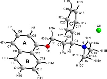
The initial molecular model, introduced as a mol file, was built with the MOPAC2016 software (Stewart, Reference Stewart2016) using the PM7 method (Stewart, Reference Stewart2013). With this model and the profile parameters obtained from the Pawley fit, the crystal structure was determined with DASH 3.4.9 (David et al., Reference David, Shankland, van de Streek, Pidcock, Motherwell and Cole2006). Initially, without considering preferred orientation, the fitting of the profile led to χ 2 = 317.760, and it was not possible to obtain a good structural model. Using the March-Dollase function as a model for the preferred orientation, in the (0 0 2) plane, the structure was determined satisfactorily with χ 2 = 65.141 and a March-Dollase parameter of 0.536. The refinement of the structure carried out with TOPAS-Academic (Coelho, Reference Coelho2016), using the same preferred orientation plane, produced a good fit. Figure 3 shows the final Rietveld refinement plot. The refinement included an overall scale parameter, the background, the peak shapes (including anisotropic broadening), absorption correction, surface roughness parameter, unit-cell parameters, atomic coordinates, three B iso parameters, and a March-Dollase parameter. The bond distances and angles were restrained based on the values of the energy-minimized structure. Three planar restraints for the molecule with a standard deviation of 0.01 Å were also established. The isotropic atomic displacement parameters for the hydrogen atoms were 1.2 times the parameter of the C or N atom to which they are attached. Figure 4, drawn with DIAMOND (Brandenburg, Reference Brandenburg1999), depicts the molecular structure of dapoxetine hydrochloride showing the atom-numbering scheme.
Rietveld plot obtained after the structure refinement of DAPHCl.

Molecular structure of DAPHCl with the labeling scheme for atoms and rings.

In total, 191 parameters were refined with 2703 data points (360 reflections), 137 restraints and 3 constraints. The final whole pattern fitting converged with good figures of merit: R e = 0.0236, R p = 0.0442, R wp = 0.0577, and GoF = 2.440. Table I shows the crystal data, experimental parameters, and the refinement parameters obtained. The DFT-D calculations of the determined structure led to an RMSCD of 0.092 Å, which is lower than the 0.35 Å value (van de Streek and Neumann, Reference van de Streek and Neumann2014), indicating that the structure determined is correct. Figure 5 shows the comparison between the determined and the DFT-D optimized structures.
Superposition of the experimentally determined (red) and the energy-minimized (blue) structure for DAPHCl.

Crystal data, experimental parameters, and refinement results for DAPHCl

C. Molecular and crystal structure
The atomic coordinates and isotropic displacement parameters for all atoms, the bond distances and angles, and torsion angles are reported in Supplementary Tables S1–S3, respectively, of the Supplementary Material. In the statistical analysis performed with the Mogul Geometry Check (Bruno et al., Reference Bruno, Cole, Kessler, Luo, Motherwell, Purkis, Smith, Taylor, Cooper, Harris and Orpen2004), all the 58 distances and bond angles in the structure have z-scores below 3, indicating that the values are reliable.
The asymmetric unit contains one chloride ion and a protonated dapoxetine moiety (DAP+) (Figure 4). The C1 carbon has an S configuration. The torsion angle N1–C1–C2–C3 is 173.6(5)°. The naphthyl unit, composed by rings A/B, makes an angle of 65.4(3)° with respect to the phenyl ring (ring C), generating an inclined arrangement. The A/B fragment makes angles with respect to the a, b , and c-axes of 34.88(12)°, 51.51(12)°, and 14.22(12)°, respectively. On the other hand, the C ring makes angles of 24.9(2)°, 28.3(2)°, and 50.6(2)°, respectively, with the a, b, and c-axes.
D. Intermolecular hydrogen bonds
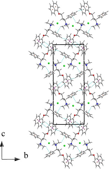
Figure 6 presents a view of the strongest interactions around the chloride anion. Cl1− is in close contact with three DAP+ moieties via five hydrogen bonds. Table II contains the geometry of the hydrogen bonds, π⋯π, and C–H⋯π interactions. As can be seen, the strongest interaction occurs between N1–H1N and Cl1 of the same asymmetric unit [2.086(13) Å, 169.0(13)°]. This molecule also interacts through C17–H17 [2.7339(12) Å, 131.1(9)°]. The rest of the interactions are weak but contribute to a lowering of the crystal packing energy.
View of the packing arrangement along the a-axis showing the hydrogen bond environment around the Cl− ions.

Geometry of hydrogen bonds, π⋯π, and C–H⋯π interactions in DAPHCl

The geometry of the contacts is defined in PLATON [Spek, 2020] by the following parameters:
a Cg(A) and Cg(B) are the centroids of rings A and B, defined in Figure 4; d = CgI⋯CgJ distance; α = dihedral angle between Planes I and J; β = angle between the Cg(I)→Cg(J) vector and the normal to plane I; γ = angle between the Cg(I)→Cg(J) vector and the normal to plane J; CgI Perp = perpendicular distance of Cg(I) on ring J; CgJ_Perp = Perpendicular distance of Cg(J) on ring I; Slippage = distance between Cg(I) and perpendicular projection of Cg(J) on ring I.
b H⋯Cg = distance from H to centroid of ring; H-Perp = Perpendicular distance of H to ring plane; γ = angle between the Cg-H vector and the ring normal; X–H⋯Cg = X–H–Cg angle (°).
E. π⋯π and C–H⋯π interactions
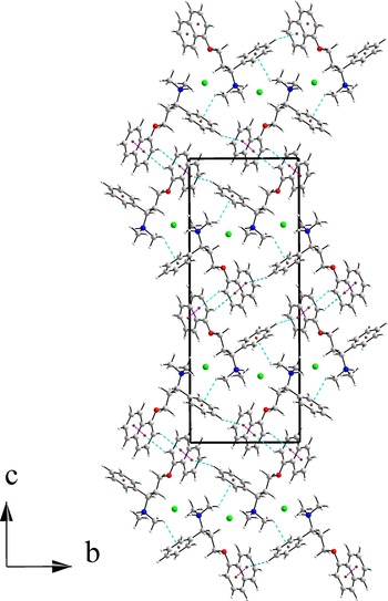
Connectivity between DAP+ moieties is achieved through π⋯π and C–H⋯π interactions (Figure 7). The shortest π⋯π interactions (d = 4.638(3) Å) occur between the centroid of a ring A (Cg(A)) with the centroid of a ring B (Cg(B)) of a molecule related by symmetry operation −1 + x, y, z. These interactions extend approximately along the a-axis. The rings are parallel to each other, but the centroids of the rings are offset by 2.864 Å.
View of the packing arrangement along the a-axis showing the sequence of π⋯π (magenta) and C–H⋯π (cyan) interactions.

Type III C–H⋯π contacts (Malone et al., Reference Malone, Murray, Charlton, Docherty and Lavery1997) connect DAPH+ units as also shown in Figure 7. The C19–H19⋯Cg(B) and C5–H5⋯Cg(A) contacts (2.814(11) and 3.021(11) Å, respectively) connect molecules related by symmetry operations x, 1 + y, z and −1/2 + x, 3/2−y, −z, respectively. They are oriented approximately along the b-axis. These interactions are common for Csp2–H phenyl groups and, in this structure, show a T-shaped approach of the phenyl rings. A weaker C–H⋯π interaction of 3.300(13) Å involves C15–H15C⋯Cg(C) related by the symmetry operation 1-x, −1/2 + y, 1/2-z.
The structure is a complex 3D arrangement of hydrogen bonds, π⋯π, and C–H⋯π interactions. It can be described in terms of layers parallel to the ab plane connected by N–H⋯Cl and C–H⋯Cl hydrogen bonds. The layers stack along the c-axis, which are connected by C–H⋯π interactions.
F. HS analysis
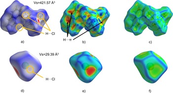
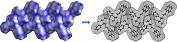
The HS mapped onto d norm, shape index, and curvedness for the DAPH+ moiety is shown in Figure 8. A very intense red spot (associated with contacts shorter than van der Waals distances) corresponding to the N–H⋯Cl hydrogen bond [Figure 8(a)] is observed, which is complemented by the most intense spot observed on the surface of the counterion [Figure 8(d)]. Additionally, three faint red spots (yellow circles) are observed that correspond to the contribution of the chlorine atom as an acceptor of a hydrogen atom, and which are complementary to the spots observed on the surface of the anion [Figure 8(d)]. The red areas in the shape index [Figure 8(b)] show the H⋯π interactions with the aromatic rings of the compound. At the same time, the curvedness plot shows flat areas where the aromatic rings of the naphthyl fragment are located [Figure 8(c)] due to the presence of the π⋯π and H⋯π interactions. This is an indication of the stacking of these groups in the structure, as shown in Figure 9. The representation of the chloride surface in terms of its curvedness results in mostly flat faces [Figure 8(f)], consistent with the packing observed in the structure.
d norm, shape index, and curvedness mapped onto the HS for DAPHCl.

Stacking of DAPHCl molecules due to π⋯π and H⋯π interactions.

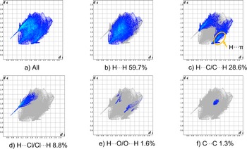
Figure 10 shows the fingerprint plots (Spackman et al., Reference Spackman, Turner, McKinnon, Wolff, Grimwood, Jayatilaka and Spackman2021) for important interactions. The H⋯H interactions contribute 59.7%, while the H⋯C/C⋯H interactions represent 28.6%, and the H⋯Cl/Cl⋯H contacts contribute 8.8%. The crystal packing is thus governed by H⋯π and H⋯Cl interactions.
Fingerprint plots and percent contributions of important interactions in the structure of DAPHCl.

V. CONCLUSION
The crystal structure of (S)-Dapoxetine hydrochloride (DAPHCl), a well-known active pharmaceutical ingredient used in the treatment of PE in men, has been determined from laboratory X-ray powder diffraction data with DASH and refined by the Rietveld method with TOPAS-Academic. The structure was evaluated and optimized using DFT-D calculations. The crystal structure of DAPHCl is based on layers of Cl− ions interacting via hydrogen bonds with DAP+ moieties which interact with other DAP+ via π⋯π and C–H⋯π interactions. Fingerprint plots show an important contribution from H⋯π interactions which, along with π⋯π interactions, produce the characteristic flat surfaces in the curvedness representation.
VI. DEPOSITED DATA
Crystallographic Information Framework (CIF) files containing the results of the Rietveld refinement and a CIF with the DFT-D geometry optimization results were deposited with the ICDD. The data can be requested at info@icdd.com.
The crystal structure data were also deposited with the Cambridge Crystallographic Data Centre (CCDC 2183908).
SUPPLEMENTARY MATERIAL
The supplementary material for this article can be found at https://doi.org/10.1017/S0885715622000380.
ACKNOWLEDGEMENTS
The authors thank the support of Vicerrectoría de Investigación y Extensión of Universidad Industrial de Santander (UIS), Colombia. Access to the Cambridge Structural Database (CSD) for Universidad de Los Andes (Venezuela) was possible through the Frank H. Allen International Research & Education Programme (FAIRE) from the Cambridge Crystallographic Data Centre (CCDC). The authors gratefully acknowledge Jim Kaduk for helpful discussions and for digitizing the pattern reported by Selvakumar (Reference Selvakumar2018).