Case 1
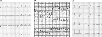
A 32-year-old female presented to hospital with hypotension and recurrent vomiting. Electrocardiogram showed sinus rhythm with prolonged QT interval (QTc 584 ms) (Fig 1a). Biochemical investigations demonstrated normal serum electrolytes except mild hypocalcaemia (Ca2+: 1.84 mmol/L, normal 2.2–2.7 mmol/l). In hospital, she developed recurrent symptomatic torsades de pointes (TdP) (Fig 1b), managed with intravenous magnesium sulphate (MgSO4). Because of persistent hypotension and recurrent unprovoked hypoglycaemia, an ACTH challenge test was performed and a diagnosis of adrenal insufficiency was confirmed (serum cortisol, both baseline and 1 hour after adrenocorticotropic hormone (ACTH) administration <28 nmol/L). Improvement in haemodynamic and biochemical parameters occurred with the initiation of corticosteroid replacement therapy (hydrocortisone and fludrocortisone). The patient received an additional diagnosis of long QT syndrome based on the observation of QT prolongation yet “normal” electrolyte status upon admission. Subsequent genetic analysis for long QT syndrome was negative. At 8 weeks follow-up, complete normalisation of the QTc interval (413 ms) was noted (Fig 1C).
Electrocardiograms of Patient 1. ( a ) On admission showing QT prolongation. ( b ) During hospital admission showing torsades de pointes (TdP). ( c ) Follow-up electrocardiogram after corticosteroid relacement showing normal QTc.

Case 2
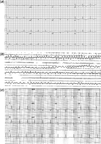
A 43-year-old female presented to the emergency department following an abrupt loss of consciousness associated with urinary incontinence. Electrocardiogram demonstrated deep T wave inversions with QT prolongation (QTc = 553 ms)(Fig 2a). Initial workup demonstrated normal serum electrolytes, an elevated cardiac troponin level, and left ventricular dysfunction (15–20%). Coronary angiography documented normal coronary arteries. During hospital admission, she developed multiple episodes of symptomatic TdP (Fig 2b). In view of QT prolongation, left ventricular dysfunction, and cardiac arrest, she received an implantable cardioverter-defibrillator. Due to recurrent, unprovoked hypoglycaemia, an ACTH challenge test was performed, resulting in a diagnosis of adrenal insufficiency (cortisol, baseline <11 nmol/L, 1 hour post-ACTH challenge: 155 nmol/L). She was treated with corticosteroid replacement and nadolol. In hospital, she received a diagnosis of Addison’s disease and long QT syndrome. At follow-up 2 weeks after discharge, electrocardiogram demonstrated a QTc of 410 ms (Fig 2c) and heart function normalised (left ventricular dysfunction: 61%). Nadolol therapy was discontinued and genetic testing for long QT syndrome was negative.
Electrocardiograms of Patient 2. ( a ) Presenting electrocardiogram with QTc of 550 ms. ( b ). Electrocardiogram during hospital admission showing torsades de pointes (TdP). ( c ). Follow-up electrocardiogram 2 weeks after discharge from hospital (QTc = 410 ms).

Case 3
A 61-year-old female was referred due to incidentally detected QT prolongation on electrocardiogram (QTc 500 ms) (Supplementary Figure S1A). Genetic testing was negative. A history of persistent dysphagia was elicited and serum biochemistry revealed hypocalcaemia (Ca2+: 1.4 mmol/L), elevated serum phosphate (2.32 mmol/l, normal 0.81–1.45 mmol/l), normal 25-OH vitamin D level with reduced serum PTH (0.64 pmol/L, normal 1.9–6.9 pmol/L). Endocrinology consultation resulted in a diagnosis of primary hypoparathyroidism and she was treated with calcitriol and calcium supplements. After 3 months of therapy, normalisation of serum calcium and phosphate levels ocurred, and electrocardiogram demonstrated normal QTc interval (QTc 390 ms) (Supplementary Figure S1B).
Case 4
A 15-year-old male experienced a syncopal event and electrocardiogram demonstrated prolonged QTc (475 ms) (Supplementary Figure S2A). He was treated with bisoprolol 5 mg daily. Genetic testing for long QT syndrome was negative. On subsequent follow-up, a history of paresthesia of the extremities was elicited, and serum biochemistry documented hypocalcaemia (Ca2+: 1.42 mmol/L), hyperphosphatemia (PO4 3-: 2.33 mmol/L), normal creatinine, and serum albumin (50 g/L) with high PTH (20.4 pmol/L). Endocrinology consultation concluded a clinical diagnosis of pseudohypoparathyroidism, and treatment with calcium carbonate and calcitriol was initiated. After 3 months of treatment, biochemical parameters normalised, and electrocardiogram demonstrated a QTc of 415 ms (Supplementary Figure S2B).
Case 5
An 11-year-old girl was diagnosed with long QT syndrome following a syncopal episode and observation of prolonged QT interval (QTc 475 ms) (Supplementary Figure S3A). Genetic testing was negative and she was considered as “gene negative” long QT syndrome and managed with atenolol 25 mg BID. Seven years after the index episode, she experienced a witnessed tonic--clonic seizure with tongue biting. Biochemistry revealed low serum calcium (1.44 mmol/L), high serum phosphate (2.33 mmol/L), and high serum PTH level (36 pmol/L). A diagnosis of pseudohypoparathyroidism was made following endocrinologic consultation and she received treatment with calcium and calcitriol. Follow-up electrocardiogram after 6 months of therapy showed a normal QTc (412 ms) and beta-blocker therapy was weaned off (Supplementary Figure S3B).
Discussion
QT prolongation on electrocardiogram is a consequence of an abnormal plateau phase (Phase 2) of the cardiac action potential caused primarily by impairment of potassium current during Phase 3 repolarization. This abnormality may be primary, caused by genetic defects in the known long QT syndrome genes, or secondary related to drug or electrolyte disturbance. Reference Adler, Novelli and Amin1,Reference Chorin, Rosso and Viskin2 Primary endocrine disease mimicking long QT syndrome is a sparsely recognised phenomena.
The first two patients in our series presented with QTc prolongation and TdP due to adrenal insufficiency. Although mild electrolyte deficiencies may have contributed to the observed QTc prolongation in these patients, glucocorticoid deficiency is known to decrease glucocorticoid inducible kinase-1 (SGK1), which controls the expression of KCNH2-encoded Ikr and directly stimulates KCNQ1-encoded Iks. Reference Busjahn, Seebohm and Maier3.Reference Lamothe and Zhang4 Accordingly, only corticosteroid hormone replacement therapy was associated with normalisation of QT interval in these two cases.
Three patients in our series had parathyroid dysfunction, one with hypoparathyroidism and two patients with pseudohypoparathyroidism. While hypocalcaemia is a known provoker of QT prolongation, elevated PTH levels independent of serum calcium is also associated with prolonged QT intervals (case 4 and 5). Reference Palmeri, Davidson, Whang, Kronish, Edmondson and Walker5 The QT prolonging effect of PTH may be due to its enhanced activation of the L-type calcium channel. Reference Ebisawa, Kimura, Nakayama, Yaginuma, Watanabe and Shimada6
Etiological diagnosis of QT prolongation is essential for prognosis and treatment strategy, that is, secondary long QT syndrome requires correction and avoidance of precipitating factors, and unlike congenital long QT syndrome, does not require lifelong medical therapy, follow-up and family screening.
Conclusion
Gene negative long QT syndrome is reported to occur in up to 30% of cases followed in long QT syndrome clinics, although this number may be variable depending on the stringency of diagnosis and search for secondary causes of QT prolongation. In our experience, over 90% of diagnosed long QT syndrome cases have a recognised genetic cause, with less than 10% of cases remaining gene elusive. Reference Adler, Sadek and Chan7
Endocrinopathies may mimic long QT syndrome and should be considered in the differential diagnosis of gene negative long long QT syndrome, particularly in the absence of family history. Non-cardiac symptoms may provide clinical clues and prompt endocrine workup, averting a diagnostic miscue.
Supplementary material
To view supplementary material for this article, please visit https://doi.org/10.1017/S1047951121004388
Financial support
This research received no specific grant from any funding agency, commercial or not-for-profit sectors.
Conflicts of interest
None.