An unrestricted pulmonary blood flow is crucial in cavopulmonary connections.Reference Girod, Fontan, Deville, Ottenkamp and Choussat 1 – Reference Senzaki, Isoda, Ishizawa and Hishi 3 Consequently, there is a significant need for sufficient image information. The echocardiographic window in this region is often limited and frequently inadequate;Reference Harris, Cosulich and Gillespie 4 therefore, another imaging modality has to be added whenever diagnostic imaging is required. In clinical routine, cardiac catheterisation, in addition to magnetic resonance imaging and multi-detector row computed tomography, is an important imaging modality in cavopulmonary connections. A routine cardiac catheterisation before partial cavopulmonary connection seems to be avoidable in most cases,Reference Brown, Gauvreau and Moran 5 but after surgically performed partial cavopulmonary connection unexpected pathological findings are frequently seen.Reference Holzer, Sisk and Chisolm 6 Cardiac catheterisation before total cavopulmonary connection is yet often recommendedReference Ba, Marini and Kammache 7 , Reference Ro, Rychik, Cohen, Mahle and Rome 8 and it remains essential for interventions.
With the introduction of flat-detector-equipped c-arms in paediatric catheterisation laboratories, there is a novel possibility of gaining flat-detector computed tomography images, mainly as angiographic computed tomography images with a high quality. These angiographic images rendered in a three-dimensional model can be used immediately during the same investigation by displaying it live on a fluoroscopy monitor to guide catheter interventions and to select the most appropriate c-arm angle.Reference Glatz, Zhu, Gillespie, Hanna and Rome 9 – Reference Kalender and Kyriakou 12 The scanning time is at least 5 seconds, and thus pulsatile motion of the heart and great vessels, as well as breathing generate artefacts. Close to the heart, these artefacts are distinctive; however, extracardial structures such as the cavopulmonary connections can be imaged by flat-detector computed tomography in a high quality.Reference Glatz, Zhu, Gillespie, Hanna and Rome 9 , Reference Glockler, Koch and Greim 10
The use of flat-detector computed tomography in catheterisation of congenital heart disease is a new imaging procedure and there are only few data about the investigative impact of this technique on cavopulmonary connections. Therefore, we evaluated and report our experience with flat-detector computed tomography as a new imaging modality in 31 consecutive cases with cavopulmonary connections.
Patients and methods
We analysed all patients with cavopulmonary connection who underwent invasive imaging in our cardiac catheterisation laboratory retrospectively in an 18-month period between January, 2010 and June, 2011. In this period, we also investigated three patients without the use of flat-detector computed tomography but excluded these cases from our study.
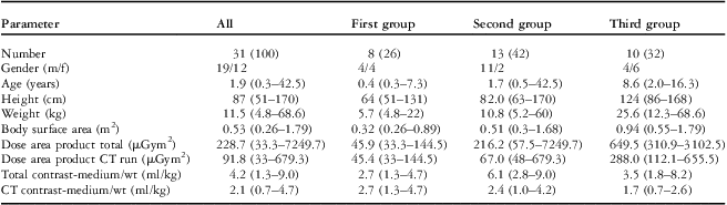
The most common congenital heart defects in the investigated patients were different kinds of double outlet right ventricle (8 out of 31 cases), double inlet left ventricle (8 out of 31 cases), and hypoplastic left heart syndromes (6 out of 31 cases), followed by pulmonary atresia with intact ventricular septum (3 out of 31 cases), tricuspid atresia (3 out of 31 cases), and others (3 out of 31 cases). We divided the studied patients in three groups (Table 1): in the first group (eight patients), we investigated all patients after partial cavopulmonary connection soon after operation; in six of these patients, the flat-detector computed tomography was performed on the first post-operative day, in two on the second post-operative day (after surgery), and all patients except one were breathing spontaneously. The administration of the diluted contrast medium was always by hand injection through the pre-existing central venous line in the right jugular vein. There were three patients with a bilateral bidirectional cavopulmonary connection. In two of these patients, contrast was administered additionally through the left external jugular vein and in one patient the left cubital vein was used. The flat-detector computed tomography acquisition by rotational angiography was used exclusively in these investigations.
The second group included 13 patients in whom flat-detector computed tomography was applied before planned total cavopulmonary connection. Routinely, the right jugular vein was used for investigations. Flat-detector computed tomography was performed by contrast dye administration through a pigtail catheter and machine-injection in the superior caval vein. An arterial access was only inserted in cases of anamnestic, clinical, or echocardiographic signs of anomalies. We administered contrast dye in the partial cavopulmonary connection several times and simultaneously in the aorta during the flat-detector computed tomography (3 out of 13 cases) to display the spatial anatomical relationship. Whenever interventional catheterisation was required, the three-dimensional vascular model was used by merging it with the fluoroscopic picture for three-dimensional navigation.
In the third group, we investigated 10 patients with failing total cavopulmonary connection requiring invasive catheter evaluation or intervention. The flat-detector computed tomography was mostly performed by simultaneous contrast administration through a pigtail catheter in the superior and the inferior caval vein simultaneously. The three-dimensional model was used for three-dimensional navigation in case of interventions.
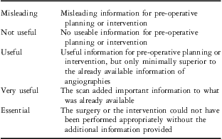
In all three groups, the ability to provide diagnostic information for surgery was jointly determined by one paediatric cardiologist and one paediatric cardiac surgeon in every case. The diagnostic contribution of flat-detector computed tomography was rated using a standardised, previously published 5-point Likert scale, see Table 2.Reference Ellis, Mulvihill, Bradley and Hlavacek 13 In the first group, the flat-detector computed tomography was used to gain early post-operative information about the anatomical structure and the function of the cavopulmonary connection. In the second and third groups, the benefit of using overlaid three-dimensional images obtained from flat-detector computed tomography during catheter-based interventions was jointly analysed by the operator and one paediatric cardiologist. Again, a classification of the Likert scale (Table 2) was used.
Modified Likert scale concerning the diagnostic utility of flat-detector computed tomography (adapted fromReference Ellis, Mulvihill, Bradley and Hlavacek 13 ).

We used flat-detector computed tomography based on rotational angiography with three-dimensional image reconstruction of a vascular model using volume-rendering technique. If transcatheter intervention was necessary, the three-dimensional model was merged with the fluoroscopic picture for three-dimensional navigation. All examinations were performed on a biplane angiography system equipped with two 20 × 20 square centimetre flat panel detectors with a 960 × 960 matrix (Axiom Artis, Siemens Medical Solutions, Forchheim, Germany). For image acquisition, the c-arm with the flat detector rotates around the patient in an array of 180° in addition to the fan angle in 5 seconds; therefore a volume data set is gained by pulsed X-rays with a frame rate of 30 per second and a dose of 0.36 microgray per image. The tube voltage was fixed at 90 kilovolt and tube current was automatically adapted. The anti-scatter grid was removed in patients less than 20 kilograms. Cross-sectional images were rendered with 0.4 millimetre thickness in a 500 × 500 matrix format using the kernel-type “HU”. Post-processing of the data set was performed on a commercially available workstation (Leonardo DynaCT, InSpace 3D software, Siemens Medical Solutions). The injected contrast agent was Imeron® 350 (Iodine 350 milligrams per millilitre; Bracco Imaging Deutschland GmbH; Konstanz, Germany); it was always diluted with saline in a median concentration of 50% (range 30–75%).
Results
During the study period of 18 months, a total of 34 patients with cavopulmonary connection were investigated in our catheterisation laboratory. Flat-detector computed tomography was applied in 31 of these patients and they were included in the study. In 14 patients (45%), an interventional procedure was performed. In the other 17 cases (55%), it was a solely diagnostic catheterisation.
The median age was 1.9 years – range 0.3–42.5 years, median weight was 11.5 kilograms – range 4.8–68.6 kilograms, and 61% of the patients were male (Table 1).
Demographic and procedural data, median (with range), number (percentage of total).

CT = computed tomography
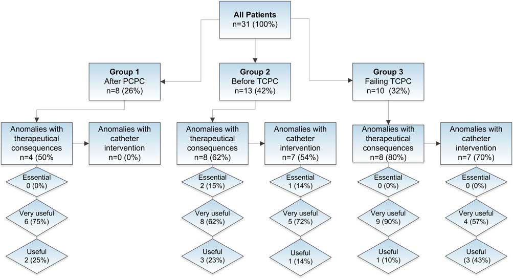
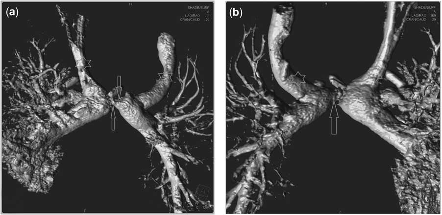
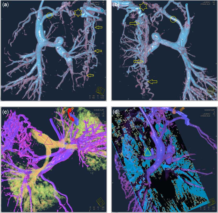
We subsumed eight (26%) patients early post-operatively after surgically performed upper cavopulmonary connection in the first group; in four cases (50%), we detected pathologies, which led to therapeutical consequences (Fig 1). In one patient, there was an inverse blood flow in the left pulmonary branch caused by huge aortopulmonary collateral arteries. The patient underwent interventional catheterisation a few days later with occlusion of these arteries. There were two patients with bilateral Glenn anastomosis who had an obstruction of the pulmonary bifurcation between the two upper caval veins. In these cases, we administered an antithrombotic therapy (Fig 2). There was one patient who had a thrombosis of the confluence of the left jugular and the left subclavian vein; thus, a thrombolysis was performed (Fig 3).
Results of investigation and rating of the diagnostic utility of flat-detector computed tomography in 31 patients with cavopulmonary connection; number (percentage in the group). PCPC = partial cavopulmonary connection; TCPC = total cavopulmonary connection.
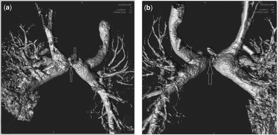
Severe stenosis of the bifurcation of the pulmonary artery. A 4-month-old boy with double outlet right ventricle, transposition of the great arteries, and sub-pulmonary stenosis, 1 day after surgery for bilateral, bidirectional Glenn anastomosis. An anticoagulative therapy was performed as consequence of thes residual pathologies. Superior caval veins marked by the stars, stenosis of the pulmonary artery by the arrows; (a) with the view from frontal and inferior and (b) from head and posterior.
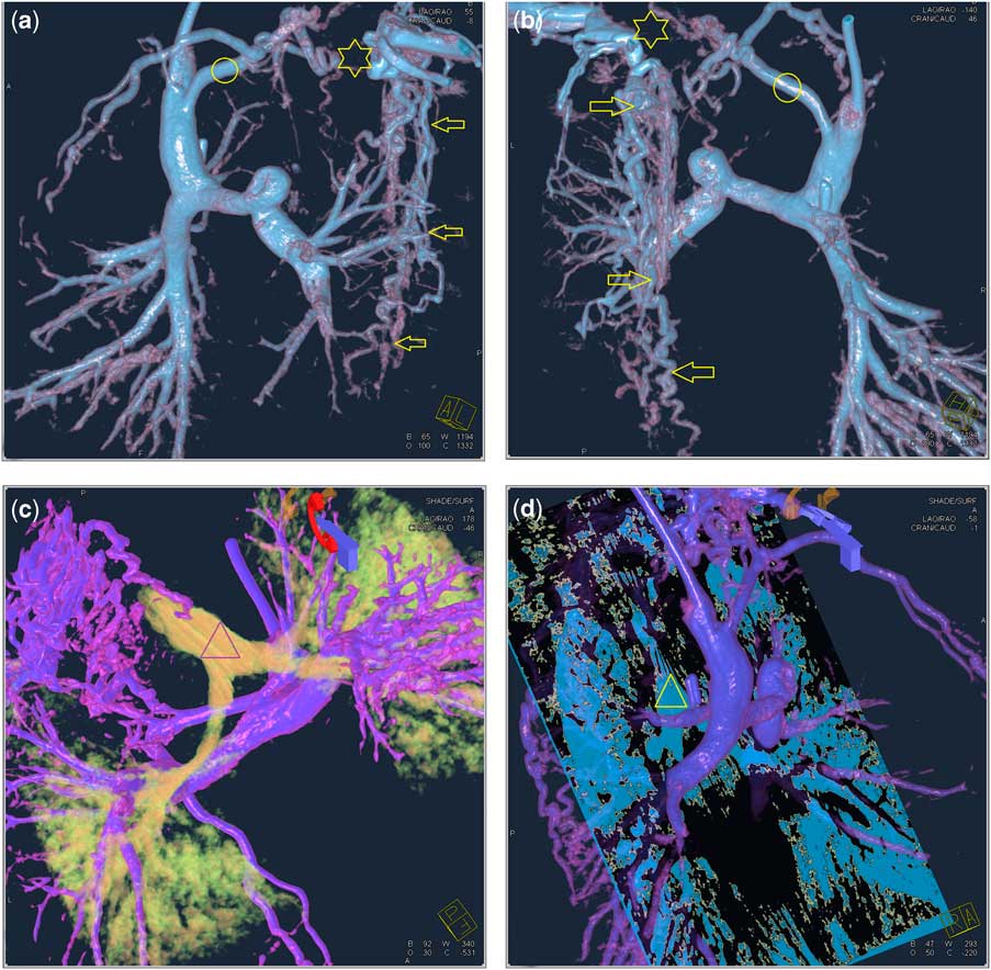
Thrombosis of the left Pirigoff angle (star). A 4-month-old baby with hypoplastic left heart syndrome 1 day after Glenn anastomosis. Huge paravertebral collateral veins (arrows) and small brachiocephalic vein (circle). Antithrombotic therapy followed. (a) shows the vascular model from anterior and (b) from posterior. In (c), the air passages are also reconstructed to demonstrate the spatial relationships; (d) shows an embedded multi-planar section with the air passages, which may help for surgical planning.
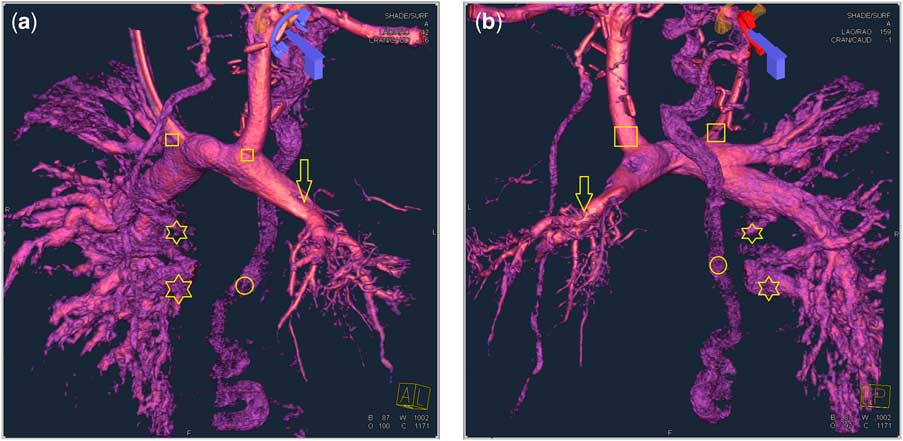
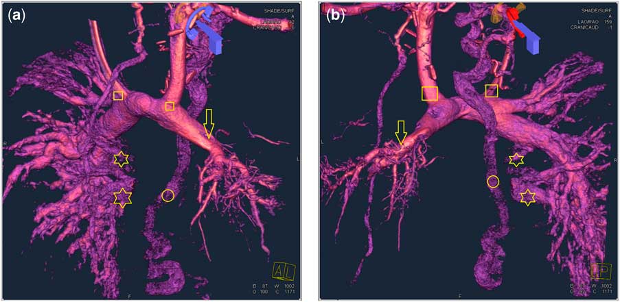
In the second group, we included 13 (42%) patients with upper cavopulmonary connection before completion of a total cavopulmonary connection (Fig 4). Of these investigations, seven (54%) were interventional catheters. In two cases, we dilated a stenosis of pulmonary branches; in four cases, the aim was an occlusion of aortopulmonary collateral arteries and additionally in one case a balloon dilatation of an aortic coarctation.
Total pulmonary vein occlusion of the left side in a 1-year-old boy with bilateral (quarters) bidirectional Glenn anastomosis with dextrocardia, heterotaxy syndrome, and an unbalanced atrioventricular septal defect. Contrast medium given through the left cubital vein and through a pigtail catheter via the right jugular vein in the left pulmonary artery. Reversal blood flow in the left pulmonary artery (seen in the dynamic rotational angiography) with small peripheral pulmonary arteries (arrow). Pulmonary veins are displayed on the right side (stars), and none on the left side. Collateral blood flow over the azygos vein (circle). (a) is the view from left anterior and (b) from left posterior.
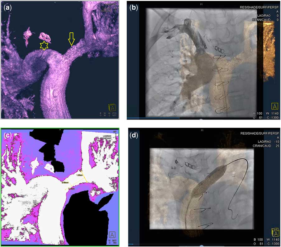
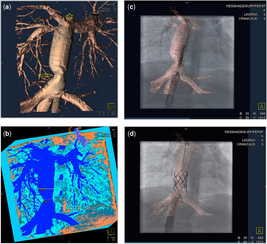
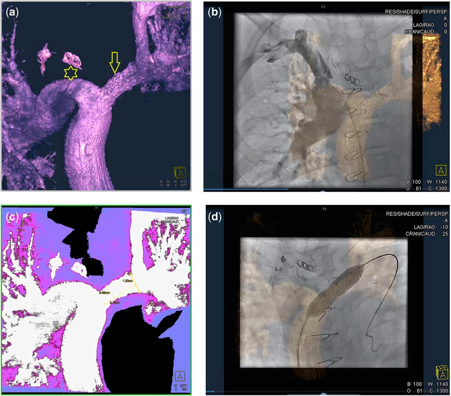
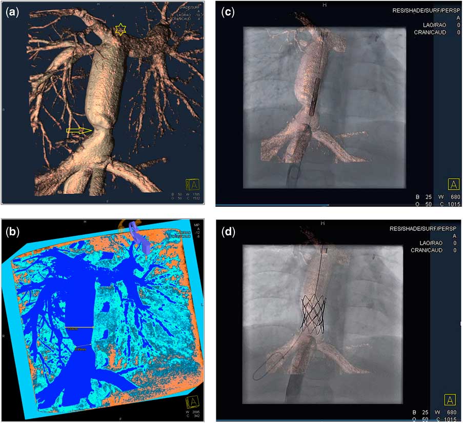
In the third group, 10 patients with total cavopulmonary connection (32%) were included. Interventions were performed in seven cases (70%): stent placement in pulmonary arteries or the extracardial tunnel (3 out of 7 cases; Figs 5 and 6) and occlusion of aortopulmonary collateral arteries (4 out of 7 cases).
Total cavopulmonary anastomosis with bilateral, bidirectional Glenn anastomosis. Nine-year-old boy with double inlet left ventricle, 3 years after surgery. Preexisting 10 millimetre stent (arrow) in the left pulmonary artery. Contrast medium given through left cupital vein by hand-injection and through a pigtail catheter by machine injection in the extracardial conduit. (a) shows the three-dimensional model; the right superior caval vein is not contrasted and the wash-out effect is seen (star). (b) shows the overlaid three-dimensional model on fluoroscopy and contrast administration in the right superior caval vein. (c) is an imaging in maximum intensity projection for measurements. In (d), the three-dimensional navigation for balloon dilatation of the stent is demonstrated.
Severe stenosis of the anastomosis between inferior caval vein and the conduit (arrow). An 11-year-old boy with hypoplastic left heart syndrome, total cavopulmonary connection, and extracardial conduit. Stent implantation in the stenosis. (a) shows the three-dimensional model in volume-rendering technique. Contrast admission over a pigtail catheter in the conduit. Owing to wash out, the superior caval vein is not contrasted (star). (b) is the same data set in major intensity projection for measurements. (c) is the overlaid three-dimensional model on fluoroscopy during the implantation procedure of a stent. Position of the stenosis and the liver veins are always displayed and three-dimensional model follows every new angulation of the c-arm. (d) shows the inflated stent.
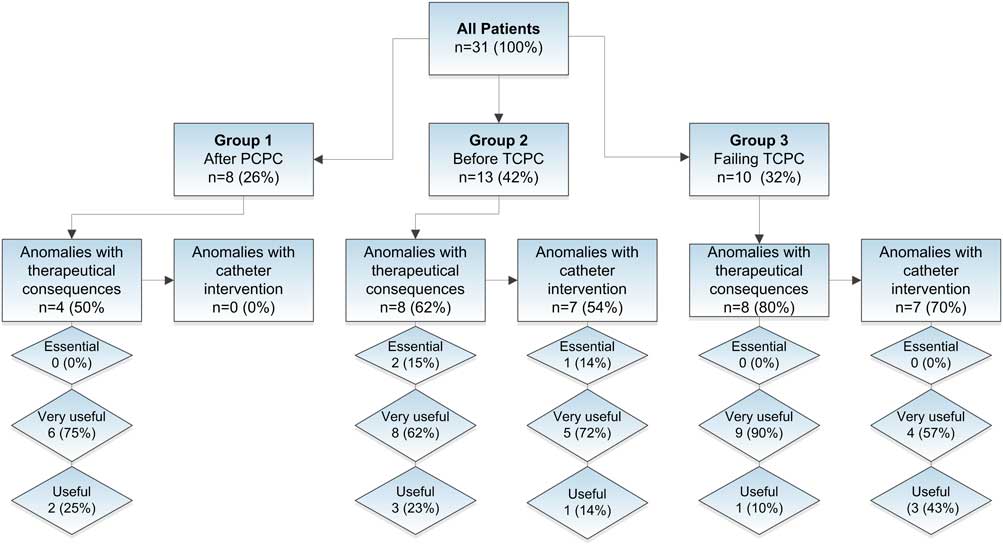
Overall, both flat-detector computed tomography images and overlaid three-dimensional reconstructions were rated superior – “essential”, “very useful”, or “useful” – to conventional angiography alone in all cases. The diagnostic value flat-detector computed tomography was classified as follows: two (7%) were rated “essential”, 23 (74%) were rated “very useful”, six (19%) were rated “useful”, and none was rated “not useful” or “misleading”.
In 14 patients in whom interventions were performed, the utility of the merged three-dimensional reconstructions of the flat-detector computed tomography with the real-time fluoroscopic image was rated as follows: One case (7%) was rated “essential”, 9 (64%) were rated “very useful”, 4 (29%) were rated “useful”, and none was rated “not useful” or “misleading” (Fig 1).
The mean required amount of contrast agent for the rotational angiography was 2.4 plus or minus 1.1 millilitres per kilogram, with a range of 0.7–4.7 millilitres per kilogram. On average, the percentage of the dose by flat-detector computed tomography of the total investigation was 49% of the entire contrast used during the procedure (range 25–100%). The median dose area product of flat-detector computed tomography was 91.8 microgray square metres (range 33.0–679.3 microgray square metres). Flat-detector computed tomography contributed an average of 43% (range 9–100%) of the entire radiation dose per procedure. Owing to the fact that tube current was automatically adapted by the X-ray imaging chain, a patient's body surface area is a predictor of the dose area product.Reference Wielandts, Smans, Ector, De Buck, Heidbuchel and Bosmans 14
Discussion
In cavopulmonary connections, residual pathologies may generate severe side effects such as reduced physical capacity, oedema, thrombosis, or protein-losing enteropathy with lymphatic malformation;Reference Feldt, Driscoll and Offord 15 – Reference Rychik 17 therefore, an optimal imaging modality is essential. Today, cardiac ultrasound is the standard procedure for pre-operative imaging of congenital heart disease. However, it has a limited sensitivity in detecting great vessel stenosis in cavopulmonary connection.Reference Ba, Marini and Kammache 7 , Reference Fraisse, Colan, Jonas, Gauvreau and Geva 18 Cardiac catheterisation cannot be performed routinely, but will be indicated if there are remaining questions after echocardiography. This can be caused by bad acoustic windows, difficult spatial relationships of cardiac and vascular structures, and if other imaging modalities such as magnetic resonance imaging or multi-detector row computed tomography are not viable.Reference Brown, Gauvreau and Moran 5 , Reference Ba, Marini and Kammache 7 , Reference Ro, Rychik, Cohen, Mahle and Rome 8 , Reference McMahon, Eidem and Bezold 19 Cardiac catheterisation is required for interventions or if an invasive haemodynamic evaluation is necessary.
Recently computed tomography imaging in congenital heart disease has been used increasingly because of improved temporal resolution and detector coverage of multi-detector row computed tomography systems and the development of low-dose imaging algorithms. High image quality can be achieved even in neonates.Reference Achenbach, Marwan and Ropers 20 – Reference Lell, May and Deak 22 The main advantage of flat-detector computed tomography imaging is the possibility of performing it during cardiac catheterisation with admission of contrast medium directly in the volume of interest, which makes it easily distinguishable from the surrounding tissue and therefore allows fast three-dimensional reconstructions. Thus, these vascular models are immediately available during the invasive investigation in the catheterisation laboratory extending hitherto available imaging modalities. Not least, in the hands of paediatric cardiologists this means an easy and fast access to a modern cross-sectional imaging technique for this profession.
In a series of 31 patients, we found that flat-detector computed tomography used as angiographic computed tomography in cavopulmonary connections provides valuable additional information, both for diagnostic purposes and for the use in interventions to conventional biplane angiographies in all of our cases. We rated the impact of flat-detector computed tomography much higher compared with other publications (70–90%);Reference Glatz, Zhu, Gillespie, Hanna and Rome 9 , Reference Glockler, Koch and Greim 10 however, compared with other anatomic regions, the cavopulmonary connections are visualised much better by this technique because of less movement artefacts and a slow blood flow. With this advanced imaging technique and an adjusted application protocol, there is an improved diagnostic accuracy in cavopulmonary connections. At the same time, the pulmonary veins can be visualised entirely. After creation of a three-dimensional vascular model, this can be viewed in any plane, enabling a demonstration in a “surgical angle of view”, and can be rotated in any desired position. Moreover, extracardial structures such as air passages, oesophagus, or bones can be displayed in addition to the vascular model, and their spatial relationship can be demonstrated. Breathing artefacts can be avoided by breath-holding; however, we investigated nearly all patients breathing normally. Breathing artefacts were generally not disturbing. The adverse effect of pulsation on imaging is most considerable in the arteries close to the heart. This can be minimised by reduction of the heart-stroke volume, for example, by rapid heart pacing, but extracardial structures such as the cavopulmonary connections can be visualised accurately under normal cardiac actions and without electrocardiogram triggering. Measurement of vascular diameters, for example before stent implantation, should not be performed in the three-dimensional model, because the result differs from the segmented data, the chosen colour, opacity, and lighting settings. In all cases, we were able to perform reliable sizing in rotational angiographies before any reconstruction and also in two-dimensional multi-planar reconstructions or in two-dimensional maximum intensity projections (see Figs 5C and 6B).
In the eight patients who underwent the partial cavopulmonary connection procedure, we detected residual anomalies in four patients (50%) by an early post-operative imaging by flat-detector computed tomography with creation of a high-resolution three-dimensional vascular model of the cavopulmonary connection. In all of the four cases, the pathological findings led to therapeutic consequences. The imaging of the partial cavopulmonary connection does not need a new vascular access, because the pre-existing central venous catheter can be used for contrast administration.
Overall, we used the new technique of flat-detector computed tomography in the 18-month period in 25% of all cardiac catheterisations of congenital heart disease successfully and without adverse effects. We mainly investigated extra-cardiac regions and used flat-detector computed tomography in questions concerning the aorta, pulmonary artery, and the cavopulmonary connections. Whenever there were questions easy to answer by a single conventional angiography, we did not apply flat-detector computed tomography.
Flat-detector computed tomography in the context of invasive imaging of congenital heart disease is new. Therefore, we have to examine the radiation exposure and the amount of the contrast material used in these cases. The radiation dose used is displayed by the dose area product. The body surface area is a predictor for the dose area product because of the automatic exposure control of flat-detector computed tomography.Reference Wielandts, Smans, Ector, De Buck, Heidbuchel and Bosmans 14 The effective dose can only be measured in phantoms. For dose assessment in conventional computed tomography, the conventional computed tomography dose index is used;Reference Shope, Gagne and Johnson 23 however, it is not practical because of the extended irritation fields in craniocaudal direction (z-direction) of the big scanners.Reference Fahrig, Dixon, Payne, Morin, Ganguly and Strobel 24 , Reference Kyriakou, Deak, Langner and Kalender 25 Thermoluminescence dosimetry measurement is complex and time consuming, and therefore its use is limited.Reference Kyriakou, Deak, Langner and Kalender 25 Dose calculations using Monte Carlo calculations provide accurate results.Reference Kalender and Kyriakou 12 , Reference Kyriakou, Deak, Langner and Kalender 25 Real dose measurements utilising acquisition programmes comparable with this type of flat-detector are rare; with an analogous acquisition programme like ours, the radiation exposure seems to be comparable to a 5-second biplane angiography.Reference Glatz, Zhu, Gillespie, Hanna and Rome 9 The flat-detector computed tomography replaced all angiographies in some cases, and therefore the percentaged radiation dose of the flat-detector computed tomography in these investigations added up to 100%. The median amount of the used contrast medium of a single rotational angiography was 2.4 millilitres per kilogram. This is more than what we usually need for a conventional angiography but not twice as much. The three-dimensional model allows unlimited views at any desired angle, whereas conventional biplane angiography requires repeated contrast injections in different c-arm positions to evaluate tortuous vascular structures entirely. In these cases, the total amount of contrast is reduced by the use of this new imaging technique. We received good images and diagnostically viable results in cavopulmonary connections even by contrast admission via a central venous line. By contrast enhancement through a peripheral venous line, sufficient imaging was achieved to visualise the venous return to an upper cavopulmonary anastomosis, but we did not examine the impact of peripheral contrast admission systematically. Concerning the long scanning time, the temporal resolution is significantly inferior to the multi-detector row computed tomography.Reference Kalender and Kyriakou 12 Therefore, moving artefacts from heart and great vessels, as well as breathing, can cause interferences, which are most evident close to the heart.Reference Glockler, Koch and Greim 10 Therefore, we do not use rotational angiography to visualise intracardial structures, because in this region additional procedures such as rapid pacing are necessary to reduce moving artefacts. Especially in cavopulmonary connections, a high-resolution imaging quality by flat-detector computed tomography can be achieved because of minimal pulsatile movement artefacts. The diagnostic value of a rotational angiography is rated above a conventional angiography in all cases and replaces repeating conventional angiographies in different angulations. Flat-detector computed tomography as an innovative and advanced imaging modality improves the diagnostic accuracy in the catheterisation of congenital heart disease and is valuable in catheter-based interventions.
Limitations
The study is solely retrospective; we included almost all patients with cavopulmonary connection and did not assign them randomly to imaging strategies with and without the use of flat-detector computed tomography. The rating of the image quality and the use in interventions by the paediatric cardiologists and the surgeon cannot be solely objective. However, best efforts to avoid bias included the use of a standardised score and the consensus of two experienced investigators in all cases.
Conclusion
Flat-detector computed tomography in cavopulmonary connections provides accurate diagnostic information by a single injection of contrast dye exceeding conventional biplane angiographies. This advanced technique can be used easily and safely during catheterisation of congenital heart disease, but also as direct post-operative three-dimensional imaging modality after surgery for partial cavopulmonary connection. It is not associated with unreasonably added radiation or contrast exposure. According to our experience, in 31 patients with cavopulmonary connections, the main advantages of this new imaging modality include the unlimited views on the high-resolution three-dimensional vascular models for surgical planning, the use of three-dimensional models in interventions, and the easy access to this cross-sectional modality for paediatric cardiologists.
Flat-detector computed tomography imaging in the paediatric cardiac catheterisation laboratory is still new, and further preferably randomised studies on a larger scale are needed to define the optimal role of this imaging modality.