Transposition of the great arteries is usually operated within the neonatal period, following balloon atrial septostomy to facilitate mixing. Although the ductus arteriosus often closes after balloon atrial septostomy and withdrawal of prostaglandin infusion, it may stay open, which is usually well-tolerated and can even optimise the mixing. Rarely, a large duct may be deleterious, compromising organ perfusion Reference Rodríguez Ogando, Planelles Asensio and de la Blanca1 and delaying surgical repair. Closure of the duct is then indicated. Transcatheter closure is difficult because the antegrade device implantation through right heart cavities impossible with the transposition anatomy. An arterial retrograde implantation through femoral artery and aorta is limited by the profile of the delivery sheath in a neonate, as most devices used for large ducts require a 6Fr sheath. Finally, dedicated devices are not suitable for retrograde closure because of their asymmetric shape. Surgical closure carries significant risk, especially with associated necrotising enterocolitis and haemodynamic instability.
We report retrograde transcatheter closure of a very short, large and poorly tolerated patent ductus arteriosus with a “single lobe” Amplatzer vascular plug (Abbott, Chicago, Il, USA), in a baby with transposition of the great arteries.
Case report
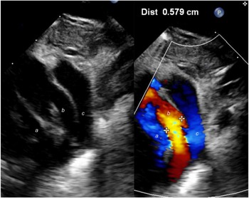
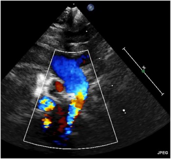
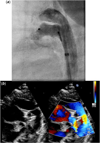
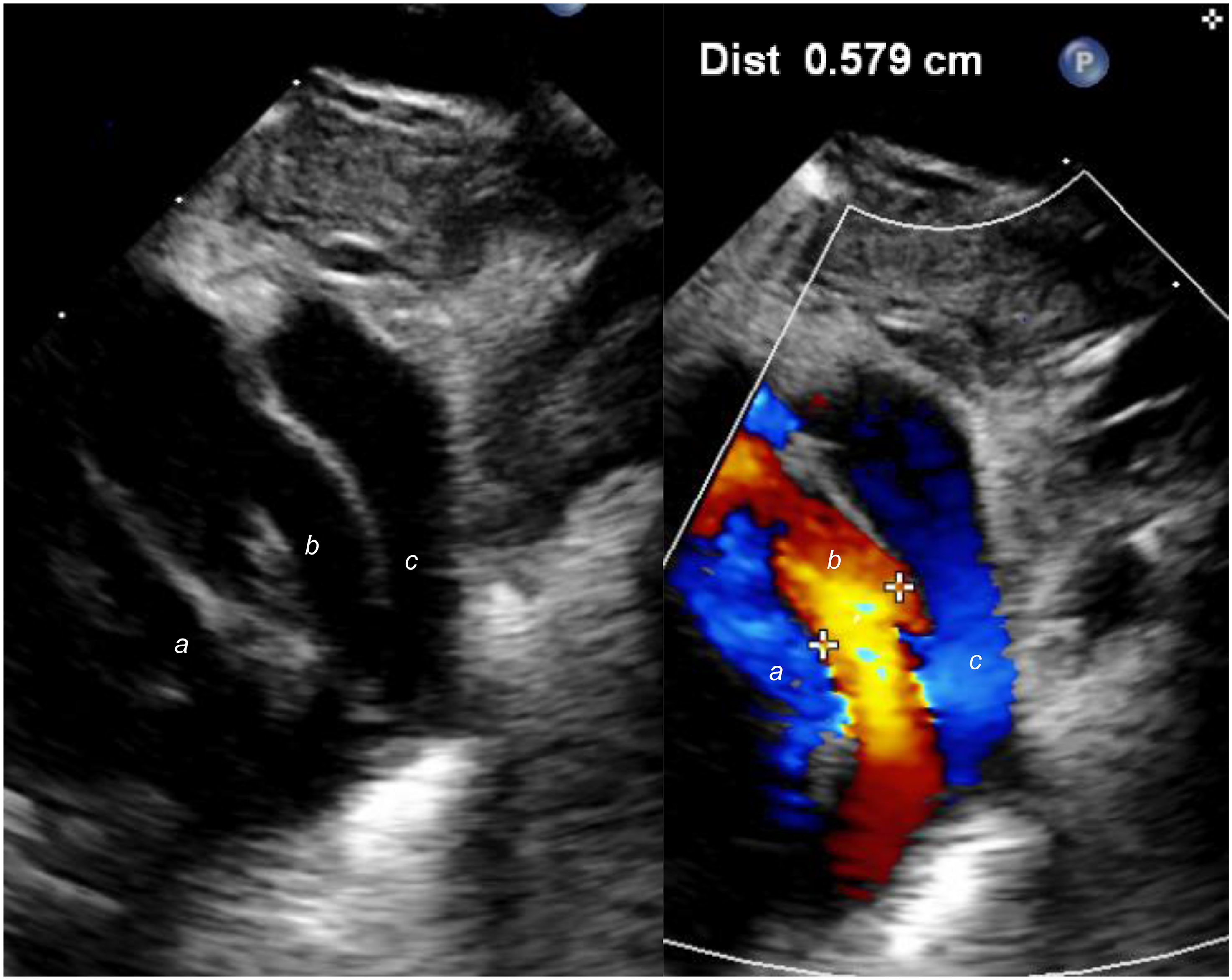
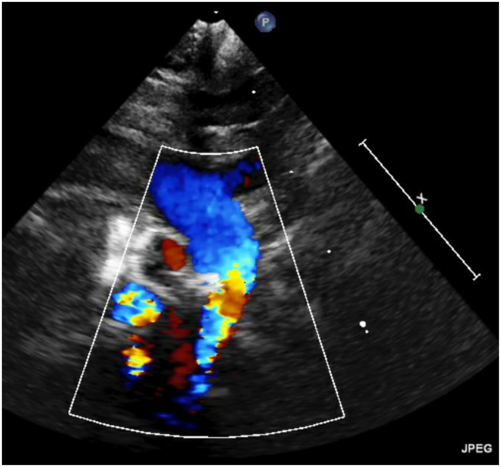
A neonate diagnosed antenatally with transposition of the great arteries, a small perimembranous ventricular septal defect and bilateral superior caval vein underwent caesarean section, at 38 weeks gestation, with a birthweight of 2.8 kg. Under prostaglandin infusion, balloon atrial septostomy was immediately performed, improving saturations to 80%. Echocardiography confirmed the aforementioned anatomy, also revealing a single coronary artery arising from the right anterior sinus. There was a large 5.8 mm ductus arteriosus (Fig 1) that remained patent after prostaglandin withdrawal. The baby experienced pulmonary over-circulation necessitating mechanical ventilation and necrotising enterocolitis which was managed conservatively. He became septic, with Klebsellia Oxytoca in blood cultures, treated with piperacillin-tazobactam, gentamicin, and linezolid. Surgical repair was delayed. At 14 days of life and 3.1 kg, he further deteriorated with liver failure. Surgical ligation of the ductus arteriosus was felt to be at too high risk due to the haemodynamic instability of the patient. Transcatheter closure of the patent ductus arteriosus was decided. Through a 5 French (Fr) sheath in right femoral artery, a lateral and right anterior oblique angiogram showed a large (5.2 mm) and short ductus, that was crossed with a 4Fr Judkins Right catheter. A Kimal Angioflex semi-rigid 150 cm × 0.035” wire (Kimal PLC, Worcester, UK) was positioned in the distal right pulmonary artery. A 5Fr (55 cm) Cook flexor sheath (Cook Medical, Bloomington, IN, USA) was advanced through the ductus over the wire. An 8 mm Amplatzer vascular plug II (Abbott, Chicago, Il, USA) was positioned. However, echocardiography revealed protrusion of the device in left pulmonary artery and aorta (Fig 2). The device was removed, and an 8 mm Amplatzer vascular plug was implanted (Fig 3a). Despite mild intra-prosthetic residual shunt, the device was efficient with increase of aortic diastolic pressure. It was successfully released. The baby dramatically improved and was extubated 2 days later.
Pre-procedural transthoracic echocardiography showing a large tubular PDA. ( a ) left pulmonary artery, ( b ) ductus arteriosus, ( c ) descending aorta.
Periprocedural transthoracic echocardiography after positioning of an 8 mm AVP II, causing aortic coarctation and left pulmonary artery obstruction.
( a ) Lateral angiogram showing implantation of an 8 mm AVP into the PDA with mild residual shunt. In ( b ), transthoracic echocardiography after implantation of the 8 mm AVP shows laminar flow in left pulmonary artery and descending aorta through colour Doppler flow.
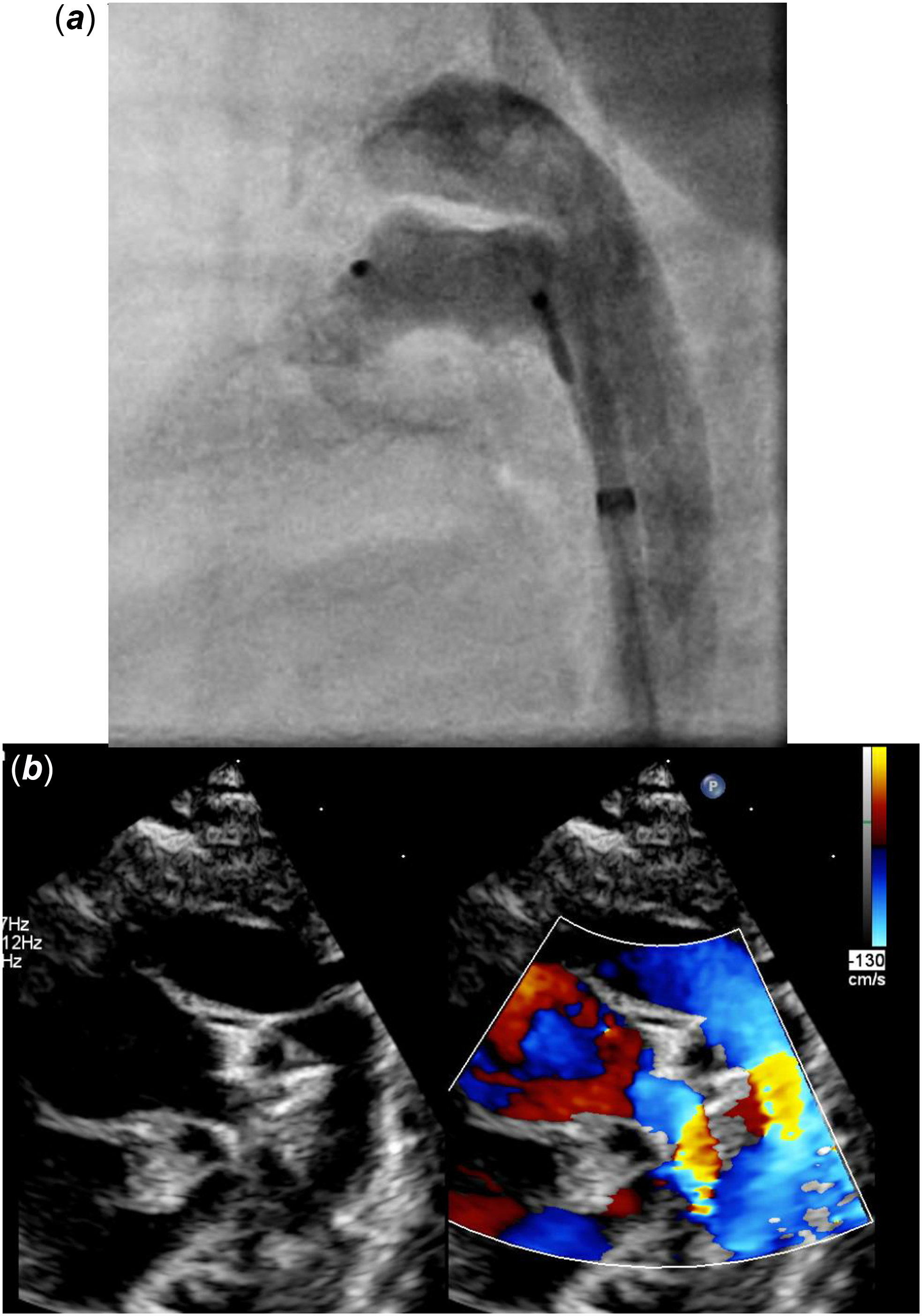
The patient continued to improve despite the residual shunt (Fig 3b) and underwent arterial switch on day 33 of life. The ductus arteriosus was ligated and divided, with the pulmonary end of the device externalised and the aortic end anchored with sutures. Post-operative course was complicated by extra-corporeal membrane oxygenation for 48 hours and delayed sternal closure for 7 days. The patient was successfully extubated after 15 days, but experienced bilateral chylothoraxes requiring parenteral nutrition and octreotide for 12 weeks. He was finally discharged home on day 156 of life. His echocardiogram showed an excellent surgical result with normal biventricular function, no residual shunt, and no aortic/pulmonary stenosis or regurgitation.
Discussion
To our knowledge, this is the first reported case of retrograde transcatheter closure of ductus arteriosus in transposition of the great arteries, using the Amplatzer vascular plug.
In babies with ductal-dependent circulation, cyanosis and decreased abdominal aortic blood flow may contribute to necrotising enterocolitis. Although this occurs more frequently in patients with single ventricle, Reference Choi, Song and Kim2,Reference Kelleher, McMahon and James3 a 2.1% prevalence is reported in transposition of the great arteries, with increased mortality. Reference Spinner, Morris and Nandi4 In our case, the alternative of surgical ligation was at very high risk because of the ongoing NEC, liver failure, and haemodynamic instability because of over-circulation through the PDA. Subsequently, the baby’s condition was dramatically improved by transcatheter closure of his patent ductus arteriosus closure, without worsening of the cyanosis.
It was not possible to use the dedicated relatively high-profile devices like the Amplatzer duct occluder (Abbott, Chicago, Il, USA) for this procedure. Additionally, the initial attempt with an 8 mm Amplatzer vascular plug II was unsuccessful despite the short 7 mm unconstrained length of the device, because of protrusion of the two proximal and distal disks after deformation of the central disk. A 6 mm vascular plug II was too small, like any Amplatzer piccolo device (Abbott, Chicago, Il, USA). Other devices like the Amplatzer vascular plug IV (Abbott, Chicago, Il, USA) that can be delivered through a 4 Fr catheter and has been used to close patent ductus arteriosus in small infants Reference Spinner, Morris and Nandi4 were too long (13.5 mm for the 8 mm diameter). Finally, the multifunction occluder device (Lifetech, Shenzhen, China) with a 4 mm waist between the two disks has been successful to close large ducts in small infants. Reference Grunenwald Gronier, Benbrik, Romefort, Prigent, Hauet and Baruteau5 However, its deployment is often associated with deformation of the right disk that could have protruded. Despite mild residual shunt due to a single layer of mesh (as opposed to six layers for the Amplatzer vascular plug II), the single lobe Amplatzer vascular plug was the best choice to close this very large and short duct in our patient. Although the unconstrained length was similar (7 mm) than for the 8 mm vascular plug II, there was minimal deformation thanks to the single lobe design. Only one other case of transcatheter closure case with the Amplatzer vascular plug has been reported in a 3 year old with a small duct. Reference Choi, Jang and Kim6 Although we were successful, the use of this device can only be recommended in very selected cases because of the risk of residual shunt and potential haemolysis.
In conclusion, we report successful transcatheter closure of a large and short patent ductus arteriosus with the Amplatzer vascular plug in a neonate with transposition of the great arteries and necrotising enterocolitis. The procedure was safe and effective. More experience is needed with such complex cases.