Background
Laryngocoeles are rare; it is estimated that one in 2.5 million individuals per year will develop a symptomatic laryngocoele.Reference Thomas and Madden1 They are most common in Caucasian males in the fifth to sixth decade of life, and are usually unilateral. Symptoms can include hoarseness, neck swelling, stridor, dysphagia, sore throat, cough and snoring, depending on the size and type of laryngocoele.Reference Cummings, Haughey, Thomas, Harker and Flint2
The laryngeal saccule is a pouch located in the most anterior portion of the roof of the ventricle, and is bordered by the false vocal fold, the inferior border of the epiglottis and the inner surface of the thyroid cartilage. It contains mucous glands which help to lubricate the vocal folds. Two main types of pathology can occur within the laryngeal saccule: laryngocoeles and saccular cysts. Both are abnormal dilations of the saccule. Laryngocoeles communicate with the laryngeal lumen, while saccular cysts do not and are filled with glandular secretions.Reference Cummings, Haughey, Thomas, Harker and Flint2 Laryngocoeles have also been described as abnormal dilations of the saccule which rise above the superior border of the thyroid cartilage.Reference Cummings, Haughey, Thomas, Harker and Flint2, Reference Close, Merkel, Burns, Deaton and Schaeffer3
Laryngocoeles are classified as internal, external or combined. Internal laryngocoeles extend postero-inferiorly within the aryepiglottic fold and the false vocal folds. External laryngocoeles protrude laterally through the thyrohyoid membrane. Combined laryngocoeles contain both internal and external components.Reference Cummings, Haughey, Thomas, Harker and Flint2, Reference Holinger, Barnes, Smid and Holinger4
Here, we report a case of combined laryngocoele which occurred in conjunction with adult-onset, recurrent respiratory papillomatosis.
Case report
A 53-year-old man presented with a four-month history of progressively worsening hoarseness. He denied odynophagia or otalgia, but complained of mild dysphagia with solids. He frequently spoke loudly, in his job as a construction electrician. He admitted drinking three to four bottles of beer per day, and had a 60 pack year history of smoking. Other relevant medical history included hypertension controlled with daily hydrochlorothiazide. There was no history of neck trauma or neck surgery.
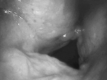
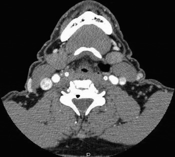
Flexible, stroboscopic laryngoscopy demonstrated masses on the true and false vocal folds, and a left laryngocoele (Figure 1). Computed tomography (CT) scanning demonstrated a combined laryngocoele (Figure 2).
Pre-operative flexible laryngeal stroboscopy image, showing papilloma and left laryngocoele.

Axial computed tomography scan showing a left combined laryngocoele.

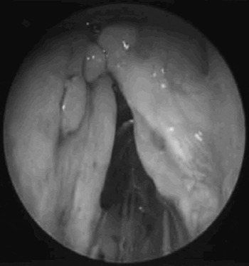
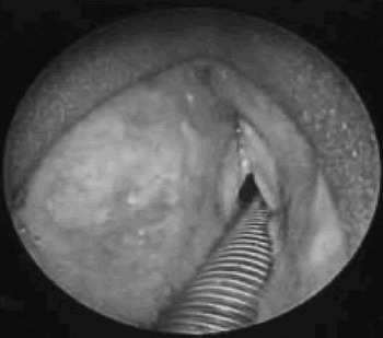
The patient was taken to the operating theatre for biopsy of a presumed squamous cell cancer causing the laryngocoele. However, upon suspension microlaryngoscopy with biopsy, the patient was found to have squamous papillomata on both true vocal folds and the left false vocal fold, plus a left laryngocoele. The papilloma on the left false and true vocal folds appeared to be blocking the left ventricle (Figures 3 and 4). Laser excision of the papillomata and marsupialisation of the left combined laryngocoele were performed.
Microlaryngoscopic view showing papillomata on the left false vocal fold and both true vocal folds.

Microlaryngoscopic view showing left laryngocoele.

The patient recovered without complication. At the time of writing, he has not presented with any recurrent papillomata or laryngocoele. His voice quality had improved and he had no further dysphagia.
Discussion
Laryngocoeles are thought to have first been described in 1826 by Larrey, a French military surgeon, who noticed these air-filled ‘tumours’ in blind men who called the hours with prayer from the Qur'an, and also in military instructors.Reference Ingrams, Hein and Merak5 In 1867, Virchow named this anomaly a laryngocele.Reference Virchow6
Laryngocoeles present in only one out of 2.5 million patients per year.Reference Thomas and Madden1 They can occur with or without cancer; however, there is a higher incidence of laryngocoele in patients with carcinoma of the larynx, compared with controls.Reference Close, Merkel, Burns, Deaton and Schaeffer3, Reference Birt7
The present case report demonstrates a unilateral combined laryngocoele occurring in conjunction with adult-onset respiratory papillomata. There are currently two other documented cases of saccular pathology accompanying papilloma in the English literature. Both cases involved younger patients who had been treated for recurrent respiratory papillomatosis for most of their childhood. Stone et al. described a six-year-old child with recurrent respiratory papillomatosis in whom a saccular cyst was drained and observed with no other surgical treatment, due to concern that removal would spread the papillomatosis.Reference Stone, Myer and Stern8 Dray et al. described a 27-year-old man with recurrent respiratory papillomatosis who displayed ventricular phonation, and in whom bilateral laryngocoeles had developed due to changes in laryngeal physiology.Reference Dray, Waugh and Hillel9
Two main theories of laryngocoele aetiology have been debated in the literature.
The first theory supports the idea of a laryngeal mass obstructing the exit of air from the ventricle during brief periods of increased intralaryngeal pressure, creating an environment conducive to laryngocoele formation.Reference Holinger, Barnes, Smid and Holinger4, Reference Ingrams, Hein and Merak5, Reference Stell and Marin10–Reference Celin, Johnson, Curtin and Barnes12 However, this theory does not explain contralateral laryngocoeles, bilateral laryngocoeles or laryngocoeles in patients with no observed laryngeal mass.
These particular presentations are better explained by the second theory, which focuses on laryngeal physiology. According to this theory, any cause of persistently increased intralaryngeal pressure, such as coughing, changes in neuromuscular mechanics or changes in phonatory mechanics, predisposes patients to develop laryngocoeles.Reference Close, Merkel, Burns, Deaton and Schaeffer3, Reference Birt7, Reference Dray, Waugh and Hillel9, Reference Harney, Patil, Walsh, Brennen and Walsh11–Reference Prasai, Phillips and Jani14 This theory also supports the formation of laryngocoeles in wind instrument and trumpet players and glass blowers.Reference Ingrams, Hein and Merak5, Reference Isaacson and Sataloff15 Dray et al. favoured this theory to explain bilateral laryngocoele formation in their recurrent respiratory papillomatosis patient displaying ventricular phonation.Reference Dray, Waugh and Hillel9
• Laryngocoeles are abnormal dilations of the laryngeal saccule, and are rare
• This report describes a case of unilateral combined laryngocoele occurring in conjunction with recurrent respiratory papillomatosis
• A papilloma can obstruct a laryngeal saccule, creating a ball valve effect which traps air and thus creates an environment conducive to laryngocoele formation
In our patient, however, no physiological laryngeal changes were observed on laryngoscopy. In addition, the papilloma located on the left false and true vocal folds appeared to obstruct the ventricle (Figure 3). Physiological laryngeal changes probably do explain the formation of many bilateral, contralateral and non-carcinomatous laryngocoeles. However, in the present case, it is likely that the patient's papilloma had the same anatomical effect as a cancer obstructing the ventricle; that is, a ball valve effect was created, resulting in blockage of the exit of air from the saccule when laryngeal pressure was increased.
Conclusion
This case report describes adult-onset, recurrent respiratory papillomatosis presenting with a concurrent combined laryngocoele. Laryngocoeles associated with papilloma are not often reported. It can be concluded that a papilloma, like a laryngeal carcinoma, may constitute an obstruction preventing air escaping from the saccule and thus increasing the likelihood of laryngocoele formation. However, it is important to note that changes in laryngeal physiology can explain other cases of laryngocoele formation.
Acknowledgements
We would like to thank Dr Christopher Chang for providing CT and pre-operative images.