The mechanical properties of np-Au, and more specifically its yield strength, have been evaluated and reported in numerous publications over the past 15 years, with a revived interest that has led to multiple studies in recent years.[ Reference Ngô, Stukowski, Mameka, Markmann, Albe and Weissmüller 1 – Reference Badwe, Chen and Sieradzki 5 ] Using scaling relations originally derived by Gibson and Ashby,[ Reference Gibson and Ashby 6 ] the yield strength of the individual interconnected ligaments, which create the three-dimensional and randomly oriented np structure, has been estimated to approach the theoretical strength of gold.[ Reference Biener, Hodge, Hayes, Volkert, Zepeda-Ruiz, Hamza and Abraham 7 – Reference Volkert, Lilleodden, Kramer and Weissmüller 9 ] However, the applicability of these scaling relations to np metals has been questioned, as they were originally developed for porous materials with ligament size in the micron range, whereas np metals typically have ligament sizes less than 100 nm. Because it is well known that the strength of metals increases as sample dimension decreases to the nanoscale, a ligament size effect is expected to contribute to the strength of np-Au. Several authors have proposed modified versions of the scaling relations using np-Au as a model material, considering ligament size effects as well as other factors such as surface effects or the np structure's randomness.[ Reference Roschning and Huber 4 , Reference Chen and Pugno 10 – Reference Hakamada and Mabuchi 14 ]
Early studies on the mechanical properties of np-Au used nanoindentation testing on bulk samples to extract Young's modulus and hardness values.[ Reference Biener, Hodge, Hayes, Volkert, Zepeda-Ruiz, Hamza and Abraham 7 , Reference Biener, Hodge, Hamza, Hsiung and Satcher 15 , Reference Hodge, Biener, Hayes, Bythrow, Volkert and Hamza 16 ] It was generally hypothesized that because of the porous nature of np-Au, the deformation of the structure would be accommodated by densification of the pores in the close vicinity of the indenter's surface, limited to a region directly below the center of the indent and within its diameter, and resulting in limited or no lateral deformation.[ Reference Hakamada and Mabuchi 14 , Reference Hodge, Biener, Hayes, Bythrow, Volkert and Hamza 16 ] In this scenario, the constraint factor typically observed for dense metals would approach 1, as the plastic Poisson's ratio becomes null, and the hardness value measured during nanoindentation could consequently be considered equal to the yield strength of the sample. In this way, the yield strength values reported for np-Au based on nanoindentation tests were taken to be equal to the hardness values.[ Reference Biener, Hodge, Hayes, Volkert, Zepeda-Ruiz, Hamza and Abraham 7 , Reference Hodge, Hayes, Caro, Biener and Hamza 8 , Reference Hakamada and Mabuchi 14 – Reference Hodge, Biener, Hayes, Bythrow, Volkert and Hamza 16 ] However, while such deformation behavior has been observed and reported for porous materials with relative densities much lower than np-Au,[ Reference Kumar, Ramachandra and Ramamurty 17 – Reference Wu, Yi, Huang, Zhang, Fang, Chang, Li, Oh, Lee, Kozlov, Chipara, Terrones, Xiao, Long, Huang, Zhang, Zhang, Lepro, Haines, Lima, Lopez, Rajukumar, Elias, Feng, Kim, Narayanan, Ajayan, Terrones, Aliev, Chu, Zhang, Baughman and Chen 20 ] its validity in the case of np-Au (and, by extension, of np metals in general), which typically have relative densities ranging from 0.25 to 0.40, is debatable.
Other studies have used more classic techniques such as tension and compression testing on millimeter-scale specimens.[ Reference Lührs, Soyarslan, Markmann, Bargmann and Weissmüller 2 , Reference Badwe, Chen and Sieradzki 5 , Reference Mameka, Wang, Markmann, Lilleodden and Weissmüller 21 – Reference Briot, Kennerknecht, Eberl and Balk 24 ] While these techniques offer the advantage of direct measurement and determination of Young's modulus, strength and Poisson's ratio, their implementation is challenging for a macroscopically brittle material such as np-Au. In addition to the cost of the raw material required for bulk np-Au specimen preparation, the required dealloying and miniature testing techniques become increasingly challenging, generally resulting in a limited number of valuable experiments from which to extract reliable, precise values.
Refining a new scaling relation for the yield strength of np-Au using experimental values necessitates the collection of a large dataset, from samples covering a wide range of parameters such as relative density and ligament size. Moreover, testing of other np metals (and potentially other types of np materials) is required to explore the universality of such scaling relations. Since np-Au fabrication is relatively straightforward, it has been used as the prototypical np material for almost all mechanical testing. However, preparation of other np metals in bulk form has had limited success, while np thin films of several metals have been produced. As a result, it appears that extracting the mechanical properties of np metal films using nanoindentation would rapidly yield a large amount of data needed to develop new scaling relations. Therefore, understanding the deformation behavior of np structures during indentation is essential, and this paper focuses on structural deformation and densification resulting from nanoindentation of np-Au.
In recent years, access to and use of focused ion beam (FIB) systems have increased, allowing for precise and direct observation of sub-surface regions. New characterization opportunities have arisen, leading to several recent studies refining our knowledge of np-Au structure.[ Reference Hu, Ziehmer, Wang and Lilleodden 25 – Reference Jeon, Kang, Gwak, Jang, Han, Hwang, Lee and Kim 27 ] In this paper, the deformed region created during nanoindentation of a bulk np-Au sample was characterized by cross-sectional imaging after FIB milling. Observations are discussed and linked to results previously obtained from mechanical testing of np-Au.
Gold-silver (AuAg) pellets with composition Au35Ag65 (Kurt J. Lesker Co.) were cut, rolled and annealed to provide large grains and compositional homogeneity. After surface polishing, the samples were dealloyed in nitric acid solutions following a process similar to that previously described by the present authors.[ Reference Briot, Kennerknecht, Eberl and Balk 24 ] Indentation tests were conducted on a NanoTest™ Vantage nanoindenter from Micro Materials Ltd., to a maximum load of 15 mN, at a loading/unloading rate of 25 µN/s. After scanning electron microscope (SEM) observation, an individual indent was selected for FIB milling. SEM imaging, platinum (Pt) growth, and FIB milling were all performed on an FEI Helios Nanolab 660. The surface of the indented region was first covered with Pt using the electron beam (2 kV accelerating voltage) to prevent subsequent exposure of the surface to the ion beam. More Pt was then grown on this initial layer using the ion beam (30 kV accelerating voltage) as additional protection for subsequent lift-out and thinning of the lamella.
It has been reported in past studies[ Reference Sun and Balk 28 ] that np-Au samples directly exposed (normal to the surface) to Ga+ ion beams in FIB systems experienced ligament coarsening and overall structure degradation. However, cross-sectioning after surface protection (with Pt) using modern FIB systems is believed to minimize such damage to materials, and observations in the current study of FIB-milled np-Au support this. Due to the nature of specimen preparation (the lamella is first extracted from the sample and re-attached to a copper grid before final thinning), only limited re-deposition from the milled portion of the lamella can occur during thinning, since the region of material removal is exposed to free space. Degradation of the np-Au structure during FIB milling has also been mitigated in other studies by impregnating the porous material with epoxy prior to ion beam exposure.[ Reference Hu, Ziehmer, Wang and Lilleodden 25 , Reference Wang and Weissmuller 29 ] Although this method was not explored in the present work, the authors believe that the FIB cross-sectioning steps performed here did not significantly alter the structure and do not interfere with characterization of the region deformed by nanoindentation.
High-resolution imaging was conducted using both secondary and backscattered electrons, to maximize visibility of the indented region and facilitate the identification of ligament edges. Relative density values in different areas under the indent were determined from cross-sectional images, using Adobe Photoshop. Each image was first converted to black and white using a threshold value set such that a (non-deformed) region away from the indent, but still within the same image, resulted in a binary image with 35% solid fraction, matching the initial relative density of the np-Au sample. Applying that same threshold to regions under the indent allowed calculation of local densification of the np-Au structure.
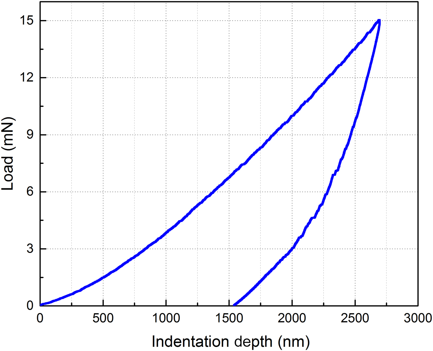
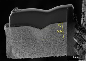
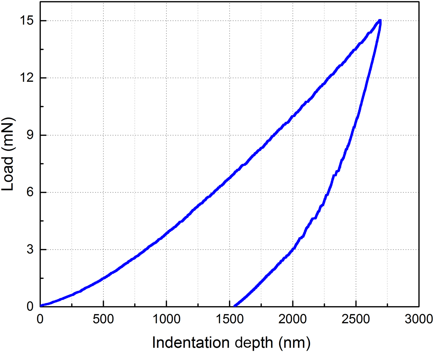
Figure 1 shows the indentation depth versus load plot for the indent analyzed during this work. A hardness of 92.2 MPa was calculated from this test, much higher than the yield strength reported during compression testing of bulk np-Au.[ Reference Bürckert, Briot and Balk 23 ] A FIB cross-section of the indented region is shown in Fig. 2. The permanently deformed region is clearly visible, and extends far ahead of the contact surface between indenter and specimen, more than three times the depth of the residual indent. In addition, no clear fully densified region is observed. Although the ligaments within this deformation zone appear to have been permanently deformed, they have not ruptured or collapsed completely. As shown in Fig. 1, a maximum depth of nearly 2.7 µm was reached at 15 mN, and a final indentation depth of just above 1.5 µm remained after load removal. The depth measured from the SEM image (annotation “a”) in Fig. 2 is in very good agreement with the final depth recorded at the conclusion of the indentation test, indicating that the FIB-milled cross-section plane is located at the center of the residual indent. This ensures that the full extent of deformation in the affected region was observed here. Because we ultimately aim to determine optimal conditions to conduct nanoindentation tests successfully on np-Au films (and np metal films in general), being able to capture the full magnitude of the deformed region is essential. The total depth of the permanently deformed region is also greater than the maximum indentation depth, providing further indication of the extent of the strain field due to indentation.
Load versus depth plot for the indent characterized during this work. The maximum depth reached during loading to 15 mN is shallower than the overall extent of remnant deformation (see Fig. 2). The residual depth measured at the indented sample surface (~1.5 µm) matches that observed in the cross-section of the indent.
Cross-sectional SEM image at the center of the indent. This region was lifted out using FIB techniques and re-attached to a copper grid for further thinning, followed by low-kV ion beam polishing. The dark, dense region above the indent is Pt deposited prior to milling, for protection of the surface during ion milling.

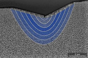
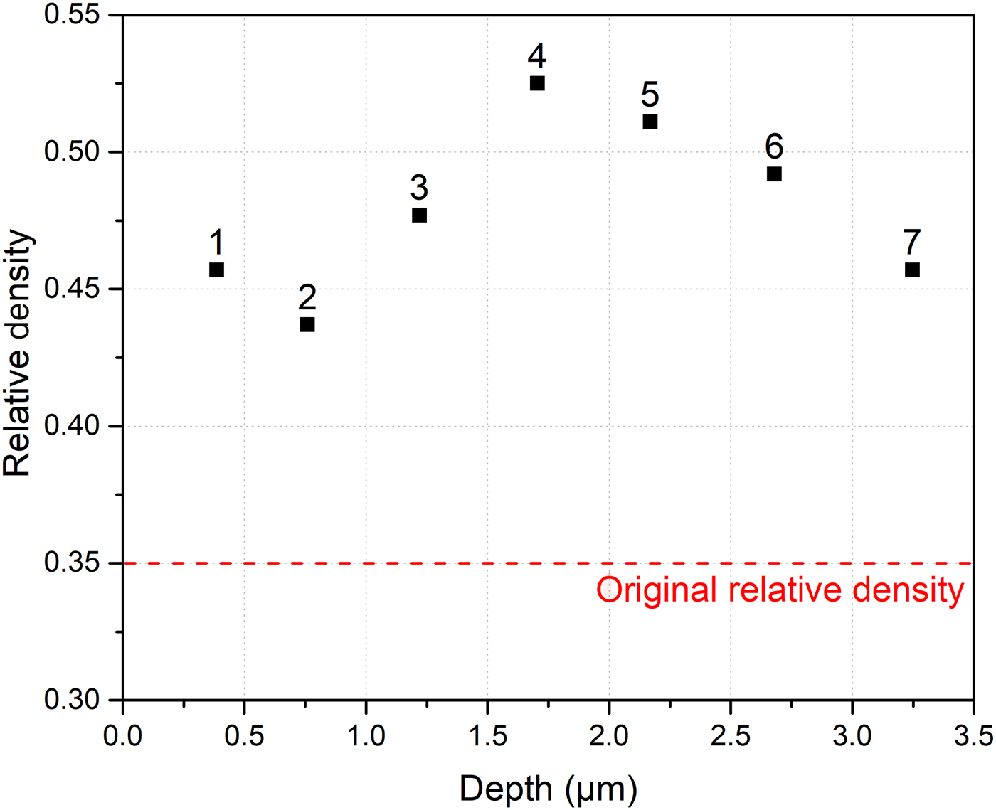
Figures 3 and 4 show the evolution of the np structure's relative density with respect to distance from the surface, directly under the center of the indent. The outermost boundary of the deformed region was identified based on SEM images of the cross-section. Concentric sub-regions were then demarcated, assuming uniform strain propagation from the center of the indent outward (zones 1–7). As described above, the relative density in each zone was determined using binarized images. In Fig. 3, the intensity of the blue overlay in each zone is proportional to the relative density, with darker shades corresponding to higher levels of densification. Very small ligament size near the sample surface, in addition to a large pore at the center of the indent, complicated the evaluation of relative density there, so this top layer region was excluded from calculations.
SEM cross-sectional image of the indented, permanently deformed region, which has been divided into seven zones for determination of relative density. The overlaid shades of blue represent the amount of densification, with darker bands corresponding to greater densification.

Distribution of np-Au relative density as a function of distance ahead of the indenter. Distances are measured on the SEM image of Fig. 3, from the tip of the residual indent to the center of each marked zone.
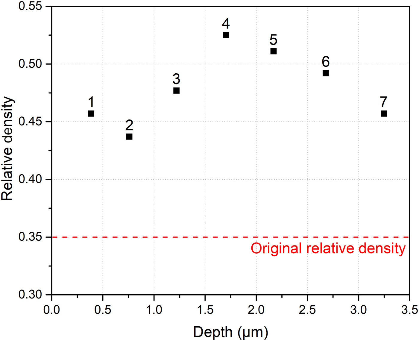
Complementing the observations made from Fig. 2, the absence of a fully densified region in the deformed structure is apparent in Fig. 3. A maximum densification of 50% was determined for zone 4, roughly 1.7 µm below the indented surface, where the relative density increased from 0.35 to 0.525. Interestingly, this zone was not directly adjacent to the indenter. Further investigations may help to better understand this distribution of deformation and its implications for np-Au mechanical behavior. Despite the noticeable increase in relative density, the porous nature of the structure still holds. Admittedly, the type of sample prepared here (cross-section revealing a single plane) does not directly offer a full view of the ligament structure or of connectivity out of the cross-section plane. In addition, this particular sample exhibited a finer ligament structure at the top surface; although a gradient in ligament size likely impacts the deformation behavior at the surface of the material, it should not significantly influence the propagation of deformation throughout the lower regions of the np structure, below the indentation as seen in Figs. 2 and 3. Nonetheless, if this variation in surface structure can be corrected, it may benefit future in-depth investigations of np metal indentation behavior.
Although measurable permanent deformation was created far ahead of the indenter, there was relatively little lateral deformation, and the deformed region was confined directly below the indentation. While this latter result agrees with hypotheses for materials with low relative density, the extent of deformation ahead of the indenter, albeit without any complete densification, is remarkable. This indicates that np-Au compressibility is limited during loading, which would imply that a constraint factor should apply. Fully compressible porous structures usually exhibit complete densification and pore collapse localized under the indenter, sometimes coupled with torn cells along punch indenter edges, for deep indents.[ Reference Kumar, Ramachandra and Ramamurty 17 ] Although the present study involved characterization of a shallow indent created with a Berkovich indenter and did not allow the observation of potential ligament tearing, full structural densification directly under the indenter was not detected. Consequently, the qualitative observations made from SEM images, combined with the hardness measured for this particular indent, indicate that the behavior of np-Au during nanoindentation aligns more closely with that of a dense metal than a low-density porous material. The latter observation implies that hardness results obtained by nanoindentation of np-Au cannot be directly taken as yield strength values, which explains the discrepancies in reported values for small-scale mechanical testing (tension/compression of bulk specimens) versus nanoindentation.
Previous bulk np-Au mechanical testing studies consistently reported strength values much lower than those extracted from nanoindentation testing, and it was recognized that applying a factor analogous to a constraint factor could reconcile the data. Jin et al. reported that values obtained from Vickers microhardness testing on bulk np-Au would agree with compression testing results on millimeter-scale bulk specimens, after dividing them by a factor of three.[ Reference Jin, Kurmanaeva, Schmauch, Rösner, Ivanisenko and Weissmüller 22 ] Likewise, the present authors harmonized values obtained from tensile testing of millimeter-scale bulk specimens with hardness values obtained from nanoindentation, by applying a factor of 2.65 (determined using experimentally measured values of Poisson's ratio).[ Reference Briot and Balk 13 ] The need for this constraint factor emerges precisely from the nature of strain propagation in np-Au during nanoindentation, revealed by the current study. The original assumption that pores would fully collapse under load, leading to complete densification of the structure directly under the indenter with the negligible constraint from the surrounding material (zero Poisson's ratio), does not agree with the observations described in the present paper.
In a recent study, the authors were able to measure Poisson's ratio beyond the initial yield point of np-Au during uniaxial compression of millimeter-scale specimens. The existence of a significant, non-zero lateral strain during plastic deformation was confirmed by calculated plastic Poisson's ratio values larger than 0.23 for structures with ligament size of ~60 nm.[ Reference Bürckert, Briot and Balk 23 ] Lührs et al.[ Reference Lührs, Soyarslan, Markmann, Bargmann and Weissmüller 2 ] reported a non-zero plastic Poisson's ratio during compression of np-Au, calculated from experimental and computational results. Interestingly, they observed an increase in plastic Poisson's ratio with increasing ligament size, although only reaching 0.2 for np-Au with ligament size larger than 120 nm, while structures with smaller ligaments (below 5 nm) indeed exhibited values of plastic Poisson's ratio approaching zero, as reported by Farkas et al.[ Reference Farkas, Caro, Bringa and Crowson 30 ] Complementary studies are underway to further understand the nature and extent of deformation during nanoindentation in np-Au, with the goal of establishing this technique for the determination of yield strength and Young's modulus of np metals, first in bulk form and then for thin films.
Acknowledgments
This work was supported by the National Science Foundation under Grant No. DMR-0847693. The authors acknowledge the Electron Microscopy Center at the University of Kentucky for access to the FEI Helios Nanolab 660 dual-beam FIB-SEM, and Ken Wu from FEI Company.