

Introduction
It is well known that microorganisms can survive in extreme environments such as the surface of glaciers or an ice sheet (for example Hodson and others Reference Hodson, Anesio and Tranter2008), or even directly in ice (Sheridan and others Reference Sheridan, Miteva and Brenchley2003). Many Archaea, bacteria, cyanobacteria, algae, fungi, ciliates and microinvertebrates (rotifers, tardigrades, arthropods etc.) have been reported from almost all investigated glaciers (for example Mieczan and others Reference Mieczan, Górniak and Świątecki2013a, Reference Mieczan, Górniak and Świątecki2013b; Sheridan and others Reference Sheridan, Miteva and Brenchley2003; Singh and others Reference Singh, Hanada and Singh2014b, Reference Singh, Singh and Dhakephalkar2014c; Takeuchi and Koshima Reference Takeuchi and Kohshima2004; Zawierucha and others Reference Zawierucha, Kolicka and Takeuchi2015). These glaciers and ice sheets should be regarded as independent biomes due to truncated food webs and distinct biogeographical structure, with the cryoconite holes as autonomous microecosystems (Anesio and Laybourn-Parry Reference Anesio and Laybourn-Parry2012; Wharton and others Reference Wharton, McKay and Simmons1985).
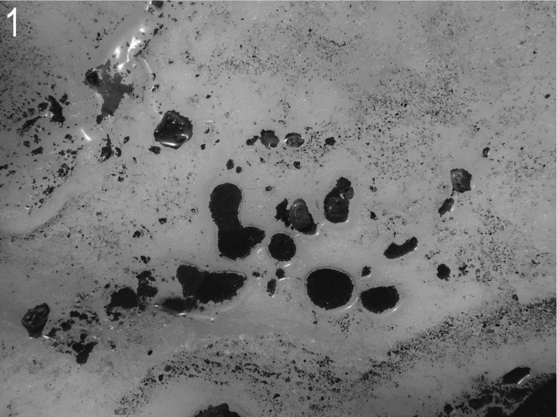
Cryoconite holes (Fig 1) are water-filled depressions situated in ablation zones on the glacier surfaces in polar and mountain regions (for example Gribbon Reference Gribbon1979; Hodson and others Reference Hodson, Anesio and Tranter2008; Wharton and others Reference Wharton, McKay and Simmons1985) and one of the most surveyed habitats in glacial ecosystems. The holes are formed due to the decrease of ice albedo in places where dark organic and inorganic debris are deposited on the glacier's surface and resultant localised melting (Gerdel and Drouet Reference Gerdel and Drouet1960; Hodson and others Reference Hodson, Anesio and Tranter2008; Wharton and others Reference Wharton, McKay and Simmons1985). Other important factors influencing the formation of cryoconite holes are the microorganisms assemblages of these unique habitats. Combined activity of primary producers with algal mats and bacteria in cryoconite holes results in the accumulation of dark-coloured organic matter and the reduction of ice albedo (Anesio and others Reference Anesio, Hodson and Fritz2009; Takeuchi and others Reference Takeuchi, Kohshima and Seko2001a). Moreover, cryoconite granules are formed by cyanobacteria (Hodson and others Reference Hodson, Cameron and Bøggild2010b) involved in the organic carbon cycles on the glaciers (Anesio and others Reference Anesio, Hodson and Fritz2009; Fig. 1).
Typical cryoconite holes in the ablation zone of the glacier.

Despite there being some inputs of microorganisms from non-glaciated habitats, cryoconite holes have distinct and independent microbial communities (Edwards and others Reference Edwards, Rassner and others2013c). Many species are directly connected with glacial habitats (Dastych and others Reference Dastych, Kraus and Thaler2003; Edwards and others Reference Edwards, Rassner and others2013c). But the impacts of local and regional conditions on the bacterial communities have been also observed (Edwards and others Reference Edwards, Mur and Girdwood2014). Despite the fact that glaciers and ice caps constitute 10% of the Earth's area and cryoconite holes cover approximately (circa) 10% of glaciers (in ablation zone) (Fountain and others Reference Fountain, Tranter and Nylen2004), the knowledge of organisms in glacial habitats is still poor. Scientific browsers such as Scopus or Web of Science reported only eight and nine papers, respectively, using the search term ‘cryoconite holes’ and ‘organisms’ and 26 and 36, respectively, using the terms ‘cryoconite holes’ and ‘bacteria (Table 1).
The number of results using the key words connected with different groups of microorganisms and cryoconite holes, cryoconite and glacier. A comparison of results from three different scientific browsers (Scopus, Web of Science (WoS) and Google Scholar (GS)). Accessed 15 December 2014).

Due to climate change, glaciers and ice sheets are among the most endangered habitats and vastly disappearing ecosystems (Hodson and others Reference Hodson, Anesio and Tranter2008; IPCC 2013). The first list of cryoconite hole organisms was presented as an MSc thesis by Mueller (Reference Mueller2001) and later published by Mueller and others (Reference Mueller, Vincent and Pollard2001). Although detailed, Mueller's list was not complete and now requires revising because many new taxa were reported from cryoconite holes in the past 14 years. Thus, in this paper an updated inventory of microorganisms (excluding data on micro-invertebrates, given in Zawierucha and others Reference Zawierucha, Kolicka and Takeuchi2015) is presented with a detailed geographical distribution and discussion on the importance of studies conducted on glacial microorganisms.
Material and methods
Literature database
The literature database incorporates all published papers on organisms (excluding invertebrates, which were published in a separate paper by Zawierucha and others Reference Zawierucha, Kolicka and Takeuchi2015) inhabiting cryoconite holes up to the end of November 2014. We are aware that it is possible we missed some papers so we ask all readers of this article to notify us about missing articles. We would be grateful for this and will add all subsequent papers and corrections to future editions of our database.
Format for presented records
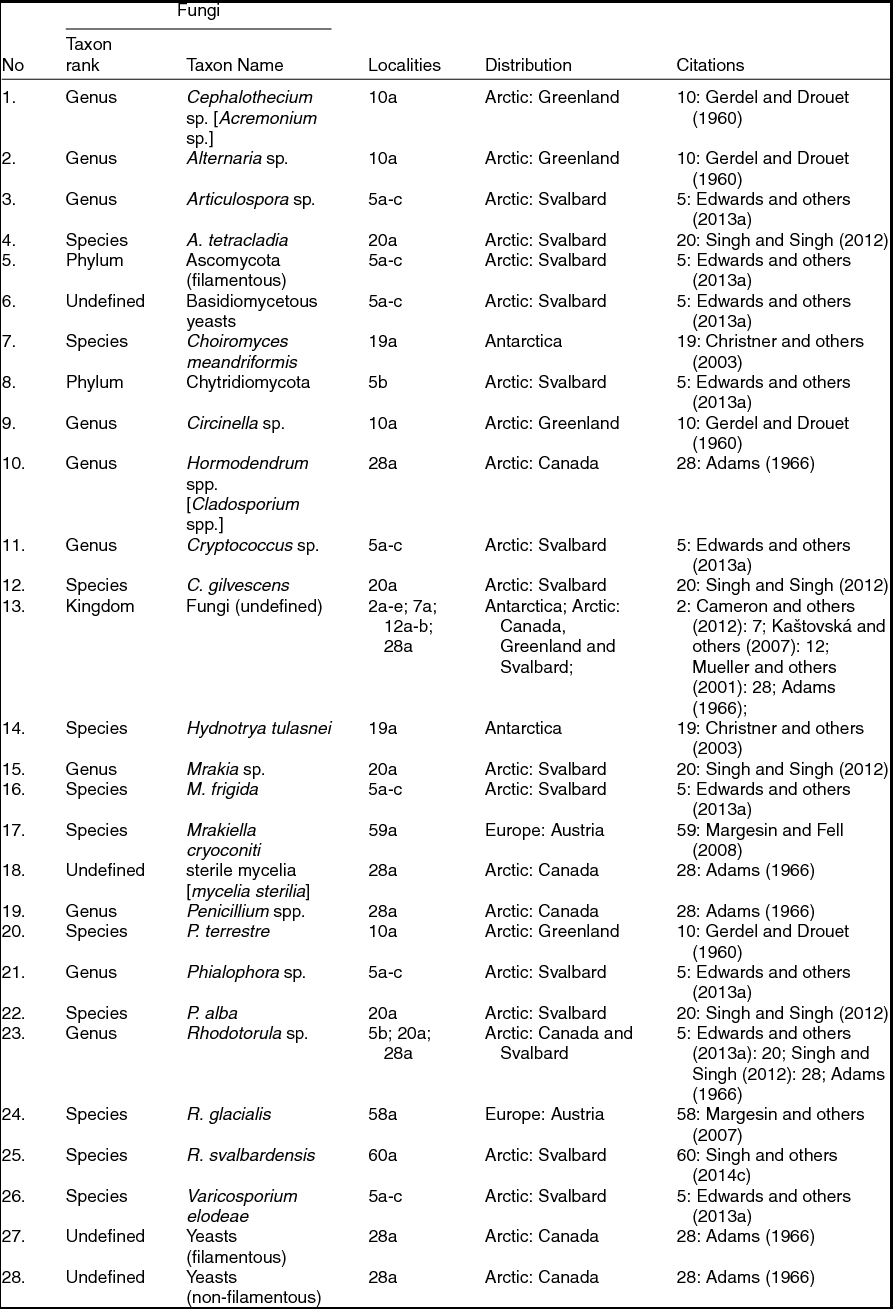
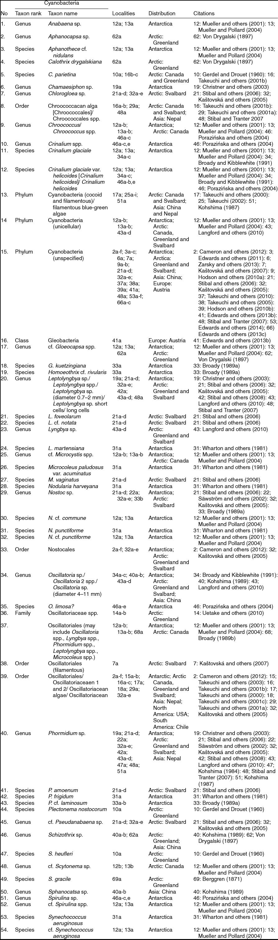
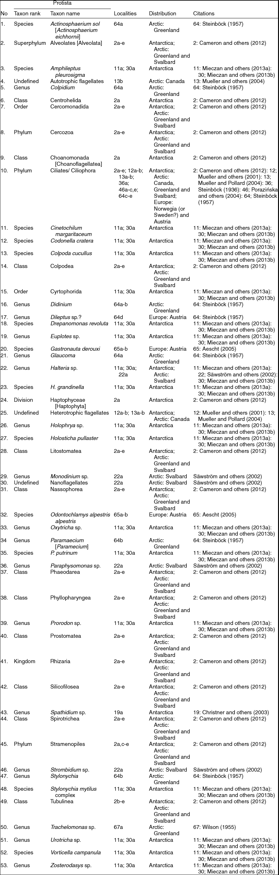
All records are presented in the form of five tables: Archaea, bacteria, and viruses (Table 2), fungi (Table 3), cyanobacteria (Table 4), algae (Table 5) and Protista (Table 6; see also Figs. 2–17). Taxa in the tables are listed in alphabetical, not taxonomical, order. Although taxonomic order is preferable, taxa presented in our tables have many different taxonomic ranks. Some taxa are lower or larger ranks of others also presented in the same table. Moreover, this presentation will be more understandable for non-biologists (for example climatologists, geochemists, glaciologists). In the tables we provide original names from the articles, even if the names were marked by question marks (?). However, if the name in the article was incorrect (for example as a result of changes in taxonomic nomenclature), we added the currently accepted name in square brackets [] behind the name appearing in the cited publication. To verify the correct taxonomic names, we used internet databases and search engines such as AlgaeBase, World Register of Marine Species (WoRMS), Fungal Databases and Protist Information Server, WoS, Scopus and Google Scholar. In some cases, if the names used by different authors were presented in different ways but referred to the same taxon (for example Chroococcus/ Chroococcus spp., Betaproteobacteria/ β-proteobacteria), we used all the names and separated them by slashes (/). In Tables 2–6, taxa without ‘scientific names’ (for example Lake Bonney clone, Freshwater lake clone, Industrial gas filter clone in Christner and others (Reference Christner, Kvitko and Reeve2003)) were deliberately omitted because they could lead to inaccurate estimates of biodiversity and confusion in future checklists and biogeographic studies.
Archaea, bacteria and viruses taxa (in alphabetic order) reported from cryoconite holes.
Fungi taxa (in alphabetic order) reported from cryoconite holes.
Cyanobacteria taxa (in alphabetic order) reported from cryoconite holes.

Algae taxa (in alphabetic order) reported from cryoconite holes.
Protista taxa (in alphabetic order) reported from cryoconite holes.
Bacteria and Fungi. 2: Janthinobacterium lividum; 3: Rhodococcus sp.; 4: Pseudomonas fluorescens; 5: conidia of Cladosporium sp. (Sources. 2: ‘Janthinobacterium lividum on TY’ by Ninjatacoshell - Own work. Licensed under CC BY-SA 3.0 (http://creativecommons.org/licenses/by-sa/3.0/) via Wikimedia Commons - https://commons.wikimedia.org/wiki/File:Janthinobacterium_lividum_on_TY.png#/media/File:Janthinobacterium_lividum_on_TY.png; 3: ‘Rhodococcus species’ by David Berd - This comes from the Centers for Disease Control and Prevention's Public Health Image Library (PHIL), with identification number #2981. Licensed under Public Domain via Wikimedia Commons - https://commons.wikimedia.org/wiki/File:Rhodococcus_species.jpg#/media/File:Rhodococcus_species.jpg; 4: ‘Pseudomonas fluorescens’ by Riraq25 - Own work. Licensed under CC BY-SA 3.0 (http://creativecommons.org/licenses/by-sa/3.0/) via Wikimedia Commons - https://commons.wikimedia.org/wiki/File:Pseudomonas_fluorescens.jpg#/media/File:Pseudomonas_fluorescens.jpg; 5: ‘Cladosporium sp conidia’. Licensed under CC BY-SA 3.0 (http://creativecommons.org/licenses/by-sa/3.0/) via Wikimedia Commons - https://commons.wikimedia.org/wiki/File:Cladosporium_sp_conidia.jpg#/media/File:Cladosporium_sp_conidia.jpg)
In the list of localities the geographic co-ordinates and altitude are provided with some modifications according to Kaczmarek and others (Reference Kaczmarek, Michalczyk and McInnes2014; Reference Kaczmarek, Michalczyk and McInnes2015) using different font formats depending on the data origin:
• Original data, where provided (also if they are presented only on the map), are in Roman and bold font.
• Where the original data were provided in decimal degrees and/or in imperial unit format, conversions to degree-minute and/or metric format are preceded by a slash (/) and in Roman and bold font.
• If the original data are inconsistent (for example co-ordinates or altitude do not correspond to the locality description or mismatched altitude for given co-ordinates), we offer values from the nearest probable site using Google Earth™ (ver. 6.2.2.6613), presented after a slash (/) and in Roman font.
• When original coordinates were not supplied, we provided estimated data in italics. In order to estimate coordinates and/or altitude, we used Google Earth™ (ver. 6.2.2.6613) and the following rules:
- where a general locality, administrative unit, or specified location was provided (for example glacier, mountain range, region, province, country), the collection site was approximated to the centre of the smallest viable unit (assisted by additional data, for example altitude);
- sample sites described as x distance in y direction from a named locality (for example a city), were approximated using Google™ Earth tools;
- the midpoint between localities A and B for sample sites reported as, ‘between locality A and B’, was used.
All collecting places in the list of localities are arranged in the following way: sequence number, author/authors of the article, year of publication, geographic region, area, locality, coordinates, and altitude (both in square brackets []). (Note: separate localities of the same author/authors are presented as a), b), c) etc.) Later, the same numbers are used in Tables 2–6 for specific taxa reported from an exact locality.
List of localities
1) Wharton and Vinyard (Reference Wharton and Vinyard1983): North America: Canada: a) Alberta State, Athabasca Mt., North Glacier [52°1 ′N, 117°13 ′W; 2,743 m asl].
2) Cameron and others (Reference Cameron, Hodson and Osborn2012): Antarctica: Signy Island: a) Tuva Glacier [60°41′S, 45°38′W;0 m asl]; Vestfold Hills: b) Sørsdal Glacier [68°39′S, 78°21′E;100 m asl]; Arctic: Greenland: c) Kangerlussuaq [67°09′N, 50°01′W;600 m asl]; Svalbard: d) Midre Lovénbreen [78°53′N, 12°03′E;250 m asl], e) Longyearbreen [78°10′N, 15°30′E;600 m asl], f) Foxfonna [78°08′N, 16°07′E;700 m asl].
3) Edwards and others (Reference Edwards, Anesio and Rassner2011): Arctic: Svalbard: a) Austre Brøggerbreen [78°55′N, 11°56′E/ 78°54′N, 11°50′E; 200 m asl], b) Midtre Lovénbreen [78°55′N, 11°56′E/ 78°54′N, 12°04′E; 100 m asl], c) Vestre Brøggerbreen [78°55′N, 11°56′E/ 78°55′N, 11°46′E; 100 m asl].
4) Lee and others (Reference Lee, Kim and Jung2011): Europe: Austria: a) Pasterze Glacier [47°04 ′N, 12°45 ′E; 2,200 m asl] b) Bankerferner [46°53′N, 11°05′E; 2,820 m asl], c) Pitztaler Jöchl Ferner [46°56′N 10°55′E; 2,875 m asl] d) Tiefenbachferner [46°55′N, 10°56′E; 2,900 m asl], e) Zugspitz Glacier [47°25′N, 10°59′E; 2,620 m asl] f) Eisgratferner - Stubaier glacier [46°59 ′N, 11°07 ′E; 2,900 m asl].
5) Edwards and others (Reference Edwards, Douglas and Anesio2013a): Arctic: Svalbard: a) Austre Brøggerbreen [78°55′N, 11°56′E/ 78°54′N, 11°50′E; 200 m asl], b) Midtre Lovénbreen [78°55′N, 11°56′E/ 78°54′N, 12°04′E; 100 m asl], c) Vestre Brøggerbreen [78°55′N, 11°56′E/ 78°55′N, 11°46′E; 100 m asl].
6) Zarsky and others (Reference Zarsky, Stibal and Hodson2013): Arctic: Svalbard: a) Aldegondabreen [77°59 ′N, 14°07 ′E; 200 m asl].
7) Kaštovská and others (Reference Kaštovská, Stibal and Sabacka2007): Arctic: Svalbard: a) Werenskioldbreen [77°05′N, 15°10′E/ 77°05′N, 15°20′E; 250 m asl].
8) Cook and others (Reference Cook, Hodson and Anesio2012): Arctic: Greenland: a) Leverett Glacier [67°04′17.1”N, 50°08′45.2”W-67°09′10.8”N, 48°22′14.6”W; 399–1,446 m asl].
9) Hodson and others (Reference Hodson, Bøggild and Hanna2010a): Arctic: Greenland: a) Kronprins Christian Land, undefined glacier [80°52′N 18°45′W], b) Kangerlussuaq [67°09 ′N, 50°01 ′W; 600 m asl].
10) Gerdel and Drouet (Reference Gerdel and Drouet1960): Arctic: Greenland: a) Thule area, undefined glacier [76°24 ′N, 68°12 ′W].
11) Mieczan and others (Reference Mieczan, Górniak and Świątecki2013a): Antarctica: a) King George Island, Ecology Glacier [62°10.226′S, 58°28.268′W/ 62°10′13”S, 58°28′16”W–62°10.404′S, 58°28.546′W/ 62°10′24”S, 58°28′33”W; 40–145 m asl].
12) Mueller and others (Reference Mueller, Vincent and Pollard2001): Antarctica: Taylor Valley: a) Canada Glacier [77°37′S, 162°55′E; 450–1,750 m asl]; Arctic: Canada: b) Axel Heiberg Island, White Glacier [79°27′N, 90°40′W; 75–800 m asl].
13) Mueller and Pollard (Reference Mueller and Pollard2004): Antarctica: Taylor Valley: a) Canada Glacier [77°37′S, 162°55′E;200 m asl]; Arctic: Canada: b) Axel Heiberg Island, White Glacier [79°27′N, 90°40′W;350 m asl].
14) Uetake and others (Reference Uetake, Naganuma and Hebsgaard2010): Arctic: Greenland: a) Qaanaaq Glacier [77°29′N, 69°14′W/ 77°30′N, 69°10′W; 276–783 m asl], b) Russel Glacier [67°09′N, 50°01′W; 510–635 m asl].
15) Takeuchi and others (Reference Takeuchi, Kohshima and Segawa2003): North America: USA: a) Alaska State, Worthington Glacier [61°10 ′N, 145°46 ′W; 550–1,110 m asl], b) Alaska State, Matanuska Glacier [61°44 ′N, 147°39 ′W; 550–1,110 m asl].
16) Takeuchi and others (Reference Takeuchi, Kohshima and Goto-Azuma2001b): Arctic: Canada: a) Buffin Island, Penny Ice Cap, G-H points, undefined glacier [67°13 ′N, 65°59 ′W; 450 and 660 m asl], b) Devon Island, Devon Ice Cap, A-C points, undefined glacier [75°21 ′N, 82°10 ′W; 300–1,065 m asl], c) Buffin Island, Penny Ice Cap, D-F points, undefined glacier [67°13 ′N, 65°59 ′W; 790–960 m asl].
17) Takeuchi and others (Reference Takeuchi, Kohshima and Yoshimura2000): Asia: Nepal: a) Langtang Region, Yala Glacier [28°14 ′N, 85°36 ′E; 5,110–5,240 m asl].
18) Takeuchi and others (Reference Takeuchi, Kohshima and Shiraiwa2001c): South America: Chile: a) Patagonian Icefield, Tyndall Glacier [51°15 ′S, 73°17 ′W; 150 m asl].
19) Christner and others (Reference Christner, Kvitko and Reeve2003): Antarctic: Taylor Valley: a) Canada Glacier [77°37 ′S, 162°59 ′E; 200 m asl].
20) Singh and Singh (Reference Singh and Singh2012): Arctic: Svalbard: a) Midre Lovénbreen [78°55′N, 11°56′E/ 78°54′N, 12°04′E; 100 m asl].
21) Stibal and others (Reference Stibal, Sabacka and Kaštovská2006): Arctic: Svalbard: a) Hansbreen [77°05′N, 15°10′E/ 77°04′N, 15°39′E; 300 m asl], b) Werenskioldbreen [77°05′N, 15°10′E/ 77°05′N, 15°20′E; 250 m asl], c) Nannbreen [77°05′N, 15°10′E/ 77°08′N, 15°18′E; 400 m asl], d) Austre Torellbreen [77°05′N, 15°10′E/ 77°10′N, 15°10′E; 200 m asl].
22) Säwström and others (Reference Säwström, Mumford and Marshall2002): Arctic: Svalbard: a) Midre Lovénbreen [78°54 ′N, 12°04 ′E; 100 m asl].
23) Säwström and others (Reference Säwström, Granéli and Laybourn-Parry2007): Arctic: Svalbard: a) Midre Lovénbreen [78°55′N, 11°56′E/ 78°54′N, 12°04′E; 100 m asl].
24) Foreman and others (Reference Foreman, Sattler and Mikucki2007): Antarctica: Taylor Valley: a) Canada Glacier [77°00′S, 162°52′E/ 77°37′S, 162°59′E; 200 m asl], b) Commonwealth Glacier [77°00′S, 162°52′E/ 77°35′S, 163°16′E; 200 m asl], c) Hughes Glacier [77°00′S, 162°52′E/ 77°44′S, 162°28′E; 400 m asl].
25) Takeuchi (Reference Takeuchi2002): Asia: Nepal: a) Langtang Region, Yala Glacier [28°N, 86°E/ 28°14′N, 85°36′E; 5,150 m asl], b) Shorong Region, AX010 Glacier [28°N, 87°E/ 27°42′N, 86°34′E; 5,000 m asl], c) Makut Region, RikhaSamba Glacier [29°N, 83°E/ unknown locality; 5,350 m asl]; China: d) Qinghai Province, Meikuang Glacier [35°N, 93°E/ 35°41′N, 94°11′E; 4,900 m asl], e) Tibet Autonomous Region, Gozha Glacier [35°N, 81°E/ 35°14′N, 81°04′E; 5,800 m asl], f) Qinghai Province, Xiao Dongkemadi Glacier [33°N, 92°E/ 33°04′N, 92°05′E; 5,600 m asl]; Arctic: Svalbard: g) Austre Brøggerbreen [79°N, 12°E/ 78°54′N, 11°50′E; 200 m asl]; Canada: h) Buffin Island, Penny Ice Cap, Greenshield Glacier [67°N, 66°W/ 67°07′N, 67°05′W; 490 m asl], i) Devon Island, Devon Ice Cap, Sverdrup Glacier [76°N, 83°W/ 75°41′N, 83°15′W; 350 m asl].
26) Anesio and others (Reference Anesio, Mindl and Laybourn-Parry2007): Arctic: Svalbard: a) Austre Brøggerbreen [78°53′N, 12°04′E/ 78°54′N, 11°50′E; 50–600 m asl], b) Midre Lovénbreen [78°53′N, 12°04′E/ 78°54′N, 12°04′E; 50–600 m asl].
27) Anesio and others (Reference Anesio, Sattler and Foreman2010): Arctic: Svalbard: a) Austre Brøggerbreen [78°53′N, 12°04′E/ 78°54′N, 11°50′E; 50–600 m asl], b) Midre Lovénbreen [78°53′N, 12°04′E/ 78°54′N, 12°04′E; 50–600 m asl]; Europe, Austria: c) Rotmoosfermer [46°50′N, 11°30′E/ 46°50′N, 11°03′E; 2,430 m asl], d) Stubacher Sonnblickkees Glacier [47°13′N, 12°60′E/ 47°08′N, 12°36′E; 2,500–3,050 m asl]; Antarctica: Horseshoe Valley: e) Patriot Hills [80°18′S, 81°21′W;1,000 m asl]; Taylor Valley: f) Canada Glacier [77°00′S, 162°52′E/ 77°37′S, 162°59′E; 200 m asl], g) Commonwealth Glacier [77°00′S, 162°52′E/77°35 ′S, 163°16 ′E; 200 m asl], h) Taylor Glacier [77°00′S, 162°52′E/77°43 ′S, 162°16 ′E; 200 m asl].
28) Adams (Reference Adams1966): Arctic: Canada: a) Axel Heiberg Island, White Glacier. b) Axel Heiberg Island, White Glacier [79°27 ′N, 90°40 ′W; 350 m asl].
29) Takeuchi and others (Reference Takeuchi, Kohshima and Seko2001a): Asia: Nepal: a) Langtang Region, Yala Glacier [28°14 ′N, 85°36 ′E; 5,100–5,750 m asl].
30) Mieczan and others (Reference Mieczan, Górniak and Świątecki2013b): Antarctica: a) King George Island, Ecology Glacier [62°10.226′S, 58°28.268′W/ 62°10′13”S, 58°28’16”W–62°10.404′S, 58°28.546′W/ 62°10′24”S, 58°28′33” W; 40–145 m asl].
31) Wharton and others (Reference Wharton, Vinyard and Parker1981): Anarctica: Taylor Valley: a) Canada Glacier [77°38′S, 162°53′E;100 m asl]
32) Kaštovská and others (Reference Kaštovská, Elster and Stibal2005): Arctic: Svalbard: a) Vestre Brøggerbreen [78°55 ′N, 11°46 ′E; 100 m asl], b) Austre Brøggerbreen [78°54 ′N, 11°50 ′E; 200 m asl], c) Vestre Lovénbreen [78°54 ′N, 11°57 ′E; 200 m asl], d) Midtre Lovénbreen [78°54 ′N, 12°04 ′E; 100 m asl], e) Austre Lovénbreen [78°53 ′N, 12°09 ′E; 200 m asl].
33) Broady (Reference Broady1989a): Antarctica: Marie Byrd Land: a) Washington Ridge [77°00′–78°30′S, 152°–154°W/ 78°06′S 154°48′W; 450 m asl], b) Mount Paterson [77°00′–78°30′S, 152°–154°W/ 78°02′S, 154°37′E; 400 m asl].
34) Broady and Kibblewhite (Reference Broady and Kibblewhite1991): Antarctica: Ganvood Valley: a) Joyce Glacier [78°02′S, 163°46′E;550 m asl], b) Garwood Glacier [78°01′S, 163°56′E;550 m asl]; Taylor Valley: c) Canada Glacier [77°37′S, 163°02′E;50 m asl].
35) Singh and others (Reference Singh, Singh and Dhakephalkar2014a): Arctic: Svalbard: a) Midre Lovénbreen [78°53′N, 12°04′E;100 m asl], b) Austre Brøggerbreen [78°53′N, 12°04′E/ 78°54′N, 11°50′E; 200 m asl], c) Vestre Brøggerbreen [78°53′N, 12°04′E/ 78°55′N, 11°46′E; 100 m asl].
36) Steinböck (Reference Steinböck1936): Arctic: Greenland: a) Disko Island, ice highland near Godhaven [Qeqertarsuaq] [69°18 ′N, 53°22 ′W; 700 m asl].
37) Takeuchi and others (Reference Takeuchi, Nishiyama and Li2010): Asia: China: a) Xinjiang Uyghur Autonomous Region, Ürümqi Glacier No. 1 [43°06′N, 86°48′E; 3,765m asl, 3,820 m asl and 3,870 m asl].
38) Takeuchi and others (Reference Takeuchi, Matsuda and Sakai2005): Asia: China: a) Gansu Province, 1st Glacier (Qiyi Glacier) [39°15′N, 97°45′E; 4,305–5,159 m asl].
39) Hodson and others (Reference Hodson, Cameron and Bøggild2010b): Arctic: Svalbard: a) Longyearbreen [78°10′49”N, 15°30′21”E;500 m asl].
40) Kohshima (Reference Kohshima1989): Asia: China: a) Tibet Autonomous Region, Chongce Ice Cap (Chongce Glacier) [35°14′N, 81°07′E; 6,120–6,160 m asl], b) Tibet Autonomous Region, Gozha Glacier [35°14 ′N, 81°04 ′E; 5,400–5,900 m asl].
41) Edwards and others (Reference Edwards, Pachebat and Swain2013b): Europe: Austria: a) Rotmoosferner [46°50′N, 11°03′E; 2,450 m asl].
42) Stibal and others (Reference Stibal, Tranter and Benning2008): Arctic: Svalbard: a) Werenskioldbreen [77°05 ′N, 15°20 ′E; 250 m asl].
43) Langford and others (Reference Langford, Hodson and Banwart2010): Arctic: Svalbard: a) Vestfonna, undefined glacier [79°56′N, 20°11′E], b) Midtre Lovénbreen [78°54 ′N, 12°04 ′E; 100 m asl], c) Longyearbreen [78°11 ′N, 15°31 ′E; 500 m asl]; Greenland: d) Kronprins Christian Land, undefined glacier [80°52′N, 18°45′W], d) Kangerlussuaq [67°09 ′N, 50°01 ′W; 600 m asl].
44) Van de Vijver and others (Reference Van de Vijver, Mataloni and Stanish2010): Antarctica: Taylor Valley: a) Commonwealth Glacier [77.57674°S, 163.27231°E/ 77°35′S, 163°16′E;200 m asl], b) Canada Glacier [77°37 ′S, 162°59 ′E; 200 m asl].
45) Margesin and others (Reference Margesin, Spröer and Schumann2003) Europe: Austria: a) Stubaier Glacier [46°59 ′N, 11°07 ′E; 3,000 m asl].
46) Porazińska and others (Reference Porazinska, Fountain and Nylen2004): Antarctica: Taylor Valley: a) Commonwealth Glacier [77°35 ′S, 163°16 ′E; 200 m asl], b) Canada Glacier [77°37 ′S, 162°59 ′E; 200 m asl], c) Howard Glacier [77°40 ′S, 163°05 ′E; 400 m asl], d) Hughes Glacier [77°44 ′S, 162°28 ′E; 400 m asl], e) Taylor Glacier [77°43 ′S, 162°16 ′E; 200 m asl].
47) Kohshima (Reference Kohshima1984): Asia: Nepal: a) Langtang Region, Yala Glacier [28°14 ′N, 85°36 ′E; 5,100–5,600 m asl].
48) Stibal and Tranter (Reference Stibal and Tranter2007): Arctic: Svalbard: a) Werenskioldbreen [77°05′N, 15°15′E; 65–600 m asl].
49) McIntyre (Reference McIntyre1984): North America: Canada: a) British Columbia, Manatee Glacier [50°35′N, 123°40′W; 1,500–2,100 m asl].
50) Teiling and others (Reference Telling, Anesio and Hawkings2010): Arctic: Svalbard: a) Midtre Lovénbreen [78°55′48”N, 11°56′59”E/ 78°54′N, 12°04′E; 100 m asl], b) Vestre Brøggerbreen [78°55′48”N, 11°56′59”E/ 78°55′N, 11°46′E; 100 m asl], c) Austre Brøggerbreen [78°55′48”N, 11°56′59”E/ 78°54′N, 11°50′E; 200 m asl].
51) Kohshima (Reference Kohshima1987): Asia: Nepal: a) Langtang Region, Yala Glacier [28°14 ′N, 85°36 ′E; 5,100–5,700 m asl].
52) Bellas and others (Reference Bellas, Anesio and Telling2013): Arctic: Svalbard: a) Austre Brøggerbreen [78°55′N, 11°55′W/ 78°54′N, 11°50′E; 200 m asl], b) Midre Lovénbreen [78°55′N, 11°55′W/ 78°54′N, 12°04′E; 100 m asl]; Greenland: c) Russell Glacier [67°09′39.7”N, 50°00′52.7”W;650 m asl].
53) Edwards and others (Reference Edwards, Mur and Girdwood2014): Arctic: Svalbard: a) Austre Brøggerbreen [78°53′54”N, 11°48′17”E; 190–310 m asl], b) Midre Lovénbreen [78°53′04”N, 12°02′17”E; 170–300 m asl], c) Vestre Brøggerbreen [78°54′45”N, 11°43′16”E; 140–270 m asl]; Greenland: d) Leverett Glacier [67°04′17”N–67°07′36”N, 49°00′36”W–50°08′45”W; 400–1,190 m asl]; Europe: Austria: e) Rotmoosferner [42°49′17”N, 11°02′47”E/ 46°49′N, 11°03′E; 2,620–2,660 m asl], f) Gaisbergferner [46°49′50”N, 11°03′59”E; 2,480–2,590 m asl], g) Pfaffenferner [46°57′42”N, 11°08′01”E; 2,780–2,880 m asl].
54) Lutz and others (Reference Lutz, Anesio, Villar and other2014): Arctic: Greenland: a) Mittivakkat Glacier [65.6848°N–37.8802°E/ 65°41′N, 37°53′W; 150–250 m asl].
55) Stanish and others (Reference Stanish, Bagshaw and McKnight2013): Antarctica: Taylor Valley: a) Canada Glacier [77°37 ′S, 162°59 ′E; 200 m asl], b) Commonwealth Glacier [77°35 ′S, 163°16 ′E; 200 m asl], c) Taylor Glacier [77°43 ′S, 162°16 ′E; 200 m asl]
56) Yallop and Anesio (Reference Yallop and Anesio2010): Arctic: Svalbard: a) Vestre Brøggerbreen [78°53′N, 12°04′E/ 78°55′N, 11°46′E; 100 m asl], b) Austre Brøggerbreen [78°53′N, 12°04′E/ 78°54′N, 11°50′E; 200 m asl]; Greenland: c) Frøya Glacier [74°24′N, 20°50′W; 800 m asl], d) Cirque Gacier [74°30′N, 20°46′W; 950 m asl]
57) Singh and others (Reference Singh, Hanada and Singh2014b): Arctic: Svalbard: a) Midre Lovénbreen [78°54 ′N, 12°04 ′E; 100 m asl].
58) Margesin and others (Reference Margesin, Fonteyne, Schinner and other2007): Europe: Austria: a) Stubaier Glacier [46°59′12”N, 11°06′53”E; 2,900 m asl].
59) Margesin and Fell (Reference Margesin and Fell2008): Europe: Austria: a) Stubaier Glacier [46°59 ′N, 11°07 ′E; 2,900 m asl].
60) Singh and others (Reference Singh, Singh and Tsuji2014c): Arctic: Svalbard: a) Midre Lovénbreen [78°55′N, 11°56′E/ 78°54′N, 12°04′E; 100 m asl].
61) Telling and others (Reference Telling, Anesio and Tranter2014): Antarctica: Taylor Valley: a) Canada Glacier [77°37 ′S, 162°59 ′E; 200 m asl].
62) Von Drygalski (Reference Von Drygalski and Kühl1897) according to Mueller and others (Reference Mueller, Vincent and Pollard2001): Arctic: Greenland: a) undefined glacier [71°42 ′N, 42°32 ′E]*.
63) Wittrock (Reference Wittrock1885) according to Steinböck (Reference Steinböck1936): Arctic: Greenland: a) south Greenland, unspecified glacier [78°00 ′N, 43°52 ′W]*.
64) Steinböck (Reference Steinböck1957): Arctic: Greenland: a) Scoresbysund (Kangertittivaq), drift ice near Liverpool coast [70°30 ′N, 25°01 ′W; 0 m asl], b) undefined locality, Scoresbysundes Glacier [70°30 ′N, 25°01 ′W]; Europe: c) Norwegia (or Sweden?): undefined locality, S Abisko, Korsawagga Glacier [68°N/68°20 ′N, 18°50 ′E); Austria: d) Sulztalerferner [47°01 ′N, 11°05 ′E; 2,530 m asl] e) Niederjochferner [46°47 ′N, 10°52 ′E; 3,000 m asl].
65) Aescht (Reference Aescht2005): Europe: Austria: a) Rotmoosferner [46°49′N, 11°02′E; circa 2,720 m asl], b) Gaisbergferner [46°50′N, 11°03′E; circa 2,600 m asl].
66) Edwards and others (Reference Edwards, Rassner and others2013c): Arctic: Svalbard: a) Austre Brøggerbreen [78°54′N, 11°50′E;200 m asl], b) Midtre Lovénbreen [78°54′N, 12°04′E;100 m asl], c) Vestre Brøggerbreen [78°55′N, 11°46′E; 100 m asl].
67) Wilson (Reference Wilson1955) according to Mueller and others (Reference Mueller, Vincent and Pollard2001): Arctic: Greenland: a) Thule Area, undefined glacier [76°24 ′N, 68°12 ′W]*.
68) Broady (Reference Broady1989b): Antarctica: Ross Island: a) Cape Bird, Mt. Bird Ice cap [77°15′S, 166°27′E;400 m asl].
69) Berggren (Reference Berggren1871): Arctic: Greenland: a) undefined glacier [71°42 ′N, 42°32 ′E].
* The authors have not had the possibility of examining these papers directly and we have only cited a list of taxa from them according to later authors.
Results and discussion
In total 370 taxa (excluding invertebrates which were listed in a previous paper by Zawierucha and others Reference Zawierucha, Kolicka and Takeuchi2015) were reported from cryoconite holes throughout the world (mostly from polar regions and European Alps) (Tables 2–6). However, most of the taxa were not identified to the species level. Up to now only 39 species or subspecies of bacteria and Archaea, 11 fungi, 17 cyanobacteria, 62 algae, and 13 Protista are known from cryoconite holes, which is approximately 38% of all taxa reported from these environments. Almost 62% of the taxa were marked as cf. (confer) or were identified only to the genera or even to the higher taxonomic units (such as families or orders) (see Tables 2–6). Additionally, many taxa (often identified to the species level) reported in the older papers need a confirmation according to modern taxonomy or molecular methods (=integrative taxonomy) (for example Casamatta and others Reference Casamatta, Vis and Sheath2003; Fenchel and others Reference Fenchel, Esteban and Finlay1997; Heger and others Reference Heger, Mitchell and Todorov2010, see also remarks in Zawierucha and others Reference Zawierucha, Kolicka and Takeuchi2015).
Most ‘cryoconite taxa’ are known from polar glaciers in the Arctic (217, circa 60%) and Antarctic regions (156, circa 43%). Fewer taxa are known from mountain glaciers in Europe (52, circa 14%), Asia (19, circa 5%), North America (15, circa 4%), and South America (5, circa 1%). This is, of course, a result of 1) the distribution of glaciated areas, which are located mainly in the polar regions, and the large number of studies conducted in these regions (Antarctica, 14 papers, circa 20%; Arctic, 39 papers, circa 57%); 2) locations of research stations, and 3) accessibility of particular glaciers. However, a relatively large number of taxa have also been reported from small central European glaciers (mainly in Austria) (3,785 square kilometers, according to WGMS report: http://www.grid.unep.ch/glaciers/), which is definitively related to the large number of studies conducted in this area (eight papers, circa 12%). A low number of cryophilic taxa have been reported from North and South America where glaciers cover 124,000 and 25,500 square kilometers, respectively (WGMS report: http://www.grid.unep.ch/glaciers/). This is probably due to the low number of studies focused on cryoconite hole organisms in these regions (only four papers, circa 6%). Surprisingly, also in Asia, with 174,000 square kilometers of ice cover areas, the number of reported taxa is also very low, although the number of papers related to this region is relatively great (eight papers, circa 12%). Here, the small number of taxa is probably due to the fact that the papers are not focused on cryoconite hole organisms but on the morphology and functioning of the ‘holes’ themselves (for example Gribon Reference Gribbon1979; Fountain and others Reference Fountain, Tranter and Nylen2004; MacDonell and Fitzsimons Reference MacDonell and Fitzsimons2008). Until now, cryoconite hole microorganisms have not been reported from New Zealand and Scandinavian (excluding invertebrates and undefined Protista) glaciers which cover respectively 1,600 and 2,940 square kilometers. Similarly, no organisms were reported from African and New Guinean glaciers, but ice-covered areas in these regions are very small (6 and 3 square kilometers, respectively) (WGMS report: http://www.grid.unep.ch/glaciers/). Hence, cryoconite organisms are rather poorly known except for in a few well-studied small areas (Figs. 2–9).
Despite the fact that first studies on the organisms inhabiting glaciers took place towards the beginning of the twentieth century (Von Drygalski Reference Von Drygalski and Kühl1897), scientific and economic potential of this unique biota is still highly unexplored. In recent years only a few papers were focused on the importance of glaciers and the organisms inhabiting them, for the astrobiology, industry and biotechnology (for example Nisbet and Sleep Reference Nisbet and Sleep2001; Tranter and others Reference Tranter, Fountain and Lyons2004; Singh and others Reference Singh, Singh and Dhakephalkar2014a, Reference Singh, Hanada and Singh2014b). Organisms inhabiting both, cryoconite holes and glacier surfaces can be treated as extremophiles (for example Zawierucha and others Reference Zawierucha, Kolicka and Takeuchi2015). As has been shown for other extremophiles (for example Singh and others Reference Singh, Singh and Dhakephalkar2014a, Reference Singh, Hanada and Singh2014b; Van den Burg Reference Van den Burg2003), organisms inhabiting cryoconite holes such as tardigrades could also be a source of novel enzymes or other useful molecules with applications in industry and medicine (for example Bradbury Reference Bradbury2001; Shill and others Reference Schill, Mali and Dandekar2009).
The earth is subject to cyclical glaciations and a significant part of its surface is covered by glaciers (Milankovitch 1941 and English translation 1998). However, at the same time, it could be concluded that at least some glaciated areas are covered by small seasonal reservoirs of liquid water. Cryoconite holes on the ice caps constitute a micrometeorite collector (Maurette and others Reference Maurette, Jehanno, Robin and Hammer1987). Micrometeorites are sources of extraterrestrial carbonaceous material on Antarctic ice. These micrometeorities are carbonaceous chondrites, which include potential catalysts. They could be chemical reactors to form molecules on the early earth (Maurette Reference Maurette1996). It is appreciated that glaciers and water reservoirs within them could constitute a model for astrobiological research because these are habitats potentially suitable for life on planets or moons covered with ice (Nisbet and Sleep Reference Nisbet and Sleep2001; Tranter and others Reference Tranter, Fountain and Lyons2004). Thus, studies on the organisms inhabiting cryoconite holes could give new insights in some aspects of astrobiology studies, as well as origin and distribution of life.
Yallop and others (Reference Yallop, Anesio and Perkins2012) have demonstrated that phototrophs growing on the ice absorb more light than dust particles, which means that organisms living on the ice have a great impact on the reducing of ice albedo. Additionally, these results, as well as previous studies (for example Takeuchi and others Reference Takeuchi, Kohshima and Seko2001a) indicated that various organisms inhabiting glaciers can influence the reduction of ice albedo and speed glacier melting. Pigmentation of cryoconite organisms protects them against high doses of ultraviolet radiation but the dark pigmentation also absorbs large amounts of heat leading to faster glacier melting (Zawierucha and others Reference Zawierucha, Kolicka and Takeuchi2015). In the context of global warming and poor knowledge on the glacier organisms, it is very important to monitor diversity and the abundance of cryoconite and ice surface inhabitants. In particular, this is because of unexplored and endangered (by glacier retreat) biodiversity (Figs. 10–17).
Cyanobacteria and Fungi. 6: Lyngbya sp.; 7: Nostoc sp.; 8: Penicillium; 9: Yeasts (non-filamentous). (Sources. 6: ‘Lyngbya’ by NASA - http://microbes.arc.nasa.gov/images/content/gallery/lightms/publication/lyngbya.jpg. Licensed under Public Domain via Wikimedia Commons - https://commons.wikimedia.org/wiki/File:Lyngbya.jpg#/media/File:Lyngbya.jpg; 7: ‘Nostoc’ by Original uploader was Gibon at cs.wikipedia - Transferred from cs.wikipedia; transfer was stated to be made by User: Vojtech.dostal. Licensed under Copyrighted free use via Wikimedia Commons - https://commons.wikimedia.org/wiki/File:Nostoc.jpg#/media/File:Nostoc.jpg; 8: ‘Penicillium’ by Y_tambe - Y_tambe's file. Licensed under CC BY-SA 3.0 (http://creativecommons.org/licenses/by-sa/3.0/) via Wikimedia Commons - https://commons.wikimedia.org/wiki/File:Penicillium.jpg#/media/File:Penicillium.jpg; 9: ‘Levure’ by Savant-fou - Own work. Licensed under CC BY-SA 3.0 (http://creativecommons.org/licenses/by-sa/3.0/) via Wikimedia Commons - https://commons.wikimedia.org/wiki/File:Levure.jpg#/media/File:Levure.jpg)
Algae. 10: Dinobryon sp.; 11: Gomphonema acuminatum; 12: Chlorella vulgaris; 13: Actinotaenium cucurbita. (Sources. 10: ‘Dinobryon sp’ by ja:User:NEON / commons:User:NEON_ja - Own work. Licensed under CC BY-SA 2.5 (http://creativecommons.org/licenses/by-sa/2.5/) via Wikimedia Commons - https://commons.wikimedia.org/wiki/File:Dinobryon_sp.jpg#/media/File:Dinobryon_sp.jpg; 11: ‘Gomphonema acuminatum’ by Philipp Gilbert (Muc) - Own work. Licensed under CC BY-SA 3.0 (http://creativecommons.org/licenses/by-sa/3.0/) via Wikimedia Commons - https://commons.wikimedia.org/wiki/File:Gomphonema_acuminatum.jpg#/media/File:Gomphonema_acuminatum.jpg; 12: ‘Chlorella vulgaris NIES2170’ by ja:User:NEON / User:NEON_ja – Own work. Licensed under CC BY-SA 3.0 (http://creativecommons.org/licenses/by-sa/3.0/) via Wikimedia Commons - https://commons.wikimedia.org/wiki/File:Chlorella_vulgaris_NIES2170.jpg#/media/File:Chlorella_vulgaris_NIES2170.jpg; 13: ‘Actinotaenium cucurbita (BREB.)’ by Oliver s. - Own work (Original text: eigene Arbeit). Licensed under Public Domain via Wikimedia Commons - https://commons.wikimedia.org/wiki/File:Actinotaenium_cucurbita_(BREB.).jpg#/media/File:Actinotaenium_cucurbita_(BREB.).jpg)
Cyanobacteria and Protista. 14: Oscillatoria sp.; 15: Halteria grandinella; 16: Vorticella campanula; 17: Holosticha pullaster. (Sources. 14: ‘Oscillatoria sp’ by ja:User:NEON / User:NEON_ja - Own work. Licensed under CC BY-SA 2.5 (http://creativecommons.org/licenses/by-sa/2.5/) via Wikimedia Commons - https://commons.wikimedia.org/wiki/File:Oscillatoria_sp.jpg#/media/File:Oscillatoria_sp.jpg; 15: ‘Halteria grandinella - 160x (14069352174)’ by Picturepest - Halteria grandinella - 160x. Licensed under CC BY 2.0 (http://creativecommons.org/licenses/by/2.0/) via Wikimedia Commons - https://commons.wikimedia.org/wiki/File:Halteria_grandinella_-_160x_(14069352174).jpg#/media/File:Halteria_grandinella_-_160x_(14069352174).jpg; 16: ‘Vorticella campanula’ by Giuseppe Vago - http://www.flickr.com/photos/giuseppevago/4938691032/. Licensed under CC BY 2.0 (http://creativecommons.org/licenses/by/2.0/) via Wikimedia Commons - https://commons.wikimedia.org/wiki/File:Vorticella_campanula.jpg#/media/File:Vorticella_campanula.jpg; 17: ‘Holosticha pullaster und Zysten - 160x (13215619384)’ by Picturepest - Holosticha pullaster und Zysten - 160x. Licensed under CC BY 2.0 (http://creativecommons.org/licenses/by/2.0/) via Wikimedia Commons - https://commons.wikimedia.org/wiki/File:Holosticha_pullaster_und_Zysten__160x_(13215619384).jpg#/media/File:Holosticha_pullaster_und_Zysten_-_160x_(13215619384).jpg)
In 1992, predictions of the effects of climate change mobilised world leaders and scientists to try to mitigate these effects. Thus, the United Nations (UN) on the earth summit in Rio de Janeiro prepared and signed a ‘climate convention’ (United Nations Framework Convention on Climate Change (UNFCCC)). Thereafter, many papers, reports, and websites focused on biodiversity loss, climate change, and glaciers melting. For instance, a CAFF website (Conservation of Arctic Flora and Fauna) focused entirely on the conservation of Arctic biota (http://www.caff.is/). In 2012, Cardinale and others published a detailed review in which they clearly indicated that biodiversity loss has a negative impact on humanity. Unfortunately, in many papers on biodiversity loss and its influence on human wellbeing, glaciers and ice sheets are most often omitted (for example Cardinale and others Reference Cardinale, Duffy and Gonzalez2012; Diaz and others Reference Díaz, Fargione and Stuart2006). Partly as a result, the diversity of organisms inhabiting glacier ecosystems remains poorly known and their loss is difficult to estimate. We probably cannot protect glaciers against melting, but understanding glacier organisms’ diversity is an urgent task. Thus, the first review published by Zawierucha and others (Reference Zawierucha, Kolicka and Takeuchi2015) (which was focused on invertebrates) together with this review are the first steps to summarize biodiversity of cryoconite holes. We hope that such wide and detailed reviews help other scientists find the gaps in our knowledge about cryobionts and indicate directions for further zoogeographical and taxonomical studies.
Acknowledgements
The authors would like to thank the anonymous reviewers for their valuable comments on the manuscript. The authors also want to thank Cambridge Proofreading LLC (http://proofreading.org/) for help in improving the English in the manuscript.
Financial support
Studies were partially supported by National Science Center, grant no. NCN 2013/11/N/NZ8/00597 for K.Z. and grant no. N N304 014939 for Ł.K. This work was also partially funded by the Prometeo Project of the Secretariat for Higher Education, Science, Technology and Innovation of the Republic of Ecuador. Studies have been partially conducted in the framework of activities of the BARg (Biodiversity and Astrobiology Research group).