Gnathostome vertebrate genomes encode a family of three tetraspan proteolipid glycoproteins, which are structurally related and expressed in the nervous system (Möbius et al., Reference Möbius, Patzig, Nave and Werner2009). While M6a/DMbeta and M6b/DMgamma are predominantly expressed in neurons, DMalpha/DM20 and its tetrapod-specific splice isoform phospholipid protein 1 (PLP1) are highly expressed in mature oligodendrocytes where they are located to the compacted myelin sheath serving as a strut to stabilize the myelin membranes (Klugmann et al., Reference Klugmann, Schwab, Puhlhofer, Schneider, Zimmermann and Griffiths1997) and also have been implicated in the intracellular transport of lipid and protein constituents in oligodendrocytes (Nave and Trapp, Reference Nave and Trapp2008). In humans, mutations at the DM20/PLP1 locus result in a diverse spectrum of central nervous system (CNS) degenerations that are classified as Pelizaeus-Merzbacher disease (PMD, OMIM#312080) and X-linked spastic paraplegia 2 (SPG2, OMIM#312920). Interestingly, the observed disease phenotypes and phenotypes from corresponding mutations in animals seem to result from multiple pathological cellular events. Recently, it has been hypothesized that a common mechanism in the etiology of human demyelinating leukodystrophies may be found in a disruption of the secretory pathway of myelinating glia by an excess of unfolded or misfolded myelin constituents (Lin and Popko, Reference Lin and Popko2009), thus leading to endoplasmic reticulum stress and subsequent apoptotic cell loss. Certain point mutations in and duplications of the DM20/PLP1 gene lead to misfolding and accumulation of DM20 or PLP1 proteins in intracellular compartments, such as the endoplasmic reticulum and endosomes/lysosomes (Gow and Lazzarini, Reference Gow and Lazzarini1996), and subsequent myelin degeneration caused by apoptotic death of oligodendrocytes (Gow et al., Reference Gow, Southwood and Lazzarini1998). Some of these point mutations seem to be toxic only in the context of the PLP1, but not the DM20 protein, e.g. the mouse rumpshaker mutant I186T, while others, such as the mouse myelin synthesis deficient mutant A242V, appear detrimental to the cell in both contexts. This illustrates the importance of the aberrant structure that a mutated protein may assume and that this toxic gain-of-function can be independent of its normal physiological function, which is clearly different between DM20 and PLP1 in mammals (Stecca et al., Reference Stecca, Southwood, Gragerov, Kelley, Friedrich and Gow2000; Shy et al., Reference Shy, Hobson, Jain, Boespflug-Tanguy, Garbern and Sperle2003), reviewed in Möbius et al. (2009). PLP1 loss-of-function mouse mutants initially myelinate normally but display reduced myelin stability, a condensed intraperiod line, and axonal swelling and degeneration (Klugmann et al., Reference Klugmann, Schwab, Puhlhofer, Schneider, Zimmermann and Griffiths1997; Griffiths et al., Reference Griffiths, Klugmann, Anderson, Yool, Thomson and Schwab1998). In humans, deletions and other functional null mutations do not impair initial myelin formation but result in reduced myelin stability and deficits in myelin maintenance. In addition, late-onset abnormalities and degeneration of axons without loss of myelin are observed (Inoue, Reference Inoue2005; Garbern, Reference Garbern2007). Thus, the clinical manifestations of DM20/PLP1 mutations are likely a combination of loss of the normal function and gain of a toxic function of DMalpha proteolipids. The cellular mechanisms that link the molecular defects to the pathological phenotypes are largely unclear.
Because of the functional importance of myelination for the vertebrate brain, the molecular composition of the myelin sheath across different vertebrates has attracted considerable attention (Zalc and Colman, Reference Zalc and Colman2000). It was found that myelin protein zero (MPZ), which in tetrapods is expressed exclusively in Schwann cells of the PNS, in fish is expressed in oligodendrocytes as well (Kitagawa et al., Reference Kitagawa, Sinoway, Yang, Gould and Colman1993; Stratmann and Jeserich, Reference Stratmann and Jeserich1995; Brösamle and Halpern, Reference Brösamle and Halpern2002). DMalpha/PLP1, in tetrapods, has acquired an additional splice variant, leading to co-expression of the original DMalpha (also called DM20) and the longer form PLP1 (Nave et al., Reference Nave, Lai, Bloom and Milner1987). In teleosts, DMalpha (and also DMbeta and DMgamma) have undergone duplication (Schweitzer et al., Reference Schweitzer, Becker, Schachner, Nave and Werner2006) in a genome-wide duplication event at the base of the teleost radiation (Postlethwait et al., Reference Postlethwait, Amores, Force and Yan1999). DMalpha/PLP1 has been extremely well conserved between various mammals with amino acid identities of more than 90%, but much less so in lower vertebrates (Kurihara et al., Reference Kurihara, Sakuma and Gojobori1997; Yoshida et al., Reference Yoshida, Shan and Colman1999). Early experiments aimed at identifying proteolipids in various fish by immunological and PCR-based methods were only partially successful and failed to detect orthologues of DMalpha for instance in zebrafish (Geltner et al., Reference Geltner, Kitagawa and Yoshida1998). These data, together with the observation that MPZ in fish is expressed in the CNS, led to the hypothesis that DMalpha may have been lost from some fish genomes and replaced by MPZ in oligodendrocytes (Jeserich et al., Reference Jeserich, Klempahn and Pfeiffer2008).
Subsequent studies, however, were able to demonstrate that DMalpha orthologues are encoded in the zebrafish genome and are expressed at mRNA (Brösamle and Halpern, Reference Brösamle and Halpern2002; Schweitzer et al., Reference Schweitzer, Becker, Schachner, Nave and Werner2006) and protein level (Morris et al., Reference Morris, Willard, Yin, Jeserich, Kinter and Trapp2004). The study by Schweitzer et al. is particularly thorough as it analyzes zebrafish proteolipids in their evolutionary context and establishes detailed developmental expression patterns for all six zebrafish proteolipid genes. They demonstrate that the two DMalpha orthologues DMalpha1 and DMalpha2 have differing expression patterns that complement each other to the DM20 expression pattern observed in mammals, suggesting a subfunctionalization of the two co-orthologues.
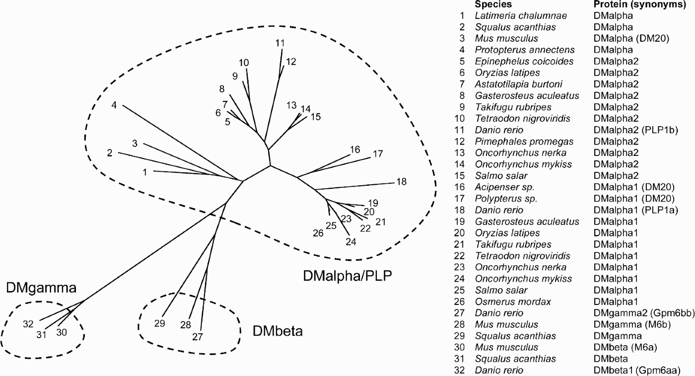
In recent years, large-scale efforts to sequence the genomes and transcriptomes of many more fish species have been initiated and a wealth of information is available now in public databases. When using mammalian DMalpha sequences to search for homologues, pairwise comparisons between sequences often make it difficult to unequivocally assign orthology. Sequence divergence during phylogeny has been sufficient to reach evolutionary distances that are almost equal between some teleost proteolipid protein sequences and either PLP1/DMalpha or M6B/DMbeta of mammals. We have therefore used the well-established zebrafish orthologues DMalpha1 and DMalpha2 (Schweitzer et al., Reference Schweitzer, Becker, Schachner, Nave and Werner2006) to search the Sanger Institute genomic databases (http://www.ensembl.org) and the Genbank collection of expressed sequence tags (ESTs) through NCBI’s BLAST web portal (http://blast.ncbi.nlm.nih.gov/Blast.cgi) for fish sequences coding for DMalpha homologous proteins. If possible, conserved synteny around the corresponding genomic loci was tested (data not shown). Our search was helped by the fact that DMalphas are relatively short transcripts, thus increasing the likelihood to find EST sequences coding for the full open reading frame or showing at least significant sequence overlap. Together with published sequences of several fish species (Tohyama et al., Reference Tohyama, Kasama-Yoshida, Sakuma, Kobayashi, Cao and Hasegawa1999, Reference Tohyama, Ichimiya, Kasama-Yoshida, Cao, Hasegawa and Kojima2000; Venkatesh et al., Reference Venkatesh, Erdmann and Brenner2001) we conducted an analysis of the phylogenetic relationships of 26 different predicted DMalpha proteins and six DMbeta and DM gamma proteins as outgroups (Fig. 1). We find that in almost all species for which significant data are available there is evidence of at least one DMalpha orthologue. All five species, whose genomes are being sequenced (Danio rerio, Gasterosteus aculeatus, Oryzias latipes, Takifugu rubripes and Tetraodon nigroviridis) possess DMalpha1 and DMalpha2 genes. Similarly, all three species of salmonids (Ocorhynchus mykiss, Oncorhynchus nerka and Salmo salar), for which large-scale EST sequencing projects are run, yield cDNAs that code for DMalpha1 and DMalpha2 orthologues. This phylogenetic preservation of both DMalpha orthologues indicates that the divergence of expression and possible subfunctionalization of two DMalpha genes is widespread in teleosts and may have occurred early in teleost evolution, shortly after the genome duplication event. Selective pressure to maintain both orthologues may then have resulted in the presence of two DMalphas in most, if not all, extant fishes. An exception is presented in the cyprinid Pimephales promegas, a close relative of the zebrafish, for which more than 100 ESTs with high homology to DMalpha2 but none to DMalpha1 was found (data not shown), possibly reflecting degeneration of the DMalpha1 gene into a no longer transcribed pseudogene or even loss of the corresponding genomic locus. If this can be confirmed, it will be interesting to examine the expression pattern of this single orthologue and compare it to the expression of DMalphas in other fishes. For species that are not highly represented in EST databases only a single representative for DMalpha was found. However, these proteins strongly clustered with either the DMalpha1 or the DMalpha2 groups (e.g. DMalpha2 of Epinephelus coicoides and Astatotilapia burtoni or DMalpha1 of Osmerus mordax), suggesting that the ‘missing’ paralogue was missed likely due to low sequencing depth of the libraries used for EST generation. The two DMalpha sequences of the basal, non-teleost actinopterygian species Acipenser sp. and Polypterus sp. appear closer to the DMalpha1 group of teleosts but still are relatively similar to teleost DMalpha2. Whether this reflects a single locus for DMalpha in this group remains to be shown.
Phylogenetic relationships of proteolipid proteins focusing of DMalpha/PLP1 in fishes. The genomes of all fish species for which significant sequence data are available encode at least one DMalpha protein. In sarcopterygian species this is the only orthologue (1–4). Actinopterygians have undergone a duplication of their genome and many if not all of these duplicate DMalpha genes have been retained and are here annotated as DMalpha2 (5–15) and DMalpha1 (16–26). For some of the more exotic species only limited data are available and the second orthologue may have been missed (e.g. 5, 7, 16, 17, 26). For comparison two outgroups of DMbeta (27–29) and DMgamma proteolipid proteins (30–32) have been included. Protein sequences were collected from publicly available databases (ensembl genbank; accession numbers are listed in Supplementary material) and a ClustalW alignment computed to infer an unrooted phylogenetic tree using DrawTree of the PHYLIP Phylogeny Inference Package. Default settings were used except that positions with gaps were excluded and the enthusiasm constant set at 0.01.

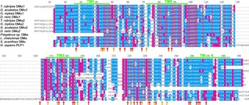
DMalphas are generally well conserved with amino acid identities of about 51% between zebrafish and mouse and 71% between zebrafish and trout (Brösamle and Halpern, Reference Brösamle and Halpern2002; Schweitzer et al., Reference Schweitzer, Becker, Schachner, Nave and Werner2006). Regions of particularly high conservation comprise the four transmembrane segments and a stretch of 21 amino acids in the second extracellular loop (residues 216–237, Fig. 2). Strikingly, most amino acids that, when mutated, are associated with severe to very severe CNS syndromes in humans (Garbern, Reference Garbern2007) are highly conserved across all vertebrates (orange and red arrows, Fig. 2). In a pairwise ClustalW alignment of human DM20 and zebrafish DMalpha2c, close to two-thirds of residues associated with severe to very severe PMD are identical, while overall only less than half are fully conserved (data not shown). This suggests that these residues critically participate in the correct folding and intracellular trafficking of the protein and that the resulting protein structures and their molecular functions have been conserved in all myelinating vertebrates. Toxic gain of function mutations of conserved residues associated with PMD that lead to ER accumulation of DM20, interruption of the secretory pathway and oligodendrocyte apoptosis will therefore probably have similar effects in non-mammalian DMalphas as well. This provides a strong justification for the development of novel PMD animal models in non-mammalian species, such as the zebrafish, which offer superior qualities for the elucidation of disease mechanisms and the development of therapeutic approaches (Lieschke and Currie, Reference Lieschke and Currie2007).
Alignment of select fish DMalpha protein sequences and human PLP1. Residues that when mutated have been shown to be associated with severe or very severe human CNS myelin pathology are indicated by orange or red arrows. Note the high degree of conservation of most disease-associated amino acids, suggesting a high relevance of these residues for myelin proteolipid folding and function. DMalpha protein sequences of a tetraodontiform (T. rubripes), a perciform (G. aculeatus), a salmonid (O. mykiss), a cyprinid (D. rerio), a primitive actinopterygian (Polypterus sp.), a sarcopterygian fish (L. chalumnae), an elasmobranch (S. acanthias) and human PLP1 were aligned using ClustalW and the degree of conservation color coded with TeXshade software. Data on point mutations and transmembrane segments (green bars) were taken from Garbern (Reference Garbern2007).

In conclusion, there is a large body of data that demonstrate the existence of DMalpha proteolipids in actinopterygian fishes. Genome duplication at the base of teleosts has led to the coexistence of two paralogues, DMalpha1 and DMalpha2, in many, if not all genomes of ray-finned fishes. In-situ hybridization, EST sequence data and mass spectroscopy of myelin membrane preparations support expression of DMalpha genes at mRNA and protein levels. DMalpha proteolipids and MPZ are co-expressed in fish oligodendrocytes, refuting the notion that MPZ has replaced DMalpha/PLP1 in fish myelin. Instead, the two likely have different functions and cannot compensate for each other. Amino acid residues that in humans are associated with severe forms of PMD are very often highly conserved in fishes. Research into the myelin biology of lower vertebrates will yield important further insights into the evolution and function of this exquisitely important vertebrate trait. We can look forward to exciting findings that will help us to develop a more complete picture of myelin and that will harness the unique properties of small aquatic vertebrates as model organisms for nervous system function and disease.
ACKNOWLEDGEMENTS
The author is supported by a grant from the German Research Foundation (DFG SFB596/N01). I thank Dr. Andrea Huber for critically reading the manuscript and the editor and reviewers for helpful suggestions.
Statement of interest
None.
Supplementary material
The supplementary material referred to in this article can be found online at journals.cambridge.org/ngb.