Introduction
Extracellular vesicles (EVs) are heterogeneous nanoparticles formed by a phospholipid bilayer and secreted by all cell types (Kumar et al., Reference Kumar, Baba, Sadida, Marzooqi, Jerobin, Altemani, Algehainy, Alanazi, Abou-Samra, Kumar, Akil, Macha, Mir and Bhat2024). EVs are present in all biological fluids and play a crucial role in intercellular communication and the regulation of the physiology of all systems in the animal organism (Jeppesen et al., Reference Jeppesen, Zhang, Franklin and Coffey2023). These nanovesicles are classically categorized based on their biogenesis into apoptotic bodies, microvesicles and exosomes (Hasan et al., Reference Hasan, Viil, Lättekivi, Ord, Reshi, Jääger, Velthut-Meikas, Andronowska, Jaakma, Salumets and Fazeli2020; Mathieu et al., Reference Mathieu, Névo, Jouve, Valenzuela, Maurin, Verweij, Palmulli, Lankar, Dingli, Loew, Rubinstein, Boncompain, Perez and Théry2021).
Microvesicles and exosomes have an important function in the extracellular transport of molecular constituents, which are delivered from a signalling cell to a target cell, enabling both short- and long-distance communication (Iannotta et al., Reference Iannotta, Amruta, Kijas, Rowan and Wolfram2024). In this way, vesicles establish intercellular communication mechanisms, facilitating the exchange of proteins, lipids and nucleic acids, which allow the activation of metabolic cascades in target cells to regulate various physiological processes (Matarredona and Pastor, Reference Matarredona and Pastor2019).
The EVs are widely used for the diagnosis, monitoring and treatment of genetic, cardiovascular, infectious, metabolic and oncological diseases (Wang et al., Reference Wang, Sun, Zhao, Yang, Cai, Xu and Cao2017). However, in recent years, these nanoparticles have been employed to enhance reproductive biotechnologies, particularly in in vitro embryo production (IVP) during maturation, fertilization and in vitro culture (IVM, IVF and IVC). They have demonstrated beneficial effects on meiotic progression, fertilization capacity and blastocyst development (Bathala et al., Reference Bathala, Fereshteh, Li, Al-Dossary, Galileo and Martin-DeLeon2018; De Ávila et al., Reference de Ávila, Bridi, Andrade, del Collado, Sangalli, Nociti, da Silva Junior, Bastien, Robert, Meirelles, Perecin and da Silveira2020a; Sidrat et al., Reference Sidrat, Khan, Joo, Xu, El-Sheikh, Ko and Kong2022).
Previous reviews, including Muñoz et al. (Reference Muñoz, Corrales, Caamaño and Rizos2022), have discussed the role of EVs in mammalian reproduction, with a particular focus on their presence and general function in reproductive fluids and tissues. Given the importance of EVs as a biotechnological tool and the scarcity of scientific articles addressing the significance of these structures in reproduction, this article aims to describe the main applications of microvesicles and exosomes, emphasizing key aspects related to biotechnological implications of EV-mediated molecular transfer in assisted reproduction systems.
The evolution of extracellular vesicles research
The first report suggesting the existence and possible function of extracellular vesicles (EVs) was made by Chargaff and West, Reference Chargaff and West1946 during their studies on blood coagulation. They hypothesized that, in addition to the thromboplastic agent, a ‘variety of minute degradation products of blood cells’ contributed to the clotting properties. In 1967, Wolf, Reference Wolf1967 provided evidence that the plasma coagulant material described by Chargaff and West was, in fact, a minute particulate substance rich in lipid content, derived from platelets, as clearly shown through transmission electron microscopy (TEM). Based on this observation, the authors initially named these particles ‘platelet dust’ (Figure 1).
Extracellular Vesicles Through Time. Schematic representation summarizing the major discoveries in the field of extracellular vesicles from the 1940s to 2020.

In the 1970s, Neville Crawford reported the presence of particles he termed ‘microparticles’, which were isolated from platelet-free plasma. In the same study, he demonstrated that such microparticles contained lipids and transported cargos such as ATP and contractile proteins (Crawford, Reference Crawford1971). This was a pioneering study in showing that these particles originated from plasma membranes. The term ‘intracellular and extracellular vesicles’ was first used by Aaronson et al. (Reference Aaronson, Behrens, Orner and Haines1971), who, using TEM, provided the first morphological evidence for the production of a wide variety of intra- and extracellular membranous structures in O. danica, a flagellated alga. From this point on, it became clear that these particles were not solely associated with platelets, but also with other cell types (De Broe et al., Reference De Broe, Wieme, Logghe and Roels1977).
Later, in 1983, researchers Pan and Johnstone observed that transferrin receptors were associated with small structures of approximately 50 nm in diameter, which were released by reticulocytes into the extracellular medium (Pan and Johnstone, Reference Pan and Johnstone1983). Four years later, in 1987, the same researcher, Rose Johnstone, was the first to assign the term ‘exosomes’ to these nanoparticles released from fragments of the plasma membrane (Johnstone et al., Reference Johnstone, Adam, Hammond, Orr and Turbide1987).
In 1996, the role of these vesicles in the immune system was discovered, demonstrating the involvement of exosomes in adaptive immune responses via secretion by B lymphocytes (Raposo et al., Reference Raposo, Nijman, Stoorvogel, Liejendekker, Harding, Melief and Geuze1996). Two years later, it was reported that dendritic cells secreted these vesicles during tumour suppression in mice, identifying them as important factors in intercellular communication (Zitvogel et al., Reference Zitvogel, Regnault, Lozier, Wolfers, Flament, Tenza, Ricciardi-Castagnoli, Raposo and Amigorena1998). This study laid the groundwork for subsequent findings in 2006 and 2007, which revealed the presence of RNA (mRNA and miRNA) and proteins as key molecules responsible for cell-to-cell transport. These discoveries confirmed not only the role of EVs in intercellular communication but also highlighted the functional value of their molecular cargo (Ratajczak et al., Reference Ratajczak, Miekus, Kucia, Zhang, Reca, Dvorak and Ratajczak2006).
In the context of reproduction, the historical background of EVs dates back to 1967, when Piko, Reference Piko1967 first demonstrated the presence of these particles in the epididymis, which are now known as epididymosomes (Piko, Reference Piko1967). In 1978, studies involving EVs from the male reproductive tract continued, with the first report describing vesicles in human prostatic fluid (prostasomes). These were identified as particles capable of interacting with spermatozoa, the epididymis and seminal secretions (Ronquist et al., Reference Ronquist, Brody, Gottfries and Stegmayr1978; Simon et al., Reference Simon, Greening, Bolumar, Balaguer, Salamonsen and Vilella2018).
However, it was not until the 2010s that research on EVs originating from the female reproductive tract began to increase significantly. Da Silveira and collaborators were the first to describe the presence of EVs in the follicular fluid (FF) of equine species, determining that the age of the animals influenced the miRNA content related to oocyte quality (Da Silveira et al., Reference da Silveira, Veeramachaneni, Winger, Carnevale and Bouma2012). One year later, the first report of EVs from oviductal fluid (OF) emerged, revealing that these vesicles participate in important reproductive events such as sperm capacitation and fertilization (Al-Dossary et al., Reference Al-Dossary, Strehler and Martin-DeLeon2013). Also in 2013, Ng et al., Reference Ng, Rome, Jalabert, Forterre, Singh, Hincks and Salamonsen2013 reported the production of EVs by endometrial cells and their presence in uterine fluid (UF), demonstrating that these nanoparticles carry miRNAs involved in embryonic implantation.
More recent studies have highlighted the importance of EV-transported miRNAs as biomarkers for predicting ovarian hyperstimulation (Gad et al., Reference Gad, Sánchez, Browne, Nemcova, Laurincik, Prochazka and Lonergan2020). Furthermore, the role of these miRNAs in the regulation of the oestrous cycle in equines has gained prominence (Gebremedhn et al., Reference Gebremedhn, Gad, Ishak, Menjivar, Gastal, Feugang, Prochazka, Tesfaye and Gastal2023; Feugang et al., Reference Feugang, Gad, Menjivar, Ishak, Gebremedhn, Gastal, Dlamini, Prochazka, Gastal and Tesfaye2024).
Given this context, review articles that present the main findings on the role of EVs and their interactions within the male and female reproductive environment are of great relevance. To this end, understanding the cellular processes responsible for the formation and release of these vesicles is essential.
Extracelullar vesicles biogenesis
The International Society for Extracellular Vesicles (ISEV) defines the term ‘Extracellular Vesicles’ based on three main criteria: (1) particles released by cells; (2) enclosed by a phospholipid bilayer; and (3) incapable of replication (Welsh et al., Reference Welsh, Goberdhan, O’Driscoll, Buzas, Blenkiron, Bussolati and Benedikter2024). Supporting this definition, Bordanaba-Florit et al. (Reference Bordanaba-Florit, Royo, Kruglik and Falcón-Pérez2021) describe EVs as heterogeneous in size, ranging from nanometres to micrometres, bounded by a lipid bilayer and carrying a cargo rich in nucleic acids, proteins, metabolites and lipids (Figure 2).
Schematic representation of the biogenesis, categorization and molecular components of extracellular vesicles. Apoptotic bodies are formed through meiosis of the plasma membrane in apoptotic cells and range in size from 50 to 5000 nm. Microvesicles (100–1000 nm) originate from outward budding of the plasma membrane of the producing cell. Exosomes (30–150 nm), in contrast, have an endocytic origin and are formed by inward budding of early endosomes, generating intraluminal vesicles (ILVs). Once formed, ILVs are transported to the plasma membrane via multivesicular bodies (MVBs), which release exosomes into the extracellular space. These vesicles participate in intercellular communication by carrying nucleic acids, proteins and lipids.

The origin of EVs varies depending on their mechanism of formation and size. They are secreted under both pathological conditions and normal physiological processes (contributing to homeostasis), and they participate in communication with target cells, inducing changes in the biochemical properties of these cells through the delivery of molecular cargo (Shifrin et al., Reference Shifrin, Beckler, Coffey and Tyska2013). The EVs can be isolated from various biological sources, including milk (Somiya et al., Reference Somiya, Yoshioka and Ochiya2018), urine (Svenningsen et al., Reference Svenningsen, Sabaratnam and Jensen2020), blood plasma (Palviainen et al., Reference Palviainen, Saraswat, Varga, Kitka, Neuvonen, Puhka, Joenväärä, Renkonen, Nieuwland, Takatalo and Siljander2020), cerebrospinal fluid (Taxiarchis et al., Reference Taxiarchis, Bellander, Antovic, Soutari, Virhammar, Kumlien, Karakoyun, Rostami and Antovic2024), bronchoalveolar lavage fluid (Bartel et al., Reference Bartel, Wolters, Noor, Rafie, Fang, Kirchner, Hoen, Pfaffl, Rutgers, Timens, van den Berge and Hylkema2024), seminal plasma (Zhang et al., Reference Zhang, Vos, Tao and Stoorvogel2020), epididymal fluid (Rowlison, Ottinger and Comizzoli, Reference Rowlison, Ottinger and Comizzoli2021), prostate (Park et al., Reference Park, Shin, Jung, Kwon, Choi, Park and Lee2016), testes (Choy et al., Reference Choy, Chan, Lam, Jin, Zheng, Law, Yu, Wang, Li, Xie, Yim, Chen and Fok2022), amniotic fluid (Matsuo et al., Reference Matsuo, Yokoi, Yoshida, Kitagawa, Asano-Inami, Miura, Yasui, Tano, Ushida, Imai, Kajiyama and Kotani2024), follicular fluid (Ferraz et al., Reference Ferraz, Fujihara, Nagashima, Noonan, Inoue-Murayama and Songsasen2020a), oviductal fluid (Mendes Júnior et al., Reference Mendes Júnior, Alcântara Neto, Tetaping, Novaes, Gonçalves, Silva-Neto, Almeida, Guedes, Leal, Souza-Fabjan, Nicolete, Padilha, Figueiredo and Rodrigues2024) and uterine fluid (Piibor et al., Reference Piibor, Waldmann, Dissanayake, Andronowska, Ivask, Prasadani, Kavak, Kodithuwakku and Fazeli2023). Based on their cellular origin, biological function or biogenesis pathway, EVs are classified into three subgroups (Figure 2): apoptotic bodies, exosomes and microvesicles (Chen and Yang, Reference Chen and Yang2024).
Apoptotic bodies
The formation mechanisms of apoptotic bodies originate from the process of programmed cell death, known as apoptosis. Apoptosis is a tightly regulated process triggered either by external stimuli (death receptor pathway) or internal signals (mitochondrial pathway) (Wong, Reference Wong2011; Aghaei-Zarch et al., Reference Aghaei-Zarch, Nia, Nouri, Mousavinasab, Najafi, Bagheri-Mohammadi, Aghaei-Zarch, Toolabi, Rasoulzadeh, Ghanavi, Moghadam and Talebi2023). These pathways activate a cascade of intracellular events that lead to characteristic morphological changes, such as chromatin condensation, DNA fragmentation, cell shrinkage and the formation of membrane blebs, ultimately resulting in the release of apoptotic bodies (Atkin-Smith et al., Reference Atkin-Smith, Tixeira, Paone, Mathivanan, Collins, Liem, Goodall, Ravichandran, Hulett and Poon2015; Battistelli and Falcieri, Reference Battistelli and Falcieri2020).
The increase in intracellular hydrostatic pressure leads to actin polymerization within the cytoskeleton, facilitating the release of apoptotic bodies into the extracellular environment. These vesicles share structural and molecular similarities with microvesicles and exosomes. However, their molecular content (DNA, RNA, proteins and lipids) is derived from dying cells, and both the cell and the apoptotic body emit chemotactic signals that culminate in phagocytosis (Xu et al., Reference Xu, Lai and Hua2019; Wen et al., Reference Wen, Creaven, Luan and Wang2023).
These chemotactic signals include soluble factors that mediate the interval between the recognition of the apoptotic cell (find me signal) and the engulfment of the apoptotic body by the phagocyte (eat me signal). This process is largely driven by the externalization of phosphatidylserine, a phospholipid typically located on the inner leaflet of the plasma membrane, which is translocated to the outer surface during apoptotic body formation. This lipid serves as a key marker for this type of vesicle due to its recognition by phagocytic cells (Segawa and Nagata, ). However, it is important to note that phosphatidylserine is also found in microvesicles and exosomes – albeit in lower abundance – since it is a common component of phospholipid membranes (Qian et al., Reference Qian, Mao, Zhao, Zhao, Liu, Lu, Zhang, Mao, Zhang, Wang, Sun and Cui2023).
Microvesicles or ectosomes
Similar to apoptotic body formation, microvesicles are generated and released directly from the plasma membrane. However, this process is tightly regulated by specific phospholipids and proteins that orchestrate membrane protrusion and, consequently, microvesicle shedding into the extracellular environment (Shu et al., Reference Shu, Tan, Miao and Zhang2019).
The biogenesis of microvesicles differs fundamentally from that of both apoptotic bodies and exosomes, involving a series of orchestrated steps that culminate in membrane budding and fission. These steps include phospholipid redistribution, actomyosin contraction and translocase-independent mechanisms (Tricarico et al., Reference Tricarico, Clancy and D’Souza-Schorey2017).
Lipid redistribution is mediated by lipid translocases – enzymes responsible for maintaining the asymmetry of the plasma membrane by facilitating lipid exchange between the inner and outer leaflets of the bilayer. These translocases are divided into three major families: flippases, floppases, and scramblases. Flippases and floppases are ATP-dependent; flippases maintain lipids on the inner leaflet, while floppases transport them to the outer leaflet (Beer and Wehman, Reference Beer and Wehman2017). Scramblases, on the other hand, are activated by increased intracellular Ca2+ and function in an ATP-independent manner to promote bidirectional lipid translocation (Aires et al., Reference Aires, Ribeiro-Rodrigues, Boia, Ferreira-Rodrigues, Girão, Ambrósio and Santiago2021).
During microvesicle formation, lipids such as phosphatidylserine and phosphatidylethanolamine (normally confined to the inner leaflet) become externalized due to flippase inhibition and activation of floppases and scramblases, triggered by ATP and intracellular Ca2+ increase, respectively (Sakuragi and Nagata, Reference Sakuragi and Nagata2023). This lipid redistribution destabilizes membrane structure, facilitating the formation of membrane protrusions that ultimately give rise to microvesicles (Kalra et al., Reference Kalra, Drummen and Mathivanan2016).
Alternatively, when translocase activity is not the primary driver, the actomyosin contractile machinery mediates membrane fission. This process involves ADP-ribosylation factor 6 (ARF6), a key regulator of microvesicle secretion. Upon activation at the plasma membrane, ARF6 initiates a signalling cascade that activates phospholipase D, which hydrolyzes phosphatidylcholine (Schwager and Reinhart-King, Reference Schwager and Reinhart-King2020). The resulting phosphatidic acid recruits extracellular signal-regulated kinase (ERK), which subsequently phosphorylates myosin light chain kinase (MLCK). MLCK then phosphorylates the myosin light chain (MLC), triggering actomyosin contraction and enabling membrane fission (Clancy et al., Reference Clancy, Schmidtmann and D’Souza-Schorey2021).
Microvesicles can also be formed through translocase-independent mechanisms that recruit elements of the exosomal biogenesis pathway to facilitate membrane outward budding (Kalra et al., Reference Kalra, Drummen and Mathivanan2016). This pathway involves components of the endosomal sorting complexes required for transport (ESCRT), specifically the recruitment of tumor susceptibility gene 101 (TSG101) and ALIX proteins. These proteins associate to recruit the ESCRT-III complex to the plasma membrane. ESCRT-III then interacts with the VSP4 ATPase, ultimately leading to membrane budding, fission, and microvesicle release. These vesicles mediate intercellular transfer of nucleic acids, proteins, bioactive lipids, and reactive oxygen species regulators to recipient cells (Taylor and Bebawy, Reference Taylor and Bebawy2019; Wang et al., Reference Wang, Li, Chen, Xu, Chang, Wang, Deng a, Gu, Zhou, Shen, Tao and Sun2022).
Exosomes
The principle behind exosome biogenesis is based on the origin of small vesicles known as exosomes. Interestingly, these structures are formed via the endocytic pathway, an intracellular membranous compartment (early endosome) undergoes membrane invagination, giving rise to a late endosome or multivesicular body (MVB), which subsequently generates particles initially termed intraluminal vesicles (ILVs). These ILVs are then released into the extracellular space via exocytosis when the MVB fuses with the plasma membrane (Ratajczak and Ratajczak, Reference Ratajczak and Ratajczak2020). This formation process is mediated by two major and often concurrent mechanisms: the ESCRT-dependent pathway and the ESCRT-independent (or alternative) pathway (Gurunathan et al., Reference Gurunathan, Kang and Kim2021).
In this context, the ESCRT-dependent pathway is composed of specialized microdomains predominantly located in the endosomal membrane, enriched in tetraspanins (CD9, CD63, and CD81), ESCRT complex components (ESCRT-0, I, II, and III), and associated proteins (TSG101, ALIX, and VSP4 ATPase) (Choi et al., Reference Choi, Kim, Kim and Gho2015). To form ILVs, this protein complex is recruited in a sequential cascade: ESCRT-0, responsible for modulating ubiquitinated proteins, interacts with TSG101 to recruit ESCRT-I and ESCRT-II. Together, ESCRT-0, I, and II select ubiquitinated cargo for inclusion in MVBs. Subsequently, ESCRT-II recruits ESCRT-III, which interacts with ALIX and VSP4 ATPase to mediate membrane budding and scission, resulting in the formation of ILVs within the lumen of the multivesicular body (Gurung et al., Reference Gurung, Perocheau, Touramanidou and Baruteau2021).
Concurrently, the ESCRT-independent (alternative) pathway involves microdomains enriched in glycoproteins, including the aforementioned tetraspanins, sphingomyelin, and enzymes embedded in the endosomal membrane. In this pathway, sphingomyelin is catalyzed by sphingomyelinase, producing ceramide. An excessive accumulation of ceramide induces membrane curvature, which facilitates ILV formation. Additionally, in both pathways, tetraspanins play a role in promoting adhesion and fusion between the MVB and the plasma membrane (Kowal et al., Reference Kowal, Tkach and Théry2014; Andreu and Yáñez-Mó, Reference Andreu and Yáñez-Mó2014).
Once formed and matured, MVBs containing ILVs are transported via two major protein groups, which direct late endosomes either to lysosomal compartments for degradation or to the plasma membrane for exocytic release: small GTPases of the Rab family and SNARE (soluble NSF attachment protein receptor) fusion machinery (Bobrie et al., Reference Bobrie, Colombo, Raposo and Théry2011). Although both protein families are involved in exosome secretion, there remains some controversy over which MVB populations are destined for lysosomal degradation versus those targeted for exocytosis. This ambiguity arises from the fact that the functional specificity of these proteins varies according to cell type and lineage (Stahl and Raposo, Reference Stahl and Raposo2019; van Niel et al., Reference van Niel, Carter, Clayton, Lambert, Raposo and Vader2022).
Nonetheless, recent studies have demonstrated that specific Rab family members, such as Rab27A, Rab11/Rab35, and Rab7, respectively play roles in: (1) MVB docking and fusion with the plasma membrane; (2) endosome formation, recycling, and fusion with the plasma membrane; and (3) MVB trafficking toward lysosomal degradation. Moreover, SNARE proteins (v-SNARE and t-SNARE) work cooperatively during the recognition and fusion of MVBs with the plasma membrane, facilitating the final release of exosomes (Krylova and Feng, Reference Krylova and Feng2023).
EVs’ mechanisms of uptake
Intercellular communication mediated by EVs occurs through different uptake mechanisms, allowing specific interaction with the recipient cell, often mediated by surface proteins. This internalization of EVs takes place through at least three main mechanisms, culminating in the release of vesicular contents into the cytoplasm: fusion, endocytosis, and surface binding (Mulcahy, Pink & Carter, Reference Mulcahy, Pink and Carter2014).
The first mechanism, known as direct fusion with the plasma membrane, consists of the ability of the phospholipid membrane of EVs to fuse with the target cell’s plasma membrane, immediately releasing the EV content into the cellular cytoplasm (Raposo & Stoorvogel, Reference Raposo and Stoorvogel2013). On the other hand, during endocytosis (receptor-mediated internalization), EVs express surface proteins such as tetraspanins (CD9, CD63, and CD81), which can bind to specific receptors on the target cell (Théry, Reference Théry2011). This binding triggers the internalization of EVs through endocytic pathways (such as pinocytosis or clathrin-mediated endocytosis), leading the EVs into an endosome (Bonsergent et al., Reference Bonsergent, Grisard, Buchrieser, Schwartz, Théry and Lavieu2021). The surface binding-mediated mechanism, in turn, allows the vesicle to act as a signalling particle, in which the interaction occurs through the plasma membrane without the need for vesicle internalization (Xie et al., Reference Xie, Hao, Li, Song, Chen, Zhou and Wang2024).
At the intracellular destination, EV lipids (such as cholesterol and sphingolipids) can follow two main pathways: they may either incorporate into the recipient cell membrane modulating its fluidity and signalling platforms (lipid rafts) or, if internalized, be directed to lysosomal degradation, where they are broken down and recycled (Ghadami & Dellinger, Reference Ghadami and Dellinger2023). The protein content of EVs, including surface proteins (such as tetraspanins) and luminal proteins (transcription factors and enzymes), is primarily released into the cytosol through endosomal escape, where functional proteins can directly participate in signalling pathways or enter the nucleus to alter gene expression. Meanwhile, non-functional proteins are subjected to proteasomal or lysosomal degradation (O’Brien et al., Reference O’Brien, Ughetto, Mahjoum, Nair and Breakefield2022). In contrast, miRNAs and other nucleic acids must escape the endosome into the cytosol, where they bind to the RNA-Induced Silencing Complex (RISC) and act in post-transcriptional gene regulation – the central mechanism by which EVs modulate cellular phenotype (Qiu et al., Reference Qiu, Yan, Hu, Yi, Liu, Lu and Xu2025).
In this context, EVs internalized by reproductive cells have gained attention for their ability to modulate oocyte maturation, fertilization, and embryonic development mechanisms (Mendes Júnior et al., Reference Mendes Júnior, Alcântara Neto, Tetaping, Novaes, Gonçalves, Silva-Neto, Almeida, Guedes, Leal, Souza-Fabjan, Nicolete, Padilha, Figueiredo and Rodrigues2024). Evidence suggests that the use of EVs supports follicular development and oocyte maturation by delivering miRNAs (such as bta-miR-494 and bta-miR-20a), which regulate the PI3K-AKT pathway, an important process in cell proliferation (Andrade et al., Reference Andrade, Meirelles, Perecin and Da Silveira2017). Additionally, recent studies using transmission electron microscopy (TEM) have shown that small vesicles can pass through transzonal projections (TZPs), participating in direct communication between the oocyte and cumulus cells (Marchais et al., Reference Marchais, Gilbert, Bastien, Macaulay and Robert2022). This finding corroborates the observations of Wydooghe et al., Reference Wydooghe, Vandaele, Heras, De Sutter, Deforce, Peelman and Van Soom2017, who reported that vesicles secreted into embryo culture media can reach the zona pellucida of pre-implantation embryos due to the presence of pores larger than 50 nm, allowing the passage of exosomes. Qu et al. (Reference Qu, Qing, Liu, Qin, Wang, Qiao and Wang2017) further reinforced that EVs derived from embryos are secreted and absorbed through exocytosis and endocytosis mechanisms, respectively.
Biotechnological potential of extracellular vesicles in animal reproduction
Extracellular vesicles (EVs) play a significant role in the reproductive tracts of both females and males and act as key regulators of various reproductive processes, including sperm maturation (Foot et al., Reference Foot, Gonzalez, Gembus, Fonseka, Sandow, Nguyen, Tran, Webb, Mathivanan, Robker and Kumar2021), oocyte maturation (Fiorentino et al., Reference Fiorentino, Merico, Zanoni, Comincini, Sproviero, Garofalo, Gagliardi, Cereda, Lin, Innocenti, Taggi, VaiarellI, Ubaldi, Rienzi, Cimadomo, Garagna and Zuccotti2024), coordination of capacitation/acrosome reaction (Xu et al., Reference Xu, Xie, Wu, Gu, Zhang, Yang, Yang, Zheng, Huang, Xu, Li, Cai, Liu, Hong and Wu2024), prevention of polyspermy (Fang et al., Reference Fang, Bang, Tanga, Seo, Zhou, Seong, Saadeldin, Lee, Cui and Cho2023), endometrial crosstalk, and embryonic development (Aguilera et al., Reference Aguilera, Wong, Gutierrez-Reinoso, Velásquez, Melo-Báez, Cabezas, Caamaño, Navarrete, Castro and Rodriguez-Alvarez2024). Consequently, the application of EVs in reproductive biotechnologies has been well established and shows promising results in the field of animal reproduction, particularly in bovine, swine, and ovine species (Machtinger et al., Reference Machtinger, Rodosthenous, Adir, Mansour, Racowsky, Baccarelli and Hauser2017; Gurunathan et al., Reference Gurunathan, Kang, Song, Kim and Kim2022).
In this context, Evans & Garcia-Gonzalez (Reference Evans and Garcia-Gonzalez2024) demonstrated that extracellular vesicles (EVs) from female reproductive tract fluids (FRF) are enriched in bioactive molecules, notably microRNAs (miRNAs), which mediate gene regulation in recipient cells. These miRNAs can modulate apoptosis, ensuring that only functionally competent spermatozoa or embryos survive under the selective pressure of the maternal environment; cell proliferation, by promoting the expansion of viable embryonic cells while inhibiting abnormal growth; and immune tolerance, by modulating the expression of cytokines and immune checkpoint molecules in the endometrium to prevent rejection of the semi-allogeneic embryo. Notably, these EVs may also enhance gamete compatibility, acting as molecular filters that favour sperm functionally suited to the female tract (Evans & Garcia-Gonzalez, Reference Evans and Garcia-Gonzalez2024).
Optimizing In Vitro embryo production (ivp) through extracellular vesicles technology
The IVP technology has been widely implemented across various species with the aim of enhancing genetic material, reducing generational intervals in animals with high zootechnical value, preserving endangered species, and deepening the understanding of embryonic development (Currin et al., Reference Currin, Baldassarre and Bordignon2021). Through this technique, physiological in vitro processes are mimicked under in vitro conditions, encompassing the steps of in vitro maturation, fertilization, and culture (IVM, IVF, and IVC, respectively) (Luedke et al., Reference Luedke, Lavach, Cassanta, do Nascimento Nunes, Schlotefeld, de Paiva, dos Santos and Neves2019).
However, IVP protocols still face several limitations, including the selection of appropriate culture systems, control of temperature and pH, management of reactive oxygen species (ROS) production, and supplementation of media with regulatory factors. These challenges often compromise the efficiency and quality of in vitro-derived embryos compared to their in vivo counterparts (Dissanayake et al., Reference Dissanayake, Nomm, Lättekivi, Ressaissi, Godakumara, Lavrits, Midekessa, Viil, Bæk, Jørgensen, Bhattacharjee, Andronowska, Salumets, Jaakma and Fazeli2020; Silva et al., Reference Silva, Barroso, Nascimento, Figueira, Azevedo, Silva and Santos2021).
In recent years, EVs have been isolated from various compartments of the female reproductive tract, such as uterine, oviductal, and follicular fluids, from different species including murine, bovine, equine, porcine, ovine, and caprine models. These investigations aim to explore molecular exchanges between uterine and embryonic cells, with a particular focus on elucidating maternal-embryonic recognition mechanisms (Hua et al., Reference Hua, Wang, Lian, Li, Xi, Xue, Kang and Lei2021; Xie et al., Reference Xie, Liu, Zang, Hu, Zhou, Li, Liu and Hong2021). Such findings contribute to a better understanding of reproductive processes and serve to enhance the outcomes of IVP protocols (Almiñana et al., Reference Almiñana, Rudolf Vegas, Tekin, Hassan, Uzbekov, Fröhlich, Bollwein and Bauersachs2021).
In reproductive biotechnology, the concentration and origin of extracellular vesicles (EVs) added to culture media critically influence oocyte and embryo development. EVs from different sources such as follicular fluid, oviduct, uterus or endometrial cells carry distinct miRNA and protein cargos that modulate gene expression, signalling pathways, lipid metabolism, cell differentiation and embryonic viability. Additionally, physiological EVs, such as those from healthy oviducts or follicular fluid, generally promote developmental competence, whereas EVs from pathological sources may impair embryonic development (da Silveira et al, Reference da Silveira, Andrade, Del Collado, Sampaio, Sangalli, Silva, Pinaffi, Jardim, Cesar, Nogueira, Cesar, Coutinho, Pereira, Perecin and Meirelles2017; Almiñana et al., Reference Almiñana, Corbin, Tsikis, Alcântara-Neto, Labas, Reynaud, Galio, Uzbekov, Garanina, Druart and Mermillod2017; Alcântara-Neto et al., Reference Alcântara-Neto, Cuello, Uzbekov, Bauersachs, Mermillod and Almiñana2022; Xue et al., Reference Xue, Zheng, Xiong and Li2024).
At the molecular level, miRNAs and mRNAs delivered by EVs are internalized by oocytes and embryos, regulating the translation of key proteins involved in development, differentiation and implantation (Almiñana et al., Reference Almiñana, Corbin, Tsikis, Alcântara-Neto, Labas, Reynaud, Galio, Uzbekov, Garanina, Druart and Mermillod2017; Alcântara-Neto et al., Reference Alcântara-Neto, Cuello, Uzbekov, Bauersachs, Mermillod and Almiñana2022; Yamei Xue et al., Reference Xue, Zheng, Xiong and Li2024). Examples include miR-148b from oviductal EVs enhancing embryonic quality via TGF-β modulation and miR-34c from follicular EVs improving oocyte competence and blastocyst development (Almiñana et al., Reference Almiñana, Corbin, Tsikis, Alcântara-Neto, Labas, Reynaud, Galio, Uzbekov, Garanina, Druart and Mermillod2017; Chen et al., Reference Chen, Liang, Qin, Zhang, Chen and Wang2022; Yamei Xue et al., Reference Xue, Zheng, Xiong and Li2024). Collectively, these findings indicate that both EV concentration and source are essential determinants of miRNA uptake and functional protein expression, directly impacting embryonic quality and developmental outcomes.
In the context of IVP, Tesfaye et al. (Reference Tesfaye, Menjivar, Gebremedhn, Salilew-Wondim, Hoelker and Schellander2021) highlighted that the miRNA cargo of extracellular vesicles (EVs) is closely associated with oocyte developmental stage, metabolic condition and maternal age, reinforcing their potential as non-invasive biomarkers of oocyte competence. The authors further demonstrated that EV supplementation in assisted reproductive technology (ART) media can significantly enhance cleavage and blastocyst formation rates, promote cellular stress resilience and improve post-thaw embryo survival. Notably, the EV-mediated delivery of stress-response molecules has been proposed as a therapeutic strategy for addressing reproductive pathologies such as polycystic ovary syndrome (PCOS), age-related fertility decline and endometritis. These findings support the dual role of EVs as diagnostic tools and as functional modulators capable of optimizing in vitro embryo development.
In Vitro maturation (IVM)
In the maternal environment, ovarian follicles are stimulated to grow until they reach the pre-ovulatory follicle stage, which is arrested at Metaphase II (Figueiredo and Lima, Reference Figueiredo and Lima2017). In vitro, alternative strategies using extracellular vesicles (EVs) as supplements in maturation media have been established to mimic oocyte maturation conditions and support the subsequent steps of in vitro embryo production (IVP) (Hung et al., Reference Hung, Hong, Christenson and McGinnis2015).
A study using EVs derived from murine blood plasma showed that supplementation of maturation medium with these nanovesicles led to an increase in cumulus-oocyte complex (COC) diameter. This resulted in a higher percentage of oocytes reaching the Metaphase II (MII) stage compared to control groups and those supplemented with epidermal growth factor (EGF), thereby demonstrating the biological effects of EVs on cell proliferation and oocyte maturation (Javadi et al., Reference Javadi, Rad, Pashaiasl, Farashah and Roshangar2022).
In bovine (de Ávila & da Silveira, Reference de Ávila and Da Silveira2020b) and equine species (da Silveira et al., Reference da Silveira, Carnevale, Winger and Bouma2014), the importance of follicular fluid-derived EVs (FF-EVs) has been highlighted in the transport of microRNAs (miR-27b, miR-372 and miR-382), which regulate follicular growth pathways during the follicular maturation stage. Additionally, Matsuno et al. (Reference Matsuno, Kanke, Maruyama, Fujii, Naito and Sugiura2019) confirmed the presence of mRNAs in EVs isolated from porcine follicular fluid that are associated with the PI3K-AKT and MAPK pathways, both essential for follicular development, steroidogenesis and oocyte maturation.
Follicular fluid-derived EVs from bovine, equine and porcine species have also been shown to play a significant role in meiotic division, cytoplasmic maturation and protection against cellular stress, all critical for oocyte developmental competence (Uzbekova et al., Reference Uzbekova, Almiñana, Labas, Teixeira-Gomes, Combes-Soia, Tsikis, Carvalho, Uzbekov and Singina2020; Ding et al., Reference Ding, Jin, Kalds, Meng, Wang, Zhong, Wang and Chen2021).
In equines, IVM media supplemented with 200 μg/mL of FF-EVs resulted in 45.7% of oocytes reaching the MII stage compared to control groups. The authors suggested that EVs secreted by follicular cells may improve IVM parameters in equine oocytes (Gábrys et al., Reference Gábrys, Kij-Mitka, Sawicki, Kochan, Nowak, Łojko, Karnas and Bugno-Poniewierska2022). In bovines, supplementation with FF-EVs during IVM increased blastocyst rates and post-thaw embryo survival (Singina et al., Reference Singina, Shedova, Uzbekov, Chinarov, Lukanina and Uzbekova2023). More recently, Gábrys et al. (Reference Gabryś, Gurgul, Szmatoła, Kij-Mitka, Andronowska, Karnas, Kucharski, Wojciechowska-Puchałka, Kochan and Bugno-Poniewierska2024) reported that EVs derived from smaller antral follicles were more readily internalized by cumulus cells, potentially enhancing cell proliferation.
Studies have also indicated that oviductal fluid-derived EVs (OF-EVs) from the bovine ampullary region, when used during IVM, had a more beneficial impact on embryonic development than FF-EVs, suggesting that EVs significantly influence oocyte maturation and subsequent fertilization capacity (Asaadi et al., Reference Asaadi, Dolatabad, Atashi, Raes, Van Damme, Hoelker, Hendrix, Pascottini, Van Soom, Kafi and Pavani2021).
Fang et al. (Reference Fang, Bang, Tanga, Seo, Zhou, Seong, Saadeldin, Lee, Cui and Cho2023) supplemented IVM and IVF media with EVs derived from porcine oviductal epithelial cells, resulting in higher cleavage and blastocyst formation rates compared to media without EVs. Notably, these EVs also reduced polyspermy rates, ultimately improving fertilization success.
During porcine oocyte IVM, EVs from seminal plasma (SP-EVs) were internalized by cumulus cells, but not by the oocytes. While the percentage of oocytes reaching Metaphase II did not differ significantly from controls, the expression levels of genes associated with oocyte maturation (HAS2) and steroidogenesis (CYP11A1 and HSD3B1) were upregulated in the presence of SP-EVs (Mateo-Otero et al., Reference Mateo-Otero, Yeste, Roca, Llavanera, Bucci, Galeati, Spinaci and Barranco2022).
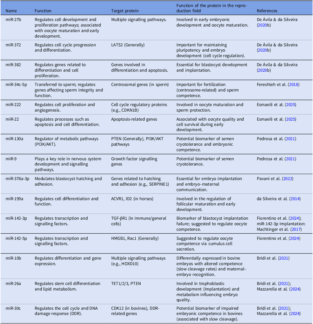
Current research is exploring the communication between cumulus cells and the oocyte, with a focus on miRNA presence and expression. Fiorentino et al. (Reference Fiorentino, Merico, Zanoni, Comincini, Sproviero, Garofalo, Gagliardi, Cereda, Lin, Innocenti, Taggi, VaiarellI, Ubaldi, Rienzi, Cimadomo, Garagna and Zuccotti2024) demonstrated that miRNAs delivered via cumulus cell-derived EVs regulate genes involved in meiotic resumption, follicular growth, fertilization and acquisition of oocyte developmental competence. The Table 1 summarizes some of these miRNAs cargo and its effects on the field of reproduction. These EV-mediated effects enhance oocyte quality and prepare it for successful fertilization (Machtinger et al., Reference Machtinger, Rodosthenous, Adir, Mansour, Racowsky, Baccarelli and Hauser2017).
MicroRNA profiles in extracellular vesicles and their role in reproductive competence

In Vitro fertilization (IVF)
The EVs play a key role in maintaining sperm viability by enhancing survival and modulating motility, a mechanism closely associated with the sperm reservoir activity in the oviductal isthmus (Alcântara-Neto et al., Reference Alcântara-Neto, Schmaltz, Caldas, Blache, Mermillod and Almiñana2020a). In bovines, studies by Franchi et al. (Reference Franchi, Moreno-Irusta, Dominguez, Adre and Giojalas2020) demonstrated that oviduct fluid-derived EVs (OF-EVs) mediate interactions between oviductal epithelial cells and sperm cells. These vesicles enhance sperm viability, stimulate tyrosine phosphorylation and increase intracellular calcium levels (Franchi et al., Reference Franchi, Moreno-Irusta, Dominguez, Adre and Giojalas2020).
In felids, supplementation of sperm capacitation media with OF-EVs improved both motility and acrosomal integrity of epididymal spermatozoa, enhancing their fertilization capacity (Ferraz et al., Reference Ferraz, Carothers, Dahal, Noonan and Songsasen2019). In bovines, incubating low-fertility semen with EVs isolated from seminal plasma (SP-EVs) significantly improved sperm fertilizing ability in vitro (Lange-Consiglio et al., Reference Lange-Consiglio, Capra, Monferini, Canesi, Bosi, Cretich, Frigerio, Galbiati, Bertuzzo, Cobalchini, Cremonesi and Gasparrini2022).
In equines, prostasomes, EVs secreted by prostate epithelial cells, were internalized by spermatozoa when exposed to capacitating conditions (rich in bicarbonate [HCO₃⁻]), mimicking in vitro processes in the uterine environment. This interaction contributes to hypermotility and promotes the acrosome reaction as sperm approach the cumulus-oocyte complex (Aalberts et al., Reference Aalberts, Sostaric, Wubbolts, Wauben, Nolte, Gadella, Stout and Stoorvogel2013).
In porcine species, OF-EVs have been shown to actively participate in the regulation of polyspermy during IVF. EV supplementation led to acceptable penetration rates and improved embryonic quality (Alcântara-Neto et al., Reference Alcântara-Neto, Fernandez-Rufete, Corbin, Tsikis, Uzbekov, Garanina, Coy, Almiñana and Mermillod2020b). Similarly, Fereshteh et al. (Reference Fereshteh, Schmidt, Al-Dossary, Accerbi, Arighi, Cowart, Song, Green, Choi, Yoo and Martin-DeLeon2018) reported that porcine OF-EVs co-incubated with spermatozoa could be incorporated into the male gametes via a fusogenic mechanism, delivering miR-34c-5p. This miRNA was subsequently transferred to blastomeres, contributing to enhanced embryo quality.
Different concentrations of SP-EVs have also been tested in porcine IVF systems. Low concentrations (100 μg/mL) maintained acrosome reaction and blastocyst rates comparable to controls (0 μg/mL), while higher concentrations (300 and 600 μg/mL) significantly impaired these outcomes (Xu et al., Reference Xu, Xie, Wu, Gu, Zhang, Yang, Yang, Zheng, Huang, Xu, Li, Cai, Liu, Hong and Wu2024). Barranco et al. (Reference Barranco, Spinaci, Nesci, Mateo-Otero, Baldassarro, Algieri, Bucci and Roca2024) further reported that SP-EV supplementation during IVF affected sperm penetration and zona pellucida binding, depending on both EV concentration and medium composition.
Recently, Javadi et al. (Reference Javadi, Gholami Farashah, Roshangar and Soleimani2024) demonstrated that EVs derived from murine blood plasma, when used in insemination media, enhanced fertilization rates and increased the number of embryos reaching the two-cell stage compared to controls.
EVs have also been applied in assisted reproduction of wild species. Ferraz et al. (Reference Ferraz, Nagashima, Noonan, Crosier and Songsasen2020b) demonstrated that oviductosomes derived from domestic cats and dogs positively influenced spermatozoa of endangered species such as cheetahs (Acinonyx jubatus) and red wolves (Canis rufus), particularly in restoring sperm function post-cryopreservation. These OF-EVs improved sperm motility and preserved acrosomal integrity.
Recent advances have highlighted the potential use of EVs as bioactive carriers to overcome cryopreservation-induced damages in sperm cells. Esmaeili et al. (Reference Esmaeili, Soleimani, Valojerdi and Moghadasian2025) reviewed the functional benefits of native and engineered EVs in restoring membrane integrity, nuclear stability, mitochondrial activity and overall sperm viability post-thaw. Importantly, EVs have been shown to deliver molecular cargos such as antioxidant enzymes (e.g., superoxide dismutase, catalase) and regulatory microRNAs, including miR‑222 and miR‑22, which maintain sperm viability and preserve ATP levels. These miRNAs play pivotal roles in maintaining mitochondrial function and reducing apoptotic signalling, thereby enhancing post-thaw motility and fertilization capacity. Furthermore, engineering strategies either indirect (e.g., modifying donor cells to overexpress specific miRNAs or membrane proteins) or direct (e.g., electroporation, sonication) have allowed the development of targeted EV-based interventions. These engineering strategies of EVs enable the customization of their cargo to meet the specific needs of cryodamaged sperm, with the aim of enhancing its viability and fertilizing potential (Esmaeili et al., Reference Esmaeili, Soleimani, Valojerdi and Moghadasian2025).
In wild boars, specific miRNAs (miR-130a and miR-9) were identified in SP-EVs, which are involved in signalling pathways related to sperm maturation. These miRNAs have been proposed as biomarkers for cryotolerance, highlighting the potential of EV-contained miRNAs in predicting sperm resilience to freezing-thawing processes (Pedrosa et al., Reference Pedrosa, Torres, Alkmin, Pinzon, Martins, da Silveira and de Andrade2021).
In Vitro development (IVD)
The application of EVs during in vitro development (IVD) has shown highly promising results in improving embryonic progression. Lopera-Vasquez et al. (Reference Lopera-Vasquez, Hamdi, Fernandez-Fuertes, Maillo, Beltran-Brena, Calle, Redruello, López-Martín, Gutierrez-Adán, Yañez-Mó, Ramirez and Rizos2016) supplemented the development medium with EVs derived from conditioned media of bovine oviductal epithelial cells, leading to enhanced embryo quality and increased cryotolerance following vitrification.
Almiñana et al. (Reference Almiñana, Corbin, Tsikis, Alcântara-Neto, Labas, Reynaud, Galio, Uzbekov, Garanina, Druart and Mermillod2017) demonstrated that oviductal fluid-derived EVs (OF-EVs) were internalized by bovine embryos during IVD, resulting in improved embryo quality due to increased embryonic survival. Mellisho et al. (Reference Mellisho, Velásquez, Nuñez, Cabezas, Cueto, Fader, Castro and Rodríguez-Álvarez2017) reported that bovine blastocysts secrete both microvesicles and exosomes into the development medium, with EV concentration varying according to embryo developmental competence.
Tesfaye et al. (Reference Tesfaye, Hailay, Salilew-Wondim, Hoelker, Bitseha and Gebremedhn2020) observed that supplementation with OF-EVs improved embryonic competence and supported pregnancy maintenance. In line with this, Marinaro et al. (Reference Marinaro, Pericuesta, Sánchez-Margallo, Casado, Álvarez, Matilla, Hernández, Blázquez, González-Fernández, Gutiérrez-Adán and Macías-García2018) showed that murine embryos co-incubated with EVs derived from endometrial mesenchymal stem cells (MSCs) exhibited higher blastocyst formation rates.
Moreover, exosomes from follicular fluid (FF) supplemented during both oocyte maturation and embryo development have been shown to mediate intercellular communication by transporting miRNAs to oocytes and early-stage embryos in bovines. This suggests a viable strategy to optimize embryo production (da Silveira et al., Reference da Silveira, Andrade, Del Collado, Sampaio, Sangalli, Silva, Pinaffi, Jardim, Cesar, Nogueira, Cesar, Coutinho, Pereira, Perecin and Meirelles2017).
Bridi et al. (Reference Bridi, Andrade, Del Collado, Sangalli, de Ávila, Motta, da Silva, Pugliesi, Silva, Meirelles, da Silveira and Perecin2021) found that in vivo-derived embryos secrete lower concentrations of EVs compared to in vitro-developed embryos. Notably, different sets of miRNAs were identified in both groups: those from in vitro embryos were associated with Ras, MAPK, oxytocin and glycerophospholipid metabolism pathways, whereas those from in vitro-developed embryos were linked to lysine degradation, HIF-1 and WNT signalling pathways.
Pavani et al. (Reference Pavani, Meese, Pascottini, Guan, Lin, Peelman, Hamacher, Van Nieuwerburgh, Deforce, Boel, Heindryckx, Tilleman, Van Soom, Gadella, Hendrix and Smits2022) reported that EVs secreted by bovine blastocysts during IVD contain miRNAs such as miR-378, which is known to regulate cell proliferation, growth and embryo implantation. This miRNA was also associated with increased blastocyst hatching rates.
More recently, Hamdi et al. (Reference Hamdi, Sánchez, Fernandez-Fuertes, Câmara, Bollwein, Rizos, Bauersachs and Almiñana2024) developed bovine embryos in the presence or absence of oviductal epithelial cells. EVs isolated from the conditioned media of these cultures expressed miRNAs involved in embryonic stem cell pluripotency and differentiation, reinforcing the role of OF-EVs as potential enhancers of embryo quality in vitro.
Mazzarella et al. (Reference Mazzarella, Cañón-Beltrán, Cajas, Hamdi, González, da Silveira, Leal and Rizos2024) identified miRNAs in EVs isolated from both oviductal fluid (OF) and uterine fluid (UF), highlighting their critical roles in modulating bovine embryo development and maternal-embryonic communication. These miRNAs are implicated in immune responses, lipid metabolism and embryo implantation processes. As reported by de Ávila et al. (Reference de Ávila, Andrade, Bridi, Gimenes, Meirelles, Perecin and da Silveira2020), EVs isolated from bovine follicular fluid, particularly from small antral follicles, were shown to modulate granulosa and cumulus cell function through molecular components of the TGF-β signalling pathway, including the activin A receptor type 1 (ACVR1) and inhibitor of DNA binding 2 (ID2), which are involved in cell proliferation and differentiation during folliculogenesis. When used to supplement IVM and embryo culture media, these EVs promoted enhanced cumulus cell expansion, an indicator of oocyte competence and significantly increased blastocyst formation rates. Additionally, embryos exposed to follicular EVs displayed altered DNA methylation patterns, indicative of epigenetic modulation and upregulation of genes associated with implantation and cell transport, such as pregnancy-associated glycoprotein 1 (PAG1) and aquaporin 3 (AQP3). Likewise, oviductal EVs, especially those derived from the isthmic region, were found to improve embryo quality and post-thaw survival, supporting the hypothesis of region-specific molecular signatures influencing embryo development and cryotolerance. Collectively, these findings reinforce the functional relevance of reproductive tract-derived EVs as bioactive supplements capable of enhancing developmental competence and viability in IVP systems (de Ávila et al., Reference de Ávila, Andrade, Bridi, Gimenes, Meirelles, Perecin and da Silveira2020).
The expanding impact of extracellular vesicles in reproductive science
As discussed in the previous sections, EVs exhibit promising applications in supporting reproductive biotechnologies across both domestic and wild species. Studies involving oviductosomes isolated from canines have demonstrated that such vesicles, when incubated with cumulus cells, induced physiological changes through the EGFR/MAPK signalling pathway. Moreover, they decreased reactive oxygen species (ROS) levels and apoptosis, thereby increasing cell proliferation and viability (Lee et al., Reference Lee, Oh, Kim and Lee2020).
In mice, EVs isolated from human umbilical cord mesenchymal stem cells (MSCs) have been used as a treatment following the in vivo induction of premature ovarian insufficiency (POI). The EVs restored ovarian function by increasing the number of primordial, secondary and antral follicles, elevating estradiol levels, reducing BAX expression and increasing the BCL-2/BAX ratio compared to animals not treated with EVs (Liu et al., Reference Liu, Yin, Jiang, Du, Wang, Liu, Li and Yang2020).
In a similar approach to alleviate POI-related damage, Li et al. (Reference Li, Fan, Liu and Liu2023) also used EVs derived from human umbilical cord MSCs. The results indicated that, upon application in mouse ovaries, the EVs promoted granulosa cell proliferation and reduced apoptosis by activating the PI3K/Akt pathway, thus mitigating POI-induced damage (Li et al., Reference Li, Fan, Liu and Liu2023).
In an experimental model using adult rats with Asherman’s syndrome, a condition characterized by intrauterine adhesions, EVs treatment derived from human bone marrow MSCs reversed adverse uterine parameters such as reduced glandular structures, thinner endometrium, increased fibrotic areas and impaired epithelial and stromal proliferation (Mansouri-Kivaj et al., Reference Mansouri-Kivaj, Nazari, Esfandiari, Shekari, Ghaffari, Pakzad and Baharvand2023).
Similarly, EVs from human umbilical cord MSCs reduced age-related damage in the testes of mice. A decrease in apoptotic markers and an increase in cell proliferation were observed, along with restored spermatogenesis and elevated testosterone levels in senescent testes (Luo et al., Reference Luo, Chen, Gao, Xiang, Deng, Xia and Gao2024).
A recent study also demonstrated that EVs from follicular fluid (FF) stimulated the proliferation of stromal cells from the bovine ovarian cortex during in vitro culture. Furthermore, these vesicles inhibited apoptotic processes by regulating BAX gene expression and promoting BCL-2 expression. In the same study, FF-derived EVs also stimulated the production of steroid hormones such as androstenedione and progesterone, highlighting their potential role in modulating ovarian stromal cell function (Ying et al., Reference Ying, Hengqin, Xiaomei, Yunqi, Yong and Fusheng2021).
Thiageswaran et al. (Reference Thiageswaran, Steele, Voigt and Dobrinski2022) investigated the role of EVs secreted by Sertoli cells in the regulation of spermatogonia during co-culture. The use of an exosome release inhibitor (GW4869) resulted in decreased spermatogonial proliferation, suggesting a critical function of EVs in spermatogonial differentiation and survival.
In sheep, it was reported that both uterine cells (endometrial glandular epithelium and luminal epithelium) and the conceptus secrete EVs that reinforce their role in maintaining key gestational events, such as embryo implantation (Burns; Brooks; Spencer, Reference Burns, Brooks and Spencer2016). Furthermore, the concentration of EVs in the uterine lumen appears to be influenced by progesterone levels, indicating that elevated hormone levels regulate EV secretion by uterine cells (Burns et al., Reference Burns, Brooks, O’Neil, Hagen, Behura and Spencer2018).
Conclusion
In light of the above, extracellular vesicles (EVs) have been widely employed across various fields of knowledge, with a clear and undeniable contribution to the advancement of animal reproductive biotechnology. Although these vesicles were first discovered in the 1940s, research focused on their application in reproduction is relatively recent and has yielded highly promising results. In the near future, it is expected that the use of EVs will be further refined in scientific research, producing outcomes with practical relevance for clinical applications in assisted reproduction in animals.
Throughout the studies compiled in this review, EVs derived from different reproductive tissues, such as follicular fluid, oviductal secretions, seminal plasma and even stem cells, have demonstrated multifaceted roles. These vesicles participate in key reproductive processes, including oocyte maturation, sperm capacitation, fertilization and embryonic development. In addition, the molecular cargo carried by EVs, especially microRNAs and proteins, has shown direct influence on gene expression, cellular communication and embryo quality. Their application has also shown therapeutic potential in the treatment of reproductive dysfunctions, such as premature ovarian insufficiency, testicular aging and uterine fibrosis.
Although conventional IVF and intracytoplasmic sperm injection (ICSI) protocols are generally successful, extracellular vesicles (EVs) enhance the efficiency of oocyte maturation and embryonic development. There is substantial evidence indicating that EVs improve embryo quality, suggesting that, although assisted reproductive technologies (ARTs) can function in the absence of EVs, the full potential of these techniques may be maximized in their presence. Even though extracellular vesicles (EVs) that interact with spermatozoa and oocytes may be partially lost or removed during assisted reproductive procedures, such as in vitro fertilization (IVF), their modulatory and supportive effects on gamete function and early embryonic development appear to persist. These residual or early-stage interactions contribute to the regulation of critical molecular pathways, ultimately enhancing embryo competence, developmental potential and overall embryonic quality (Almiñana et al., Reference Almiñana, Corbin, Tsikis, Alcântara-Neto, Labas, Reynaud, Galio, Uzbekov, Garanina, Druart and Mermillod2017; Alcantara-Neto et al., Reference Alcântara-Neto, Fernandez-Rufete, Corbin, Tsikis, Uzbekov, Garanina, Coy, Almiñana and Mermillod2020b; Alcantara-Neto et al., Reference Alcântara-Neto, Cuello, Uzbekov, Bauersachs, Mermillod and Almiñana2022).
Given their natural bioactivity and capacity to modulate reproductive pathways, EVs represent a powerful and versatile tool with significant potential to improve the efficiency of in vitro embryo production systems and conservation programmes for domestic and wild species. However, despite current advances, challenges remain regarding the standardization of EV isolation techniques, characterization of their molecular cargo and validation of species-specific responses. Nonetheless, the consistent progress and growing scientific interest in this field strongly indicate that EVs are not just a promising innovation, but a central component in the future of assisted reproductive technologies.
In summary, extracellular vesicles (EVs), particularly exosomes, are emerging as transformative agents in animal reproductive biotechnology and human IVF. Their unique ability to deliver functional proteins, miRNAs and other bioactive molecules enables precise modulation of oocyte maturation, fertilization and embryonic development, surpassing what conventional assisted reproductive technologies alone can achieve. Studies in bovine, porcine and ovine models demonstrate that supplementing culture media with EVs can enhance embryo quality, implantation potential and post-transfer viability. Successful application requires careful titration, as miRNA cargo exerts dose-dependent regulation of specific mRNAs and their corresponding proteins. Timing is also critical, since delayed miRNA effects, analogous to late preimplantation maternal-effect transcripts, can significantly influence embryogenesis. Consequently, EVs represent a revolutionary tool for animal reproduction, offering novel avenues to improve reproductive efficiency and embryo developmental competence, while their controlled application holds substantial promise for both biotechnological and therapeutic interventions.
Funding
Roberto Mendes Júnior is a recipient of a grant from Coordination for the Improvement of Higher Education Personnel (CAPES: 88887.902418/2023-00). Ana Paula Ribeiro Rodrigues is the recipient of a grant (number of the process: 309113/2020-2) from The National Council of Technological and Scientific Development (CNPq).
Competing interests
There is no conflict of interest in this article.
Ethical standards
Not applicable.