Introduction
Artificial insemination (AI) in dairy goats is less commonly used than in other domestic animals. Nevertheless, the number of does inseminated has progressively increased during the last two decades, encouraged by sanitary reasons and by the introduction of genetic selection in breeding programmes (Ponsart et al., Reference Ponsart, Gerard and Caplin2004; Verberckmoes et al., Reference Verberckmoes, Van Soom and de Kruif2004). Both multiparous and nulliparous goats are used in AI programmes but fertility in nulliparous does is currently limited, being irregular and low compared with multiparous females (Leboeuf et al., Reference Leboeuf, Manfredi, Boue, Piacère, Brice, Baril, Broqua, Humblot and Terqui1998), often leading breeders to prefer natural mating for first conceptions. A recent study in nulliparous dairy heifers emphasized that most variations in conception rates was attributable to oestrus detection, pelvic size and breeding season (Donovan et al., Reference Donovan, Bennett and Springer2003). The farm, the inseminator and semen deposition site also contributed to variations of fertility in ruminants (Ron et al., Reference Ron, Bar-Anan and Wiggans1984; Donovan et al., Reference Donovan, Hanrahan, Kummen, Duffy and Boland2004; Verberckmoes et al., Reference Verberckmoes, Van Soom and de Kruif2004; Paulenz et al., Reference Paulenz, Soderquist, Adnoy, Nordstoga and Andersen Berg2005). In goats, a recent report by Salvador et al. (Reference Salvador, Viudes-de-Castro, Bernacer, Gomez and Silvestre2005) clearly indicated that the depth of semen deposition is a determining factor for pregnancy rates, with higher fertility achieved when the insemination gun reached the uterus, as observed in the past by Ritar and Salamon (Reference Ritar and Salamon1983). However, intrauterine AI remains problematic in nulliparous goats due to the difficulty of traversing the cervix with the insemination gun. Also, excessive genital stimulation in an attempt to pass the cervix can reduce fertility. For instance, experiments conducted in ewes showed that vaginal dilation with the speculum may considerably increase uterine motility, through an inadequate sequence of autonomic nerve reflexes compared with natural mating (Houdeau et al., Reference Houdeau, Raynal, Marnet, Germain, Mormede, Rossano, Monnerie and Prud’homme2002; Raynal and Houdeau, Reference Raynal and Houdeau2004), and previous studies emphasized the deleterious effects of intensive uterine activity on fertility rates in the ewe (Lightfoot and Restall, Reference Lightfoot and Restall1971) and sow (Langendijk et al., Reference Langendijk, Soede and Kemp2005), as in woman (Leyendecker et al., Reference Leyendecker, Kunz, Wildt, Beil and Deininger1996; Kunz and Leyendecker, Reference Kunz and Leyendecker2002). However, current AI procedures in nulliparous goats are performed using a speculum designed for parous animals. This equipment increases genital stimulation in addition to movement of the insemination gun through the cervix, both having a negative impact on pregnancy rates to first AI.
There is a paucity of information for breeders on management factors associated with fertility in nulliparous goats after AI. The present study aimed to examine the effect of duration of the AI procedure with frozen semen on pregnancy rates in Alpine dairy nulliparous goats. We first conducted an observational study to determine the average duration of genital stimulations associated with the current AI protocol, and its relationship with pregnancy rates after insemination. Based on this information, a second experiment was designed to study, in a large-scale field assessment, the effects of short- and long-time duration for the complete achievement of semen deposition by AI on the fertility of does.
Material and methods
Animals
Experiments were conducted before the breeding season from May to September in 2001 to 2004. In all, 372 Alpine dairy goats, 240 ± 24-days old, and mean body weight 34.0 ± 3.2 kg were used. All were nulliparous at the time of experiments.
Oestrus induction and artificial insemination
Hormonal treatment for induction/synchronization of oestrus and ovulation consisted of vaginal fluorogestone acetate (40 mg FGA) sponges (Chronogest®; Intervet, Angers, France) inserted for 11 days. At 48 h before sponge removal, goats were treated with 50 μg i.m. of cloprostenol (Hoechst, France) and 250 IU of eCG (Folligon®, Intervet). Goats were inseminated once (exocervicaly way) at 45 h after sponge removal with 100 × 106 of total frozen spermatozoa contained in 0.25 ml straws.
Experiment 1
This experiment was conducted to determine whether the duration of the AI procedure affected the fertility rate. In all, 52 goats from three herds were assigned to standard AI, with one technician, and the total duration of AI, from insertion to withdrawal of the speculum, was recorded for each goat. For AI, the speculum (IMV, L’Aigle, France) was introduced and carefully opened to visualize the entry of the cervix. Cervical deposition of the semen dose was performed using an insemination gun (IMV). When the cervix was not sufficiently open to permit the introduction of the gun, the semen was deposited at the entry of the cervical canal.
Experiment 2
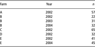
In all, 338 goats, from five herds, were allocated to this experiment conducted from 2002 to 2004 (Table 1). In each herd, goats were randomized in two parallel groups according to their body weight and age at the time of AI. Each group was allocated by drawing lots for either short- or long-duration AI . The insemination procedure was performed as described in Experiment 1, with two operators. In each group of AI duration, the goats were assigned by drawing lots to one or other of the technicians, who were instructed to deposit the semen alternatively according to a short- or a long- duration AI, as defined with results from Experiment 1. For the short AI treatment, goats were subjected to an insemination duration of 20 s: the insemination gun was inserted into the cervix where the semen was quickly deposited (intracervical). When the cervix was not sufficiently open to permit the introduction of the AI gun, the semen was deposited at the entry of the cervical canal (exocervical) (mean time ± s.e. to perform short AI from insertion to withdrawal of the speculum was 25.2 ± 6.8 s). For the long AI treatment, goats were subjected to an insemination duration of 60 s: the insemination gun was inserted into the cervix and moved into the cervical canal for approximately 40 s for cervical deposition of the semen dose as deep as possible, without harming the cervical epithelium. When the inseminator failed to insert the gun into the cervix within 50 s, the semen was deposited at the entry of the cervical canal (mean time ± s.e. to perform long AI from insertion to withdrawal of the speculum was 63.3 ± 5.0 s). Over the whole experiment, an intrauterine passage of the insemination gun was observed in 13 goats only, from three herds, and mostly observed (n = 9) after long-duration AI, so these animals were excluded from the study.
Number of nulliparous goats and year of AI in each farm from experiment 2
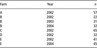
AI = artificial insemination; n = number of goats.
Pregnancy diagnosis
Trans-abdominal ultrasonography for pregnancy diagnosis was made at day 40 to 45 after insemination (AI). The pregnancy rate per AI was defined as the percentage of goats confirmed pregnant.
Statistical analysis
Pregnancy rates were analysed by the generalized linear model (GENMOD procedure of SAS; SAS Institute Inc., Cary, NC, USA) using the logit link function and the binomial distribution. Deviance analysis was used to test the effect of farm, AI duration, inseminator and deposition site of semen. In Experiment 2, only factors with probabilities (P) less than 0.10 were retained in the final model. Interactions between AI duration and main factors were then tested. All least square means were estimated with weighted coefficient for the deposition site of semen such as the size of modalities intracervical and exocervical were 85% and 15%, respectively. These modalities were considered as observed factors, the weighted coefficient matched the achievement in this experiment (n = 281 intracervical AI, and n = 42 exocervical AI).
Results
Experiment 1
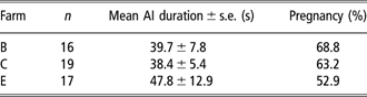
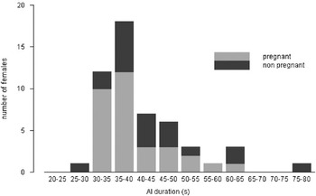
The total pregnancy rate was 61.6%, with pregnancy values ranging from 52.9% to 68.8% between the three herds (n = 16 to 19 goats per farm) (Table 2). The farm did not affect pregnancy rates (P = 0.64), but had a significant effect on the total duration of the AI procedure (P < 0.01), which varied from 38.4 ± 5.4 to 47.8 ± 12.9 s between herds (Table 2). Global duration of AI measured was 41.9 ± 9.9 s, with a median of 39 s (Figure 1). There was a marked trend for a significant effect of AI duration on pregnancy rates (P = 0.06). Indeed, AI performed in less than 39 s resulted in a mean pregnancy rate of 75% (mean AI duration: 35.4 ± 2.8 s, n = 28 goats), while only 46% of inseminated females were pregnant when the AI procedure lasted more than 39 s (mean AI duration: 49.7 ± 9.9 s, n = 24 goats) (Figure 1).
Variation of duration of AI procedure and pregnancy rate between herds
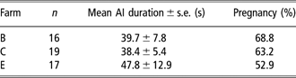
AI = artificial insemination; n = number of goats.
Pregnancy in relation to the duration of AI.

Experiment 2
This experiment was conducted to determine whether short- or long-duration AI affected pregnancy rates. Two different modalities were defined on the basis of the median time (39 s) for completion of the procedure in Experiment 1. Goats were assigned to short- (20 s) or long-duration AI (60 s).
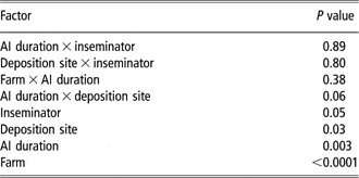
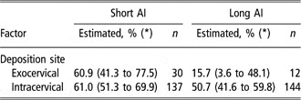
During the 3 years of this experiment, 325 goats were inseminated, and 179 became pregnant. The levels of significance for the effects of different factors accounting for variation in pregnancy rates are presented in Table 3. Estimated pregnancy rates after short- and long-duration AI are listed in Table 4. The estimated pregnancy rates for short and long AI duration were 61.2% and 44.2%, respectively. There was no effect of age and body weight between short and long AI groups of animals. The AI duration × deposition site interaction (P = 0.06) was mainly due to the lower pregnancy rate in long AI duration when the semen was deposited exocervically (15.7%) in comparison with intracervical deposition (50.7%). By contrast, there was no evidence of difference between the two deposition modalities in short AI duration (61.0% v. 60.9% for intracervical and exocervical semen deposition, respectively) (Table 4).
Levels of significance of various factors accounting for variation of pregnancy rates after first AI
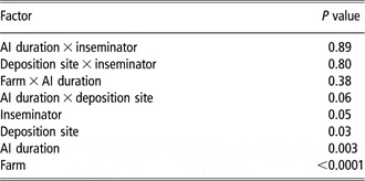
AI = artificial insemination.
Effect of semen deposition site on pregnancy rates of nulliparous goats after short (20 s) and long (60 s) AI procedures
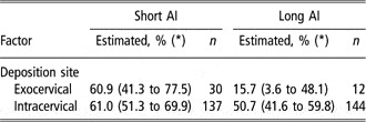
AI = artificial insemination; n = number of goats.
(*) = 95% confidence interval.
A significant difference between inseminators was found (P = 0.05), but no significant interaction was observed between inseminators for either AI duration or deposition site (Table 3). There was no difference between inseminators for the duration of AI for each protocol (27.8 ± 7.8 s v. 22.8 ± 4.4 s for short AI, and 64.2 ± 6.0 v. 62.3 ± 3.5 s for long AI).
Discussion
This study showed that the duration of the AI procedure, as defined by the time spent from insertion to withdrawal of the speculum, affected fertility in nulliparous goats inseminated with frozenthawed semen. Fertility rate when the semen was deposited exocervically was significantly improved after a rapid AI procedure. The cervix constitutes the initial barrier to the ascent of spermatozoa after exocervical insemination in small ruminant species. The site and depth of semen deposition are important factors that influence fertility in goats (Ritar and Salamon Reference Ritar and Salamon1983; Corteel et al., Reference Corteel, Leboeuf and Baril1988; Ritar et al., Reference Ritar, Ball and O’May1990; Salvador et al., Reference Salvador, Viudes-de-Castro, Bernacer, Gomez and Silvestre2005). A relatively small proportion of the cells deposited exocervically during insemination penetrate the cervical canal. During insemination in the goat, unlike in the sheep, the semen can be deposited into the uterus. In this case, the fertility rate is better than for the adult does inseminated into the cervical canal (Corteel et al., Reference Corteel, Leboeuf and Baril1988). However, an intrauterine passage of the insemination gun is infrequently obtained in nulliparous goats. Indeed, in the current study, an intrauterine semen deposition was observed only in 13 of the 325 goats inseminated, and none became pregnant (data not shown). Such animals were removed from our experiment. The depth of insemination mainly depends on the anatomical structure of the cervix of each animal and the skill of the inseminator. In this study, short- and long- duration AI were performed by the same operators but the site of semen deposition was not controlled.
The negative effect of long AI duration on fertility was highlighted by the fertility after exocervical deposition of semen. In this case, it is not possible to deposit the semen into the cervix. It may be that these females were inseminated too early in relation to the ovulation time, which is known to decrease fertilily in goats (Maurel et al., 1992). On the other hand, we have previously shown in the ewe that genital stimulation during AI enhanced uterine motility (Houdeau et al., Reference Houdeau, Raynal, Marnet, Germain, Mormede, Rossano, Monnerie and Prud’homme2002; Raynal and Houdeau, Reference Raynal and Houdeau2004), and previous studies highlighted a negative correlation between increased uterine motility at the time of AI and fertility rates in small ruminants (Lightfoot and Restall, Reference Lightfoot and Restall1971; Toutain et al., Reference Toutain, Marnet, Laurentie, Garcia-Villar and Ruckebusch1985), and also in women (Kunz et al., Reference Kunz, Beil, Deininger, Wildt and Leyendecker1996; Leyendecker et al., Reference Leyendecker, Kunz, Wildt, Beil and Deininger1996). From the perspectives of the AI technique, the longer the duration of insemination, the more mechanical stimulation is experienced by the vagina and cervix by the AI apparatus, often due to the difficulty of reaching the cervical canal with the insemination gun. In the current study, we found that fertility after exocervical deposition decreased using long AI duration in comparison with short AI duration (15.7% v. 60.9%, respectively). In the sow, Huhn et al. (Reference Huhn, Fritzsch and Dahms1977) also reported a decrease in pregnancy rate with increasing time needed for insemination. Our results showed that short duration of AI improved the performance of exocervical insemination, with fertility rates within the same range than those observed when the semen was placed into the cervical canal. It is also interesting to note that short AI duration tended to improve fertility rates after intracervical deposition of semen in comparison with long AI duration (61% v. 50.7%, respectively; P = 0.15). Further experiments are needed to determine whether long AI duration is a limiting factor during intracervical insemination in nulliparous goats.
Although uterine activity was not recorded in the present study, it is probable that uterine activity increased during insemination, as reported in ewes (Lehrer et al., Reference Lehrer, Schindler, Brown and Fischler1979; Houdeau et al., Reference Houdeau, Raynal, Marnet, Germain, Mormede, Rossano, Monnerie and Prud’homme2002), mares (Troedsson et al., Reference Troedsson, Liu and Crabo1998) and women (Sahmay et al., Reference Sahmay, Atasu and Karacan1990). In the ewe, by analysing the AI operation gesture step-by-step, it appeared that both introduction and opening of the speculum in the vagina, then searching of the cervical canal for the correct site of semen deposition evoked autonomic nerve-mediated uterine contractions that considerably differed from the normal motor pattern observed during natural mating (Raynal and Houdeau, Reference Raynal and Houdeau2004). In domestic mammals and humans, sperm transit towards the oviducts occurs rapidly after mating (Overstreet and Cooper, Reference Overstreet and Cooper1978a and Reference Overstreet and Cooper1978b; Hawk, Reference Hawk1983; Kunz et al., Reference Kunz, Beil, Deininger, Wildt and Leyendecker1996), and is thought to be the result of spontaneous contractions that facilitate the transport of spermatozoa along the uterine horns (Hawk, Reference Hawk1983; Kunz and Leyendecker, Reference Kunz and Leyendecker2002; Suarez and Pacey, Reference Suarez and Pacey2006). Studies in sows, ewes and women suggest that stimulation of contractions or abnormal propagation of contractions along the uterus may increase the reflux of semen and/or impair the passive infusion of semen from the cervix towards the uterine lumen (Zerobin and Spörri, Reference Zerobin and Spörri1972; Leyendecker et al., Reference Leyendecker, Kunz, Wildt, Beil and Deininger1996; Langendijk et al., Reference Langendijk, Bouwman, Kidson, Kirkwood, Soede and Kemp2002). Using cloprostenol to increase uterine contractions in sows, Langendijk et al. (Reference Langendijk, Bouwman, Kidson, Kirkwood, Soede and Kemp2002) reported a sharp diminution in the number of spermatozoa recovered along the genital tract after insemination, associated with reduced fertility. They concluded on a probable obstruction of the uterus by successive contractions, thereby limiting the flow of spermatozoa towards the oviducts.
In conclusion, a short time of insemination improves fertility rate after AI in nulliparous dairy goats, and a high fertility rate can be obtained if the time spent for semen deposition is about 20 s. Of interest, equivalent fertility rates between exocervical and intracervical inseminations were only observed when AI was performed quickly. It is suggested that rapid semen deposition may limit the reflex activation of uterine contractions provoked by speculum insertion and the movement of the insemination gun and ameliorate the fertility of nulliparous goats after their first AI. This result obtained under field conditions opens the possibility to engage nulliparous dairy goats in AI programmes for sanitary reasons or genetic selection improvement.
Acknowledgements
The authors would like to thank H. Coutineau from the Lycée Agricole de Melle (79), breeders for available animals to this experiment (Mr G. Dupuis, M. Georges, D. Guillot, SCEA Coynault-Mercier) and E. Bruneteau, F. Dugué, N. Riquet, E. Santy and F. Bouvier for their technical assistance.