INTRODUCTION
The protistan parasite Bonamia ostreae, the causative agent of bonamiosis, has had a detrimental effect on the production of the European flat oyster, Ostrea edulis in Europe since the 1970's (Comps et al. Reference Comps, Tigé and Grizel1980; Balouet et al. Reference Balouet, Poder and Cahour1983; Bucke and Feist, Reference Bucke, Feist and Ellis1985; Elston et al. Reference Elston, Farley and Kent1986; Montes and Figueras, Reference Montes and Figueras1987; Van Banning, Reference Van Banning1990, Reference Van Banning1991; McArdle et al. Reference McArdle, McKiernan, Foley and Hugh-Jones1991; Boudry et al. Reference Boudry, Chatain, Naciri-Graven, Lemaire and Gerard1996; Lynch et al. Reference Lynch, Armitage, Wylde, Mulcahy and Culloty2005). The presence of another parasite of the genus Bonamia, B. exitiosa, has been recorded in O. edulis in Spain and more recently in Italy (Abollo et al. Reference Abollo, Ramilo, Casas, Comesaña, Cao, Carballal and Villalba2008; Narcisi et al. 2010). Prior to its discovery in Europe, B. exitiosa was considered a southern hemisphere species whose hosts were Ostrea chilensis and Ostrea angasi (Dinamani et al. Reference Dinamani, Hine and Jones1987; Hine and Jones, Reference Hine and Jones1994; Hine, Reference Hine1996; Corbeil et al. Reference Corbeil, Arzul, Robert, Berthe, Besnard-Cochenned and Crane2006).
B. ostreae can be transmitted directly from infected oysters to uninfected oysters (Elston et al. Reference Elston, Kent and Wilkinson1987; Culloty et al. Reference Culloty, Novoa, Pernas, Longshaw, Mulcahy, Feist and Figueras1999); however, it has not been definitively determined whether the parasite's life cycle is fully represented by this direct transmission. A similar situation exists with B. exitiosa and the transmission by infective stages carried passively on currents between oyster beds is suspected (Hine, Reference Hine1996; Cranfield et al. Reference Cranfield, Dunn, Doonan and Michael2005; Corbeil et al. Reference Corbeil, Arzul, Robert, Berthe, Besnard-Cochenned and Crane2006). The resilience of B. ostreae infections has previously been described, with one study indicating that infection remains present on the beds and that O. edulis become infected when relayed to areas that have remained fallow for a number of years (Van Banning, Reference Van Banning1988).
Although B. ostreae is characterized by direct transmission, another characteristic of haplosporidians is the presence of a spore stage (Burreson and Ford, Reference Burreson and Ford2004) that has not been observed in B. ostreae but has been observed in Bonamia perspora (Carnegie et al. Reference Carnegie, Burreson, Hine, Stokes, Audemard, Bishop and Peterson2006). As a result, questions still remain as to what constitutes the complete life cycle of B. ostreae or other Bonamia spp. The possibility that other invertebrate species may act as carriers or reservoirs for B. ostreae cannot be ruled out.
In previous studies, transmission trials have been undertaken investigating various shellfish species as potential carriers or reservoirs for B. ostreae. The Pacific oyster Crassostrea gigas, mussels Mytilus edulis and M. galloprovincialis, and clams Ruditapes decussatus and V. (=Ruditapes) philippinarum could not be infected either in field or in laboratory trials, either naturally or via experimental infection, and these bivalves did not appear to act as either vectors or intermediate hosts for B. ostreae (Figueras and Robledo, Reference Figueras and Robledo1994; Renault et al. Reference Renault, Cochennec and Grizel1995; Culloty et al. Reference Culloty, Novoa, Pernas, Longshaw, Mulcahy, Feist and Figueras1999). The cockle, Cerastoderma edule, was screened for B. ostreae in one Dutch study, but no infection was found (Van Banning, Reference Van Banning1987). However, all of these studies employed cytology and histology to screen for the parasite. More recent studies, using molecular techniques, have demonstrated the presence of B. ostreae DNA in other invertebrate species: results from a preliminary study over a 14-month period showed that 8 benthic macro-invertebrate species and 19 grouped zooplankton samples taken from a B. ostreae-endemic area gave positive PCR results for B. ostreae DNA (Lynch et al. Reference Lynch, Armitage, Mulcahy and Culloty2007). In that study, B. ostreae was visualized in the heart smears of 2 naïve O. edulis, co-habiting with the brittle star, Ophiothrix fragilis, brought to the laboratory from a B. ostreae-endemic area. The basis of that transmission is unclear but demonstrates that various marine organisms may at least act as incidental carriers of B. ostreae.
In Ireland, C. gigas spat are imported from France and the Channel Islands with very limited supply from Irish hatcheries. C. gigas is an important commercial species (F.A.O., 2006) and has been transplanted worldwide by various means (www.ices.dk); however, it is a host to several parasites (Bower et al. Reference Bower, McGladdery and Price1994). Some of these pathogens have been transferred with movements such as the protistan Haplosporidium nelsoni which transferred from the Pacific oyster to Crassostrea virginica (Friedman et al. Reference Friedman, Cloney, Manzer and Hedrick1991; Friedman, Reference Friedman1996; Burreson et al. Reference Burreson, Stokes and Friedman2000; Renault et al. Reference Renault, Stokes, Chollet, Cochennec, Berthe, Gérard and Burreson2000). Another issue related to movements can be epiflora and epifauna in a C. gigas consignment and the presence of other organisms held within the shell cavity (Minchin et al. Reference Minchin, Duggan, Holmes and Neiland1993; O'Mahony, Reference O'Mahony1993; Minchin, Reference Minchin1996; Wasson et al. Reference Wasson, Zabin, Bedinger, Diaz and Pearse2001; Minchin and Rosenthal, Reference Minchin, Rosenthal, Leppäkoski, Gollasch and Olenin2002; Ruesink et al. Reference Ruesink, Lenihan, Trimble, Heiman, Micheli, Byers and Kay2005).
The European Food Safety Authority considers C. gigas to be a low risk carrier for B. ostreae (www.efsa.europa.eu). There is no evidence available to date to suggest that C. gigas is a natural host for this pathogen. Carriers or reservoir hosts are rarely included in the pathogen screening process and the effects of such transfers are unpredictable. Here, we define a ‘carrier’ as an incidental host which harbours the disease organism in their body or within the shell without manifesting the symptoms, thus acting as a distributor of infection. A ‘reservoir’ is defined as one or more epidemiologically connected populations or environments in which the pathogen can be permanently maintained and from which infection is transmitted to the target population (Haydon et al. Reference Haydon, Cleaveland, Taylor and Laurenson2002). As O. edulis and C. gigas can both be grown in adjacent beds the transfer of pathogens between these species could potentially occur.
The objectives of this study were to determine whether C. gigas could act as a carrier or a reservoir for B. ostreae. Some experiments were performed in Ireland involving (a) natural exposure of C. gigas to the parasite in the field and (b) co-habitation experiments in the laboratory to evaluate if B. ostreae could be transferred from infected O. edulis to naïve C. gigas and from field-exposed (17 months) C. gigas to naïve O. edulis. Other experiments were performed in Spain to evaluate whether C. gigas can become infected by B. ostreae through (a) field exposure and (b) experimental injection of the parasite in the laboratory.
MATERIALS AND METHODS
Field transmission trial in Ireland
One thousand half-grown C. gigas (16 months old, mean weight=60 g) from Oysterhaven, Co Cork, a B. ostreae-free site (51°84′N, 8°8′W), were deployed to the field site at Rossmore, Co Cork a B. ostreae-endemic site (51°85′N, 8°26′W) (McManus, Reference McManus1988; McArdle et al. Reference McArdle, McKiernan, Foley and Hugh-Jones1991; Lynch et al. Reference Lynch, Armitage, Wylde, Mulcahy and Culloty2005). The temperature range in Rossmore varies between 5°C and 20°C. The coldest temperatures occur in February-March and the warmest temperatures between June and August. The surface salinity varies between 26‰ and 33‰ but varies depending on rainfall and on tide (Lynch et al. Reference Lynch, Armitage, Mulcahy and Culloty2007). The oysters were relayed (150 oysters/bag) onto the sediment on the mid-lower intertidal shore from March 2006 to January 2007. B. ostreae has been consistently detected in flat oysters in this area with prevalence of infection varying from 2% to 33% in 4+ oysters screened throughout 2005 (personal observations and The Marine Institute Ireland).
An initial sample of C. gigas (n=150) was prepared and screened for B. ostreae prior to commencement of the field trial, to ensure that the oysters were uninfected. Following relaying, 2-month, 6-month and 10-month samples of 142, 128 and 122 C. gigas individuals respectively were sampled and screened for B. ostreae.
Field transmission trial in Spain
In December 2005, 230 Pacific oyster Crassostrea gigas spat, that had been removed from spat collectors set in Brittany (France) and exported to Galicia, (NW Spain) were set in trays and hung from a raft, along with flat oysters from Cambados, in the Ría de Arousa (Galicia) which has been affected by bonamiosis since the early 1980s (Polanco et al. Reference Polanco, Montes, Outón and Meléndez1984; da Silva et al. Reference Da Silva, Fuentes and Villalba2005).
From January to December 2007, 15–20 oysters were taken randomly, monthly and transported to the laboratory, where they were cleaned and all fouling organisms were carefully removed. A total of 213 Pacific oysters (107·90 mm in mean length; 151·0 g in mean weight) were processed for diagnosis of bonamiosis by histology, PCR using gill and heart tissue and in situ hybridization. Environmental conditions were not measured during the study. Nevertheless, data recorded weekly at one sampling station (A7) of the Net of Oceanographic Conditions of the Galician Bays (Controlled by the Instituo Tecnolóxico para o Control do Medio Mariño de Galicia, INTECMAR), located close to the raft, showed that temperature and salinity measured at a depth of 5–10 m (at which oysters were deployed) ranged from 12°C to 18°C and from 33·6‰ to 35·7‰, respectively.
Laboratory transmission trial I in Ireland
The laboratory trial ran for a period of 6 months from May 2007 to November 2007. Three hundred naïve C. gigas, that had been relayed to a B. ostreae-endemic area, Rossmore, Cork Harbour, for the field transmission trial for a 17-month period, were returned to the laboratory to be held in tanks with naïve O. edulis sourced from Loch Ryan, Scotland (5 years old with a mean whole weight of 50–60 g). An initial sample of Loch Ryan O. edulis (n=150) was screened for B. ostreae using heart smears and PCR, prior to commencement of the trial, to confirm that the oysters were uninfected. Prior to being placed into the system, epifauna and epiflora on the C. gigas shells were removed and the oysters were washed with freshwater, to remove any traces of mud and other organisms. Three control tanks (600 L), each containing 200 naïve Loch Ryan O. edulis, and 3 experimental tanks (600 L), each containing 100 naïve Loch Ryan O. edulis together with 100 exposed C. gigas, were used. Seawater from Bantry Bay, Co Cork, (salinity 26‰) a B. ostreae-free designated area, (51°42′N 9°28′) was used in the experimental tanks and in the control tanks. Half of the seawater from each tank was replaced every 3 weeks. The laboratory transmission trial was carried out at ambient temperatures, from 6°C to 30°C. The animals were fed a heteromorphic phytoplankton, Nannochloropsis sp., Tetraselmis sp. and Isochrysis sp. mix (6 billion cells per 10 ml of Kent Marine Phytoplex) twice weekly.
The oysters were checked on a daily basis and dead or dying oysters were removed and screened by heart-smear screening and by PCR. Histology and ISH were carried out on any individuals found to be positive for B. ostreae in the heart-smear screening and in the PCR. At the end of the experiment, all of the O. edulis and C. gigas remaining in both control and experimental tanks were screened by heart-smear screening and by PCR for B. ostreae. Histology and ISH was carried out on individuals found to be positive for B. ostreae in the heart smears and in the PCR.
Laboratory transmission trial II in Ireland
The trial ran for a period of 6 months from March 2006 to September 2006. Naïve C. gigas (n=1100), 16 months old and with a mean whole weight of 16·0 g, were obtained from Dungarvan, Co Waterford a B. ostreae-free area (51°59·6′, 7°34·6′), (originally sourced from Satmar, France). An initial sample of animals was screened for B. ostreae, prior to the commencement of the trial, to ensure that the oysters were uninfected. Flat oysters, O. edulis, exposed to B. ostreae, were sourced from Rossmore, Cork Harbour and were approximately 5–6 years old. An initial sample of these flat oysters (n=150) was screened by heart-smear screening and by PCR, to ensure that they were infected with B. ostreae, and to determine the prevalence and intensity of infection. Three control tanks (600 L), each containing 200 naïve C. gigas, and 3 experimental tanks (600 L), each containing 100 naïve C. gigas and 100 Rossmore O. edulis, were used.
Half of the seawater from each tank was replaced every 3 weeks. The laboratory transmission trial was carried out at ambient temperatures, from 6°C to 30°C. The animals were fed a heteromorphic phytoplankton, Nannochloropsis sp, Tetraselmis sp. and Isochrysis sp. mix (6 billion cells per 10 ml of Kent Marine Phytoplex) twice weekly. The trial was completed after a 6-month period.
The oysters were checked on a daily basis, and dead or dying oysters were removed and screened for B. ostreae by heart-smear screening and by PCR. Histology and ISH were carried out on any individuals found to be positive for B. ostreae in the heart-smear screening and in the PCR. At the end of the 6-month experiment, all of the O. edulis and C. gigas, remaining in both control and experimental tanks, were screened by heart-smear screening and by PCR for B. ostreae. Histology and ISH was carried out on individuals found to be positive for B. ostreae in the heart smears and in the PCR.
Laboratory trial in Spain
Ninety Pacific oysters (102·62 mm in mean length; 117·16 g in mean weight) were collected from Oysterhaven, Co Cork (Ireland) (51°84′N, 8°8′W), from a bed where O. edulis do not co-occur, in March 2006, and transported to the laboratory of Centro de Investigacións Mariñas (Galicia, Spain). Oysters were acclimated in tanks with 0·2 μm-filtered, aerated seawater at 18°C for 1 week before injection. Before and after injection, the oysters were fed with a mix of cultured algae Isochrysys T-ISO and Skeletonema costatum continuously added to the tank. The tanks were cleaned and the seawater was changed daily to avoid opportunistic infections.
Isolation of B. ostreae from infected O. edulis
A batch of flat oysters O. edulis (n=89) (64·21 mm in mean length; 46·93 g in mean weight) deployed in an area of the Ría de Arousa (Galicia, NW Spain) were used as source of B. ostreae cells. To estimate infection intensity, haemolymph cell monolayers were performed by cytocentrifugation according to the method of da Silva and Villalba (Reference Da Silva and Villalba2004). The percentage of haemocytes infected by microcells in the haemolymph cell monolayers was estimated with light microscopy at 1000× magnification. Heavily infected oysters, accounting for 5–25% of haemocytes infected by B. ostreae, were used for the isolation of parasite cells, which was performed according the protocol of Mialhe et al. (Reference Mialhe, Bachère, Chagot and Grizel1988). The purified B. ostreae cells were resuspended in filtered seawater and viability was assessed with the trypan blue exclusion test. B. ostreae cells were then counted using a Malassez haemocytometer.
Injection of B. ostreae into C. gigas
Before the injection, the Pacific oysters were anaesthetized using MgCl2 (3·5%) according to the method of Culloty and Mulcahy (Reference Culloty and Mulcahy1992). Two experimental groups of 45 individuals were established. Oysters belonging to the infected group were inoculated with 135 000 cells of the purified parasite resuspended in 50 μl of filtered seawater, and the oysters belonging to the control group were inoculated with the same volume of filtered seawater. The oyster was injected in the adductor muscle using a 1 ml syringe with a G26-gauge needle. After injection, the oysters were kept out of water for 45 min before re-immersing in seawater. Ten days after injection, a first subsample of 40 oysters (20 infected and 20 control) was processed for diagnosis of bonamiosis by histology and PCR using gill tissue. After 80 days, the remaining live individuals (19 infected and 22 control) (second subsample) were also processed.
Diagnostic methods-Ireland
Heart/gill smears
Ventricular heart or gill smears were prepared immediately, screened and the intensity of infection determined according to the protocol of Bachére et al. (Reference Bachére, Durand and Tigé1982) and Culloty et al. (Reference Culloty, Novoa, Pernas, Longshaw, Mulcahy, Feist and Figueras1999) under a light microscope (400× magnification).
Histology
C. gigas tissues (including mantle, gill, digestive gland and gonad) were fixed in Davidson's solution and preserved in 70% ethanol for histology (Shaw and Battle, Reference Shaw and Battle1957). Tissues were embedded in paraffin wax, sectioned at 5 μm, stained with Harris' haematoxylin and eosin (Howard and Smith, Reference Howard and Smith1983) and tissue sections were observed under a light microscope (400× and 1000× magnification) for diagnosis.
PCR
One small sample of heart or gill tissue (5 mm2) from each oyster was frozen for DNA extraction. Fluid (100 μl) from the internal shell cavity of each oyster was also taken, using a pipette, for PCR screening for B. ostreae DNA. The Chelex-100 resin extraction method was used to extract the DNA (Walsh et al. Reference Walsh, Metzger and Higuchi1991; Goff and Moon, Reference Goff and Moon1993).
PCR screening for B. ostreae DNA using BO BOAS primers (Cochennec et al. Reference Cochennec, LeRoux, Berthe and Gerard2000) was carried out. Positive and negative controls were used in each PCR reaction. Positive controls were obtained from O. edulis, heavily infected with B. ostreae. Negative controls consisted of double distilled water (ddH20). The Carnegie c primers (c_for, c_rev) were used in a repeat PCR on C. gigas individuals found to be positive following use of Bo Boas primers (Carnegie et al. Reference Carnegie, Barber, Culloty, Figueras and Distel2000). PCR products were checked on 2% agarose gels after electrophoresis. PCR results were graded as either positive or negative. Expected product size was 300 bp in the BO BOAS PCR and 760 bp in the Carnegie c PCR.
In situ hybridization (ISH)
This procedure was carried out using a digoxigenin (DIG)-labelled probe using the Cochennec primers (Bo Boas) and all solutions and buffers were made up according to the protocol described by Sambrook and Russell (Reference Sambrook and Russell2001). This probe targets the SSU rDNA gene (Cochennec et al. Reference Cochennec, LeRoux, Berthe and Gerard2000). Tissue sections were prepared by the following procedure. Slides were immersed in xylene (2×10 min), absolute ethanol (2×10 min), air dried, and then 200 μl of proteinase K in Tris-EDTA (TE) buffer was placed on the tissue sections. Slides were incubated in a moist chamber for 20 min at 37°C, then dehydrated in absolute ethanol (2×1 min), air dried and covered with a gene frame. Hybridization of the tissue sections was carried out by placing the mixture of probe (2 μl of probe+1000 μl of hybridization buffer) onto the tissue sections on the slides. Denaturation of the probe was carried out by placing the sections into a Hybaid™ thermo cycler for 1 cycle for 5 min at 94°C. The slides were placed on ice for 2 min and incubated overnight at 42°C in a moist chamber. The following day the slides were placed in 2× saline-sodium citrate (SSC) buffer (2×5 min), 0·4× SSC (at 42°C) (10 min) and DIG 1 buffer (1 min). Then 200 μl of diluted conjugate were added to the tissue and incubated in a moist chamber at room temperature for 1 h. The slides were washed with DIG 1 buffer (2×1 min), DIG 3 buffer (10 min) and 200 μl of nitroblue tetrazolium and 5-bromo-4-chloro-3-indolyl phosphate (NBT/BCIP) were added to the tissue sections and incubated in the dark for at least 30 min. The staining was stopped with DIG 4 buffer. The slides were washed in distilled water and placed in Bismarck Brown Y solution for 2 min. Tissue sections were dehydrated in 95% ethanol (1 min), absolute ethanol (1 min) and placed in xylene prior to being cover-slipped with Eukitt (SigmaAldrich TM).
Sequencing
To ensure that the PCR products, generated in the positive controls and in the C. gigas and O. edulis DNA samples, were specifically B. ostreae, the positive products, using a Qiagen Qiaquick gel extraction kit to clean the DNA, were sequenced by MWG BIOTECH AG, Martinsried, Germany. Samples from the same source i.e. either tissues or shell cavity fluid were pooled with bands from 3 individuals combined to increase the concentration of DNA. The purified DNA was directly sequenced using the BO BOAS primers (Cochennec et al. Reference Cochennec, LeRoux, Berthe and Gerard2000) and the Carnegie c primers (Carnegie et al. Reference Carnegie, Barber, Culloty, Figueras and Distel2000).
Diagnostic methods-Spain
Histology
Histology was carried out as described in the previous section.
PCR
The heart and a fragment of gills were excised using sterile scissors and were fixed in 96% ethanol for DNA isolation. Before and after using the scissors, they were washed in sterile seawater and dried in filter paper. DNA extractions were performed employing the DNAzol reagent® (Invitrogen Life Technologies™) according to the manufacturer's instructions and quantified on a SmartSpec™ Plus Spectrophotometer (Bio-Rad). Generic primers for Bonamia named Bo-Boas (Cochennec et al. Reference Cochennec, LeRoux, Berthe and Gerard2000) and CF-CR (Carnegie et al. Reference Carnegie, Barber, Culloty, Figueras and Distel2000), which amplify a fragment of the 18S rDNA gene, were used for molecular diagnosis of Bonamia spp.
Sequencing
PCR products were ligated into cloning vector pCR2.1 at 14°C overnight and transformed into Escherichia coli One Shot Top 10F′ Chemically Competent (Invitrogen Life Technologies™). Transformed cells were screened by PCR using the vector's M13 forward (5′ GTA AAA CGA CGG CCA G 3′) and reverse (5′ CAG GAA ACA GCT ATG AC 3′) primers. The positive clones were cleaned for sequencing using the ExoSAP-IT reagent (USB Corporation) according to the manufacturer's instructions. The sequencing reaction was performed on an ABI PRISM™ 3100 (Applied Biosystems) using a BigDye® Terminator v3.1 Cycle Sequencing Kit (Applied Biosystems) as supplied by the manufacturer. All amplified fragments were sequenced with M13 commercial primers and the chromatograms were analysed using ChromasPro version 1.41 Technelysium Pty Ltd. All sequences generated were searched for similarity using BLAST (Basic Local Alignment Search Tool) through web servers of the National Center for Biotechnology Information (USA).
In situ hybridization (ISH)
Two consecutive tissue sections of each sample were mounted on AAS slides (Silane-Prep Slides; Sigma) for ISH. PCR clones of Bonamia sp. were used as template DNA to prepare a DIG-labelled probe following the manufacturer's protocol (Roche PCR DIG Probe Synthesis Kit). ISH was performed as described above for Ireland.
RESULTS
Field transmission trial in Ireland
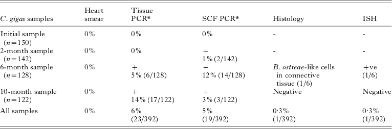
In the field trial, the initial sample of C. gigas, screened prior to relaying to Rossmore, Cork Harbour, was found to be negative for B. ostreae both by heart-smear screening and PCR, confirming that the oysters were uninfected with B. ostreae. In total, during the 10-month trial, 392 naturally exposed C. gigas were screened for B. ostreae using both heart smears and PCR. In the 2-, 6- and 10-month samples, post-relaying to Rossmore, Cork Harbour (Table 1), no B. ostreae cells were visualized when the heart smears were screened. However B. ostreae DNA was detected by PCR in either the shell cavity fluid from 2 months or in both the tissue or the shell cavity of the animals from 6 months, with the highest prevalence in the tissues observed after 10 months.
Results of screening of heart smears, histology, PCR and ISH for Bonamia ostreae in Crassostrea gigas prior to, and following, relaying to the North Channel, Cork Harbour
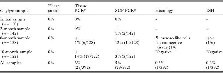
+ Positive for B. ostreae, SCF Shell cavity fluid, -not screened.
* (Primers BO, BOAS, Cochennec et al. Reference Cochennec, LeRoux, Berthe and Gerard2000; Carnegie c for & rev, Carnegie et al. Reference Carnegie, Barber, Culloty, Figueras and Distel2000.)
Histology and ISH were carried out on the 23 individuals that generated positive PCR results from tissue samples in the 6- and 10- month samples. Among the 6 positive individuals screened from the 6-month sample, B. ostreae-like cells were detected in the connective tissue of one individual by histology (Fig. 1A and B). The microcells observed were uninucleate and binucleate, 2–3 μm in size with both central and eccentric nuclei. The B. ostreae-like cells were located in the connective tissue. According to the classification system for assessing intensity of B. ostreae infection (Bachère et al. Reference Bachére, Durand and Tigé1982; Culloty et al. Reference Culloty, Novoa, Pernas, Longshaw, Mulcahy, Feist and Figueras1999) a Class 2 level of intensity was observed (more than 10 but less than 100 microcells present). Screening of the sections by ISH yielded positive staining in that same individual. No B. ostreae-like cells or other pathogens were detected in the 10-month sample, either by histology or ISH. The sequences from PCR products, generated from DNA extracted from pooled (groups of 3) C. gigas shell cavity fluid samples (n=6) and pooled (groups of 3) C. gigas tissue samples (n=7), were identical to the original B. ostreae rDNA subunit as published by Cochennec et al. (Reference Cochennec, LeRoux, Berthe and Gerard2000) (Accession no. AF192759) and Carnegie et al. (Reference Carnegie, Barber, Culloty, Figueras and Distel2000) (Accession no. AF262995).
(A) Field trial: Ireland. Bonamia ostreae-like cells in the connective tissues of Crassostrea gigas naturally exposed to B. ostreae in the field 6 months post-T0 (H&E staining). (B) Field trial: Ireland. ISH staining of B. ostreae-like cells (dark blue staining) in the tissues of C. gigas naturally exposed to B. ostreae in the field 6 months post-T0.

Field transmission trial in Spain
No infection was observed by histopathological analysis in any of the examined oysters. Positive PCR results were observed for template obtained from gills (7/213) and heart (14/213). Of them, only 4 oysters were PCR-positive in both organs. No seasonal trend for positive results was observed. Cloning was performed with PCR products from 11 oysters; it was not possible for the other 6 oysters with positive results because of the scarcity of the amplification product. After cloning and sequencing of the PCR products, BLAST search showed sequence identity values of 100% with B. ostreae (4 clones of 4 oysters) and with B. exitiosa (7 clones of 7 oysters). Both protozoan species were detected in the DNA purified from gill and heart. ISH assays were performed with those samples that were positive by PCR (N=17) and negative results were obtained for all samples analysed, in spite of the fact that they were repeated 3 times.
Laboratory transmission trial I in Ireland
The initial sample of naïve Loch Ryan O. edulis (n=60) was negative for B. ostreae prior to the trial. All control O. edulis (n=251) screened using heart or gill smears and PCR remained negative for B. ostreae throughout the trial using heart-smear screening and PCR. All animals, both experimental O. edulis and experimental C. gigas, screened by heart smear were negative for B. ostreae infection throughout the trial.
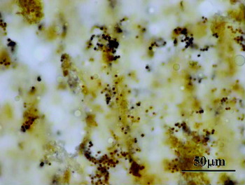
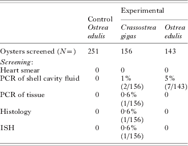
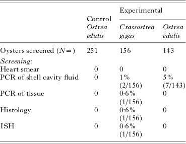
B. ostreae DNA was detected by PCR in the shell cavity fluid (n=2) and tissues of C. gigas (n=1) between weeks 14 and19 and in the shell cavity fluid of O. edulis (n=7) between weeks 6 and 14 (Table 2). The sequences from PCR products, generated from 1. DNA extracted from C. gigas shell cavity fluid, 2. O. edulis shell cavity fluid samples and 3. C. gigas tissue samples from the 1 positive individual, were identical to the original B. ostreae rDNA subunit as published by Cochennec et al. (Reference Cochennec, LeRoux, Berthe and Gerard2000) (Accession no. AF192759) and Carnegie et al. (Reference Carnegie, Barber, Culloty, Figueras and Distel2000) (Accession no. AF262995). Histological examination of the PCR-positive C. gigas revealed B. ostreae-like cells in the connective tissue that were positive when screened by ISH (Fig. 2). The microcells were 2–3 μm in size, uninucleate and binucleate with eccentric and central nuclei and the microcells were located in the connective tissue and near the male gonad. According to the classification system for assessing B. ostreae intensity of infection a Class 2 infection (more that 10 microcells but less than 100 microcells present) was observed.
ISH staining of Bonamia ostreae-like cells (dark blue cells) in the connective tissue of Crassostrea gigas, held in the laboratory, following natural exposure in a B. ostreae-endemic area.

Summary of PCR results of laboratory transmission trial (I) in Ireland in which Crassostrea gigas naturally exposed to Bonamia ostreae in the field were held with naïve Ostrea edulis
Laboratory transmission trial II in Ireland
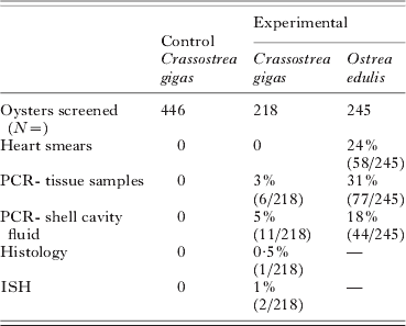
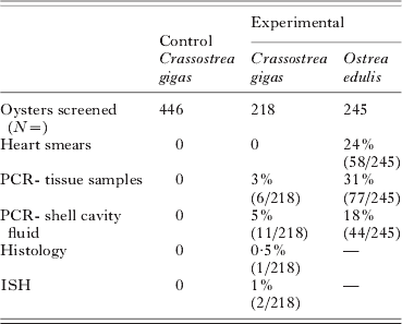
Screening of the initial sample of naïve C. gigas (n=150) by heart smears and PCR prior to the trial commencing confirmed that the oysters were negative for B. ostreae. B. ostreae was not detected in any of the 446 control C. gigas held independently over the 6-month period (Table 3). Overall a prevalence of infection of 31% was detected in the naturally exposed O. edulis by screening tissues using PCR. When the heart smears were screened prevalence of infection for this group was assessed at 24%. In the experimental C. gigas, B. ostreae was not detected by heart smears but positive results were obtained in tissue samples (n=6) and shell cavity fluid (n=11) by PCR. Three (1%) Pacific oysters were positive in both the shell cavity fluid and tissues. The PCR-positive C. gigas results from tissue samples and shell cavity fluid were obtained 98–117 days, post-exposure. The sequences from PCR products, generated from DNA extracted from pooled C. gigas shell cavity fluid samples (n=5) and pooled C. gigas tissue samples (n=2), were identical to the original B. ostreae rDNA subunit as published by Cochennec et al. (Reference Cochennec, LeRoux, Berthe and Gerard2000) (Accession no. AF192759) and Carnegie et al. (Reference Carnegie, Barber, Culloty, Figueras and Distel2000) (Accession no. AF262995).
Summary of PCR results of laboratory transmission trial II in which naïve Crassostrea gigas were held with Ostrea edulis naturally infected with Bonamia ostreae
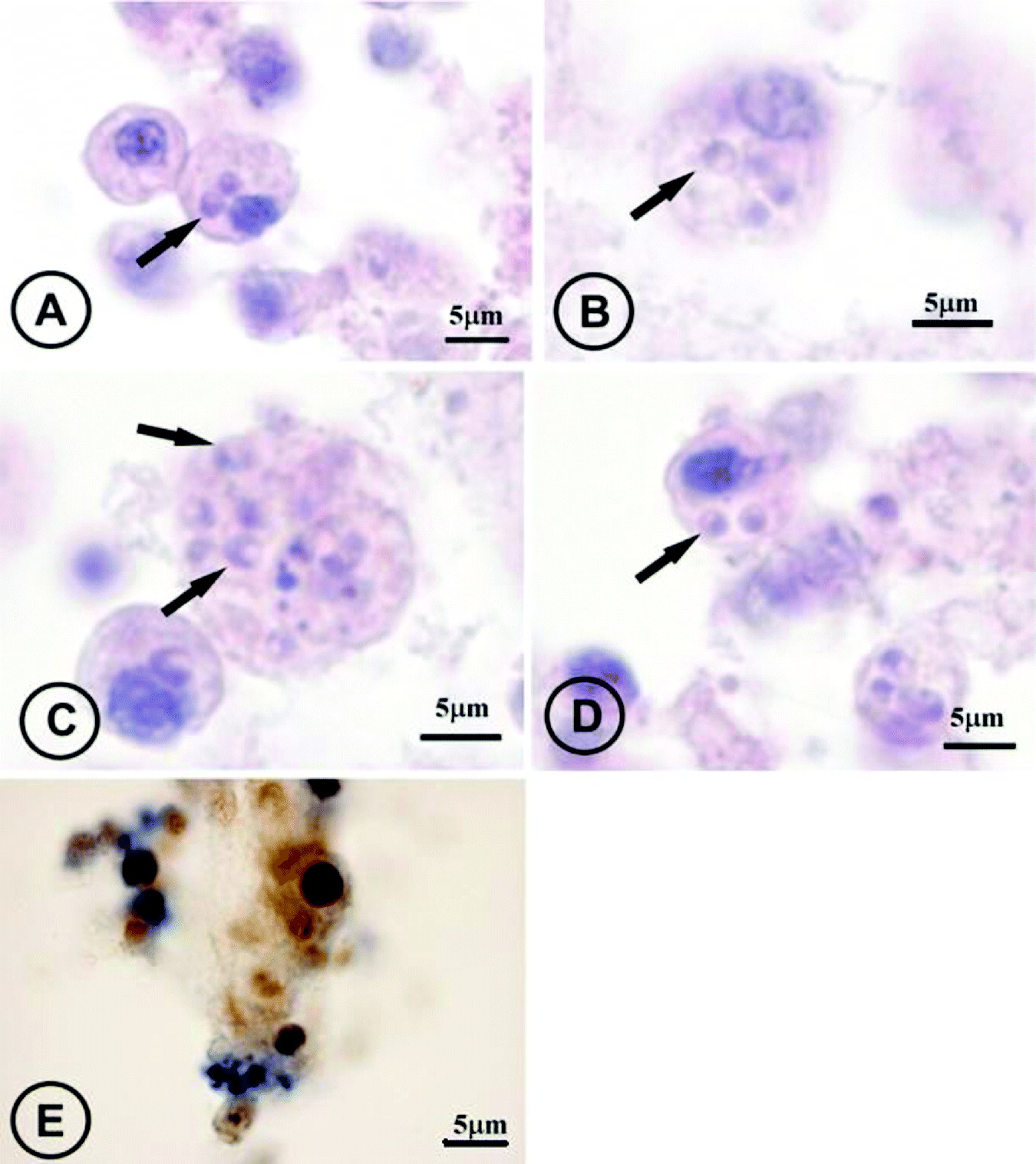
In the 6 tissue-positive individuals, 1 C. gigas had B. ostreae-like cells (microcells) both intracellularly and extracellularly (Fig. 3A) and positive staining was observed in that individual using ISH (Fig. 3B). In a second C. gigas, B. ostreae-like cells were detected extracellularly in the connective tissue and positive staining of the cells occurred using ISH also in that 1 individual. The microcells were 2–4 μm in size, uninucleate and binucleate with eccentric nuclei and the microcells were located in the connective tissue. According to the classification system for assessing B. ostreae intensity of infection a Class 2 infection was observed.
(A–D) Microcells in the haemocytes of naive Crassostrea gigas held with infected Ostrea edulis in the laboratory (H&E staining). (E) Microcells stained dark blue in the haemocytes of naive C. gigas held with infected O. edulis in the laboratory (DIG-labelled probe).

Laboratory trial in Spain
Experimental injection of B. ostreae into C. gigas
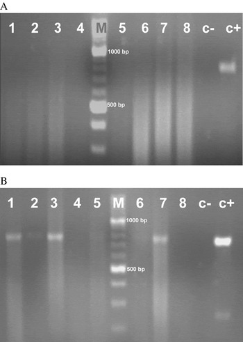
No infection was observed by histopathological analysis in any of the examined oysters. Negative PCR results were obtained for control and infected individuals from the first subsample (10 days post-injection), and for control individuals from the second subsample (80 days post-injection). However, positive PCR results were observed for inoculated individuals (11/19) from the second subsample with the 2 primer pairs (Fig. 4). BLAST search showed identity values of 100% with B. ostreae (11 clones of 11 oysters) for the sequence of the PCR-amplified products.
Detection of Bonamia ostreae in Crassostrea gigas injected with the parasite by PCR using CF /CR primers, 10 days (A) and 80 days (B) after injection. Lanes 1–8, DNA isolated from C. gigas gill pieces; lane c−, negative control; lane c+, positive control; lane M, 100 bp molecular marker.

DISCUSSION
The results of this study have demonstrated that B. ostreae DNA can be found in the tissues and shell cavity fluid of C. gigas following exposure of this oyster both in the field, in the laboratory and long term after injection. Along with B. ostreae DNA, B. exitiosa DNA was also detected in C. gigas in the field trial in Spain. B. ostreae-like cells were observed in the haemocytes of C. gigas and extracellularly when tissue sections were screened. These results indicate that C. gigas could act as a potential carrier or reservoir of Bonamia spp.
The field results from Ireland and Spain were comparable. In the Cork Harbour field study, B. ostreae DNA was initially detected in the C. gigas shell cavity fluid but in subsequent months positive PCR results were also observed in the tissues, possibly indicating a progression of the parasite within the animal. In Spain, positive PCR results were obtained from the gill and heart tissue indicating a similar scenario as exposure time increased. Along with B. ostreae DNA, B. exitiosa DNA was also detected as this parasite is also present at that site (Abollo et al. Reference Abollo, Ramilo, Casas, Comesaña, Cao, Carballal and Villalba2008).
In the laboratory in Ireland, when naïve C. gigas were held in close proximity with naturally infected O. edulis, B. ostreae-like cells and DNA were transferred from infected O. edulis to naïve C. gigas. These observations may indicate B. ostreae infection in C. gigas. One other explanation for the observation of B. ostreae-like cells in C. gigas tissues is that O. edulis mortalities occurred, due to an increase in the ambient temperature, possibly releasing large numbers of B. ostreae cells into the water column. These cells may have been taken up by the Pacific oysters via respiration and or feeding; however, it does still demonstrate the ability of the Pacific oyster to act as even an accidental carrier. In the Spanish study, experimental injection in the adductor muscle of oysters with B. ostreae cells resulted in positive PCR results from gill tissue in samples taken after 80 days, which could be the result of the progression of infection within a considerable percentage of animals, although the possibility that haemocytes had taken the injected cells from the adductor muscle and then distributed DNA fragments of the parasite to different organs cannot be discarded.
Of concern in this study is the fact that when naïve O. edulis were held in close proximity with exposed C. gigas, B. ostreae DNA was detected both in the tissue and shell cavity fluid of C. gigas and in the shell cavity fluid of O. edulis. Due to mortalities within the tanks it was only possible to screen 48% of the experimental O. edulis which may have resulted in an underestimation of the infection level in this group. The integrity of these B. ostreae cells is unknown, and similarly it is not known if bonamiosis would have developed in these animals if observed for a longer period of time. In previous studies, 6 months was sufficient time to allow naïve flat oysters to be exposed to infected flat oysters or to be infected with parasites and to develop infection (Culloty and Mulcahy, Reference Culloty and Mulcahy1996; Culloty et al. Reference Culloty, Cronin and Mulcahy2001; Lynch et al. Reference Lynch, Armitage, Wylde, Mulcahy and Culloty2005). Other studies have used varying numbers of parasites to induce experimental infection. It has been concluded from these studies that the rate of experimental bonamiasis development is dose dependent (Hervio et al. Reference Hervio, Bachère, Boulo, Cochennec, Vuillemin, Le Coguic, Cailletaux, Mazuri and Mialhe1995; Culloty et al. Reference Culloty, Novoa, Pernas, Longshaw, Mulcahy, Feist and Figueras1999). Martin et al. (Reference Martin, Gérard, Cochennec and Langlade1993) used a dose of 50 000 parasites per oyster. Hervio et al. (Reference Hervio, Bachère, Boulo, Cochennec, Vuillemin, Le Coguic, Cailletaux, Mazuri and Mialhe1995) concluded that a dose of 80 000 parasites per oyster caused a prevalence of infection of 50%. In this study, the integrity of the DNA cannot be definitely determined and it is clear from infection levels in the flat oysters that infection pressure was low within the system. However, the fact that positive results were obtained over several months and from the shell cavity fluid of O. edulis may indicate that B. ostreae cells were replicating in the system.
Deagle et al. (Reference Deagle, Eveson and Jarman2006) stated that the ability to recover large fragments of DNA via PCR indicates relatively low levels of DNA damage. In both the Irish and Spanish studies, a PCR product of 760 bp was detected in the Carnegie cPCR. Recent research by Arzul et al. (Reference Arzul, Gagnaire, Bond, Chollet, Morga, Ferrnad, Robert and Renault2009) has shown that B. ostreae may remain viable for up to 1 week outside of its host when held in seawater. In this study, B. ostreae DNA was transported to the laboratory with 300 C. gigas which may have significant implications when considering movements of C. gigas for ongrowing.
It is known that the haemocytes of C. gigas can become infected with B. ostreae, but in vitro studies demonstrated that the parasite is quickly compartmentalized in a parasitophorous vacuole and destroyed; in comparison, in the haemocyte of O. edulis the parasite begins to multiply by binary fission (Chagot et al. Reference Chagot, Boulo, Hervio, Mialhe, Bachère, Mourton and Grizel1992). In the field studies, it is not known how long individual C. gigas were harbouring B. ostreae DNA, from the time of exposure and prior to being sampled. However, in the laboratory samples, particularly in the oysters experimentally injected, B. ostreae DNA was detected 80 days post-exposure. Like O. edulis, individual C. gigas may show more or less ability to destroy B. ostreae.
Parasites can persist in animals that are not their normal host. Itoh et al. (Reference Itoh, Tun, Komiyama, Ueki and Ogawa2004) reported that 3 protistan parasites, an unidentified haplosporidian in the connective tissues, Marteilia sp. in the digestive gland and Marteilioides sp. in the oocytes, were discovered in the Manila clam, Venerupis philippinarum. Histopathological observations suggest that Marteilia sp. and Marteilioides sp. were not pathogenic to the host and that the prevalence of these parasites was low. B. ostreae cells may infiltrate C. gigas tissues or may be incidentally taken in during feeding and respiration and be destroyed, and traces of B. ostreae DNA may remain amongst the C. gigas tissues, thus resulting in positive detection by PCR. A PCR-positive signal does not necessarily indicate an infection or a viable parasite (Burreson, Reference Burreson2008; Ford et al. Reference Ford, Allam and Xue2009). Positive detection of the parasite in individuals using PCR and lack of detection in the histological examination of those same individuals may indicate that the environment is not favourable for infection to develop or that the PCR is detecting a stage of the parasite which is not infective to the host (Ford et al. Reference Ford, Allam and Xue2009). However, the detection of intact B. ostreae-like cells within C. gigas haemocytes may indicate that at least some viable B. ostreae cells are present for an unknown time-period within C. gigas.
This study raises questions in relation to the role of C. gigas, at least, as a carrier or reservoir of B. ostreae and B. exitiosa. Shell cavity fluid and tissues may retain B. ostreae and B. exitiosa cells and due to the ability of Bonamia sp. to survive outside its host may allow this protistan to be transferred to new sites with movements of the Pacific oyster. Similarily, in areas where both O. edulis and C. gigas are cultured and B. ostreae or B. exitiosa is present, Pacific oyster populations could act as a reservoir of infection for these pathogens. Even if the probability of C. gigas acting as an effective carrier or reservoir is low, that possibility should be taken into account in risk assessment and disease management. Current results would justify further research to characterize more accurately the role of C. gigas as a reservoir or carrier of B. ostreae and B. exitiosa.
ACKNOWLEDGEMENTS
The authors would like to acknowledge Ms Deborah Cheslett (Shellfish Health Manager, Marine Institute Ireland), Mr Micheal O Cinneide (Marine Instiute), Mr Mark Norman (Taighde Mhara Teo) and Mr Iarlaith Connellan (\Caarton Point Shellfish Ltd) for their guidance and advice throughout the Irish study. The authors are grateful to Pilar Comesaña, M. Isabel Meléndez and Elena Penas for their technical assistance. Many thanks to the shellfish growers who provided the shellfish material for this study and, in particular, David and Tristan Hugh Jones, Atlantic Shellfish, Ireland.
FINANCIAL SUPPORT
The Irish component of the study (grant-aid Agreement No. ST/05/25) was carried out with the support of the Marine Institute and the Marine RTDI Measure, Productive Sector Operational Programme, National Development Plan 2000–2006. The work in Spain was supported by Xunta de Galicia, under the projects PGIDIT05RMA50101PR and PGIDIT-CIMA06/01. Thanks are also due to the Ministerio de Ciencia e Innovación of the Spanish Government for financial support to E.A. under the Programme Ramon y Cajal.