Introduction
Witchweeds of the Striga genus threaten the livelihood of at least 300 million people in sub-Saharan Africa by limiting the production of the region’s staple grains, namely maize (Zea mays L.), millets, grain sorghum [Sorghum bicolor (L.) Moench ssp. bicolor), upland rice (Oryza sativa L.), and cowpea [Vigna minima (Roxb.) Ohwi & H. Ohashi] (Ejeta Reference Ejeta, Ejeta and Gressel2007). Although the Striga genus has about 28 recognized species, purple witchweed [Striga hermonthica (Delile) Benth.] and Asiatic witchweed [Striga asiatica (L.) Kuntze], which parasitize cereals, and cowpea witchweed [Striga gesnerioides (Willd.) Vatke], which infects cowpea, are the most pernicious (Parker Reference Parker2009). Crop losses due to this parasite range between 30% and 100%, with the loss estimated to cost at least US$7 billion per year (Ejeta Reference Ejeta, Ejeta and Gressel2007).
Peculiarities of Striga’s parasitic lifestyle make the weed exceptionally difficult to manage (Runo and Kuria Reference Runo and Kuria2018). The parasite’s dust-like seeds remain dormant in the soil and only germinate in response to chemical stimulants called strigolactones, which are produced by the roots of host and some non-host plants (Matusova et al. Reference Matusova, Rani, Verstappen, Franssen, Beale and Bouwmeester2005). Germinated seeds rapidly develop and attach to a host, channeling nutrients, water, and macromolecules into the parasite, leading to severe retardation and frequently death of an infected plant (Dörr Reference Dörr1997). As Striga eventually emerges from the soil, it becomes photosynthetic, flowers, and produces up to 50,000 seeds per plant that can endure in the soil seedbank for up to 10 yr (Atera et al. Reference Atera, Itoh and Onyango2011). To minimize infestations, farmers use agronomic and cultural practices such as crop rotation and intercropping with non-hosts, hand weeding, and resistant or tolerant crops (Kanampiu et al. Reference Kanampiu, Makumbi, Mageto, Omanya, Waruingi, Musyoka and Ransom2018). However, these strategies have only achieved moderate success, because Striga continues to expand its geographic and host range (Ejeta Reference Ejeta, Ejeta and Gressel2007).
An integrated approach that capitalizes on host resistance may achieve practical success in Striga management. From a population genetics and evolutionary perspective, natural Striga resistance is more likely to be found in cereals that coevolved with the parasite (Bellis et al. Reference Bellis, Kelly, Lorts, Gao, DeLeo, Rouhan, Budden, Bhaskara, Hu, Muscarella, Timko, Nebie, Runo, Chilcoat and Juenger2020; Lopez et al. Reference Lopez, Bellis, Wafula, Hearne, Honaas, Ralph, Timko, Unachukwu, dePamphilis and Lasky2019). Importantly, sorghum and millets, whose centers of domestication overlap with Striga’s natural range (Musselman and Hepper Reference Musselman and Hepper1986), are good reservoirs of Striga resistance, but of particular interest are wild relatives of these grasses, because they may harbor resistance sources that have not been bred out by domestication. To this end, sources of Striga resistance from wild sorghum [Sorghum leiocladum (Hack) C.E. Hubb] have been identified (Mbuvi et al. Reference Mbuvi, Masiga, Kuria, Masanga, Wamalwa, Mohamed, Odeny, Hamza, Timko and Runo2017; Mohamed et al. Reference Mohamed, Ellicott, Housley and Ejeta2003).
There are two general categories of Striga resistance: pre-attachment resistance, which can occur due to the parasite’s failure to germinate, and, post-attachment resistance, which is due Striga’s inability to successfully attach to and grow on host tissue. Mechanistically, pre-attachment resistance is due to production of less-potent strigolactone-based germination stimulants, which are part of host root exudates, as described in the Striga-resistant sorghum variety ‘SRN39’ (Gobena et al. Reference Gobena, Shimels, Rich, Ruyter-Spira, Bouwmeester, Kanuganti, Mengiste and Ejeta2017), rice (Jamil et al. Reference Jamil, Rodenburg, Charnikhova and Bouwmeester2011), and the Kenyan maize landrace KSTP'94 (Yoneyama et al. Reference Yoneyama, Arakawa, Ishimoto, Kim, Kisugi, Xie, Nomura, Kanampiu, Yokota, Ezawa and Yoneyama2015). Post-attachment resistance can be caused by biochemical or physical barriers that hinder parasite attachment and growth, as demonstrated in wild sorghum (Mbuvi et al. Reference Mbuvi, Masiga, Kuria, Masanga, Wamalwa, Mohamed, Odeny, Hamza, Timko and Runo2017), or activation of the host immune response leading to programmed cell death at the point of attachment, as indicated by a hypersensitive reaction (HR) following infection, for example, in sorghum (Mohamed et al. Reference Mohamed, Ellicott, Housley and Ejeta2003) or rice (Gurney et al. Reference Gurney, Slate, Press and Scholes2006). At the genetic level, pre-attachment resistance has been well elucidated in sorghum and found to be caused by deletion of a sulfotransferase gene. This deletion alters the stereochemistry of 5-deoxystrigol, a powerful germination stimulant, to orobanchol, a poor gemination inducer (Gobena et al. Reference Gobena, Shimels, Rich, Ruyter-Spira, Bouwmeester, Kanuganti, Mengiste and Ejeta2017). Post-attachment genetic resistance mechanisms have been elucidated in S. gesnerioides resistance against cowpea (Li and Timko Reference Li and Timko2009). This typical gene-for-gene resistance response is due a canonical coiled-coil leucine-rich repeat gene termed RACE SPECIFIC STRIGA GESNERIODES 3 (RSG3) that elicits an HR response upon recognition of the parasite (Li and Timko Reference Li and Timko2009). Other post-attachment Striga-resistance responses are quantitative, and correspondingly, various quantitative trait loci associated with Striga resistance have been described in sorghum (Haussmann et al. Reference Haussmann, Hess, Omanya, Folkertsma, Reddy, Kayentao, Welz and Geiger2004), maize (Badu-Apraku et al. Reference Badu-Apraku, Adewale, Paterne, Gedil and Asiedu2020), and rice (Swarbrick et al. Reference Swarbrick, Scholes, Press and Slate2009).
This study sought to harness Striga resistance from wild, noncultivated pearl millet [Pennisetum glaucum (L.) R. Br.]. Highly preferred because of its ability to withstand harsh growing conditions, pearl millet is an important food security crop for millions of smallholder households in the arid and semiarid tropics (Jukanti et al. Reference Jukanti, Gowda, Rai, Manga and Bhatt2016). West and Central African regions are the world’s largest pearl millet producers, with annual production of over 16.8 million ha and an average yield of up to 880 kg ha−1 (ICRISAT 2021). However, most cultivated pearl millet varieties are susceptible to Striga (Kountche et al. Reference Kountche, Hash, Dodo, Laoualy, Sanogo, Timbeli, Vigouroux, This, Nijkamp and Haussmann2013). In contrast, some wild pearl millet genotypes have proven to be useful sources of Striga resistance (Wilson et al. Reference Wilson, Hess, Hanna, Kumar and Gupta2004, Reference Wilson, Hess and Hanna2007). Investigations in this study hinged on the wild pearl millet line 29Aw, following an assertion that the genotype demonstrated resistance and tolerance to Striga under natural infestations (International Crops Research Institute for the Semi-Arid Tropics, personal communication, June 2017). The aim was to determine whether the observed resistance of 29Aw was due to low Striga seed germination stimulation (pre-attachment resistance) or inhibition of parasite attachment and growth after germination (post-attachment resistance).
Materials and Methods
Plant Materials
Two pearl millet genotypes with contrasting resistance responses (resistant vs. susceptible) to S. hermonthica were investigated. ‘SOSAT-C88-P10’ (susceptible), a high-yielding, open-pollinated, improved Malian variety derived from a cross between the landraces Sauna and Sanio (Izge and Song Reference Izge and Song2013; Omanya et al. Reference Omanya, Weltzien-rattunde, Sogodogo, Sanogo, Hanssens, Guero and Zangre2007) and 29Aw, a wild pearl millet line anecdotally reported to be resistant and tolerant to S. hermonthica (Institute for the Semi-Arid Tropics, Niger). Seeds of S. hermonthica were collected from two Striga-infested regions in western Kenya, Alupe (0.499°N, 34.121°E) and Kibos (0.067°S, 34.812°E), in 2016.
Surface Sterilization of Pearl Millet Seeds
Pearl millet seeds were washed and dehusked in running tap water for an average of 20 min, then transferred to a clean fume hood for surface sterilization with 70% (v/v) ethanol for 3 min. Seeds were then rinsed three times using sterile distilled water, followed by soaking in commercial bleach (10%) containing 2 to 5 drops of 0.5% Tween-20 for 20 min. Surface-sterilized seeds were rinsed again with double-distilled water and transferred to 90-mm-diameter petri plates lined with moisturized filter paper (Whatman Paper No. 1, Whatman, Maidstone, UK) and incubated at 27 C in the dark for 48 h.
Preparation of Striga Seeds
Striga hermonthica (25 mg, approximately 5,000 seeds) were preconditioned to break their dormancy as described by Gurney et al. (Reference Gurney, Grimanelli, Kanampiu, Hoisington, Scholes and Press2003), with modifications as follows: continuous immersion in 70% (v/v) ethanol for 30 s, followed by soaking in 10% (v/v) commercial bleach and gentle agitation for 10 min, then being rinsed three times with distilled, autoclaved water. Seeds were then spread in 90-mm-diameter petri dishes lined with moisturized filter papers and incubated at 30 C in the dark for 11 d. Preconditioned (nongerminated) seeds were used for pre-attachment (agar gel) assays, while for post-attachment (rhizotron) assays, preconditioned seeds were pre-germinated by treatment with 0.34 µM of the artificial germination stimulant GR24 (Chiralix, Amsterdam, Netherlands) and overnight incubation at 30 C before use.
Agar Gel Assays to Determine Pre-attachment Resistance of Pearl Millet against Striga
Agar gel assays were performed as described in Hess et al. (Reference Hess, Ejeta and Butler1992). Briefly, a gel of 8 g L−1 agar (Phyto Agar, Duchefa Biochemie B.V., Haarlem, Netherlands) was prepared, autoclaved, and poured into 90-mm petri plates just before setting (approximately 50 C). About 5,000 preconditioned Striga seeds were then dispensed into the semisolid agar by pipetting. To do this, the tip of a 1-ml pipette was cut and used to pipette 200 μl of seeds suspended in sterile distilled water. The solution was dispersed into the semisolid agar with gentle agitation to allow even seed distribution. Striga hermonthica seeds from Kibos were used for the agar gel assay, as this ecotype is known to be more virulent (Mbuvi et al. Reference Mbuvi, Masiga, Kuria, Masanga, Wamalwa, Mohamed, Odeny, Hamza, Timko and Runo2017).
When the agar media set, three emerging pearl millet seedlings were placed at the edge of each plate with the tips of their roots pointing across the plates. The gel-embedded host−parasite mixture was then incubated at 30 C in the dark for 2 d to allow establishment of pearl millet seedlings in the media. Subsequently, pearl millet seedlings were thinned to one per plate, and the host−parasite mixture was incubated for a further 2 d to induce germination of Striga seeds. Thereafter, images of plates containing germinated Striga seedlings were taken using a Leica MZ10F microscope fit with a DFC310FX camera (both Leica, Wetzlar, Germany). In all imagery, the Leica MZ10F zoom lens was set at 1.6× magnification.
Resulting images of germinated Striga and pearl millet were analyzed for germination efficiency and germination distances. ImageJ v. 1.50i (https://imagej.nih.gov/ij) was used to measure the maximum germination distance (MGD) as mean of the distance, in millimeters, between a pearl millet root and the three furthest-germinated Striga seeds. The experiment was conducted in three biological and two technical replicates. Parasite germination activity was considered high if MGD was ≥10 mm and low if MGD was <10 mm (Hess et al. Reference Hess, Ejeta and Butler1992). Germination efficiency was determined using photographs from three random fields from each of the six individual plates as a percentage of germinated to nongerminated seeds.
Rhizotron Assays to Determine Post-attachment Resistance of Pearl Millet against Striga
Pearl millet seeds were pre-germinated for 10 d in 10 by 10 by 7 cm plastic planters filled with vermiculite and were watered with 40% Long Ashton solution (Hudson Reference Hudson1967). Germinated pearl millet seedlings were then transferred to rhizotrons (root observation chambers) as described by Mbuvi et al. (Reference Mbuvi, Masiga, Kuria, Masanga, Wamalwa, Mohamed, Odeny, Hamza, Timko and Runo2017). In brief, the base of a bioassay plate (25 by 25 by 5 cm) was filled with vermiculite and lined with a foam strip at the bottom to soak up excess water. A nylon mesh fabric was laid over the vermiculite. A germinated pearl millet seedling was placed onto the nylon mesh fabric, the chamber was closed, and the lid was fastened with PVC tape. Chambers were then wrapped in aluminum foil to block out light. Plants in rhizotrons were maintained inside a greenhouse for 11 d under a 12-h light/ 12-h dark photoperiod, with a moderate temperature regime (28 C/24 C, day/night), while being drip-fed with 20 ml of 40% Long Ashton solution (Hudson Reference Hudson1967) twice a day. After 10 d in the rhizotrons, pearl millet plants were infected with 25 μg (˜5,000 seeds) of pre-germinated Striga seeds by aligning parasite seedlings on host roots using a fine-tipped paintbrush. Two S. hermonthica ecotypes (Alupe and Kibos) were evaluated in three replicates of five plants per pearl millet line (single experimental run) in a completely randomized design. The entire experiment was repeated twice.
After infection, rhizotron chambers were closed, and plants were maintained in the greenhouse while being irrigated twice a day with 25 ml of 40% Long Ashton nutrient solution (Hudson Reference Hudson1967). Data were collected at 21 d after infection (DAI). All attached Striga seedlings were harvested and placed on 90-mm-diameter petri plates for photo-documentation. Obtained images were then used to determine the number and length of Striga attachments using ImageJ software v. 1.50i. Additionally, total Striga biomass attached to the roots of pearl millet was determined by drying harvested Striga seedlings at 45 C for 7 d and then weighing them.
Histological Analysis to Determine Mechanisms of Pearl Millet–Striga Interaction
The extent of S. hermonthica parasitism in pearl millet was determined by analysis of (1) the frequency of vascular connections between Striga and pearl millet using safranin O staining (Yoshida and Shirasu Reference Yoshida and Shirasu2009) and (2) parasite ingression into host tissue using histological analysis (Mbuvi et al. Reference Mbuvi, Masiga, Kuria, Masanga, Wamalwa, Mohamed, Odeny, Hamza, Timko and Runo2017).
For frequency of vascular connection analysis, small fragments of roots with S. hermonthica attachments were harvested from rhizotrons at 9 DAI and fixed in ethanol and acetic acid (4:1) for 10 min, followed by staining in 1% safranin O dissolved in 30% ethanol for 5 min. Stained tissues were cleared with chloral hydrate (2.5 g ml−1) for 12 h before being photographed using a Leica MZ10F stereomicroscope fit with a DFC 310FX camera (both Leica, Germany). These images were used to determine the frequency of vascular connection between host and parasite.
The same tissues were processed for histological analysis using a Technovit 7100® kit following the manufacturer’s instructions (Heraeus Kulzer GmbH, Hanau, Germany) and protocols described in Mutinda et al. (Reference Mutinda, Masanga, Mutuku, Runo and Alakonya2018). Briefly, fixed tissues of Striga attached to pearl millet were first pre-infiltrated in Technovit® 1 and ethanol (1:1) for 2 h and then in Technovit® 1 and ethanol (1:1) for 15 min. Afterward, tissues were infiltrated in 100% Technovit® 1 for 3 d. For embedding, tissues were placed in an upright position in hardener 2 and Technovit® 1 (1:15) in eppendorf tube lids and left to dry completely at room temperature for at least 2 wk before being mounted onto wooden blocks using Technovit® 3040 kit as per the manufacturer’s instructions. Transverse sections (5-microns thick) at the point of Striga–pearl millet attachment were cut using a Leica RM 2155 microtome (Leica). Sections were collected in water droplets, dried on a hot plate at 65 C for 30 min, then stained with 0.1% toluidine blue O dye in 100 mM phosphate buffer (pH 7.0) for 2 min. Dried slides were overlaid with coverslips using DePex (BDH, Poole, UK) and photographed using a Leica DM100 microscope fit with a MC190 HD camera (both Leica).
Data Analysis
Data obtained for pre-attachment resistance (means of MGD and germination frequency [GF]), post-attachment resistance (means of Striga attachments, length, and biomass of the parasite seedlings), and means of frequency of vascular formation were compared with ANOVA performed using the SAS v. 9.1 (SAS Institute, Cary, NC). The results were presented as box plots, prepared using the ggplot2 package in R software (v. 3.5.1). Significant differences between the means were evaluated using Tukey’s honest significant difference test at a 95% confidence interval.
Results and Discussion
Pre-attachment Striga Resistance in the Wild Pearl Millet Line 29Aw
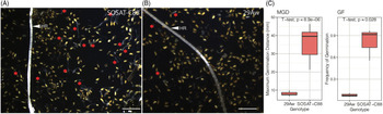
The agar gel assay is used to determine the ability of a host’s root to induce the germination of Striga seeds. It also determines the furthest distance from which a host root can stimulate the parasite’s seed germination. This distance is the MGD. Resistant hosts have low germination stimulation effects and low MGD (Hess et al. Reference Hess, Ejeta and Butler1992). Agar gel assay results (Figure 1A and B) showed that the wild pearl millet line 29Aw had significantly lower GF (8.14%) compared with 85.04% SOSAT-C88-P10. The MGD of 29Aw was also significantly lower (7.9 ± 2.75 mm) compared with the susceptible genotype SOSAT-C88-P10 (35.9 ± 2.88 mm) (Figure 1C), indicating that 29Aw did not efficiently induce germination of Striga seeds in culture.
Agar gel assay for pearl millet pre-attachment resistance against Striga hermonthica resistance. (A) Susceptible SOSAT-C88-P10 induced germination of numerous parasite seeds that were far from the host root. (B) Resistant 29Aw induced low germination of parasite seeds. Most germinated parasite seeds were in close proximity to the host root. HR, host root; red asterisks, germinated S. hermonthica seedlings. Scale bar: 2 mm. (C) Analysis of Striga germination stimulation activity using germination frequency (GF) and maximum germination distance (MGD). Susceptible SOSAT-C88 had significantly higher GF and MGD compared with the resistant 29Aw.

According to Hess et al. (Reference Hess, Ejeta and Butler1992), sorghum genotypes with MGDs of ≤10 mm are resistant, and those with MGDs of ≥10 mm are susceptible. Following that classification, the MGD of the 29Aw line falls into the category of low inducers, comparable to sorghum cultivars ‘IS7777’ (3.8 ± 1.7 mm; Gobena et al. Reference Gobena, Shimels, Rich, Ruyter-Spira, Bouwmeester, Kanuganti, Mengiste and Ejeta2017) and ‘IS9830’ (3 mm; Hess et al. Reference Hess, Ejeta and Butler1992). However, an MGD of 7 mm is considerably higher than that of the Striga-resistant sorghum cultivar ‘SRN39’ with only 1 mm, as reported by Gobena et al. (Reference Gobena, Shimels, Rich, Ruyter-Spira, Bouwmeester, Kanuganti, Mengiste and Ejeta2017). This finding suggests that the pre-attachment resistance in SRN39 is higher than that of 29Aw.
From a physiological and biochemical standpoint, SRN39 produces low levels of the less potent strigolactone 5-deoxystrigol relative to orobanchol because of a gene deletion on the low-germination loci (Gobena et al. Reference Gobena, Shimels, Rich, Ruyter-Spira, Bouwmeester, Kanuganti, Mengiste and Ejeta2017). In maize, the resistant line ‘KSTP’94’ produces more sorgomol relative to 5-deoxystrigol produced by the susceptible ‘Pioneer 3253’ (Yoneyama et al. Reference Yoneyama, Arakawa, Ishimoto, Kim, Kisugi, Xie, Nomura, Kanampiu, Yokota, Ezawa and Yoneyama2015). For this reason, it is reasonable to assume that pre-attachment resistance in the line 29Aw is due to low relative amounts or less potent Striga germination stimulants. However, a detailed chemical characterization of the 29Aw root exudates remains to be performed. So far, biochemical studies on pearl millet’s root exudate indicate that a majority of the genotypes produce 5-deoxystrigol (Awad et al. Reference Awad, Sato, Kusumoto, Kamioka, Takeuchi and Yoneyama2006). Of note, in a study by Wilson et al. (Reference Wilson, Hess and Hanna2007), wild pearl millet showed Striga resistance under field infestation, but such resistance could not be attributed to pre-attachment resistance as deduced from agar gel assays, leading the authors to conclude that alternative resistance mechanisms were responsible for the Striga resistance observed.
Post-attachment Striga Resistance in the Wild Pearl Millet Line 29Aw
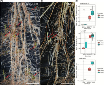
Rhizotron experiments determine host–Striga interactions after the parasite has germinated. Resistant germplasm is characterized by a low number, short length, and reduced biomass of attached Striga seedlings (Mbuvi et al. Reference Mbuvi, Masiga, Kuria, Masanga, Wamalwa, Mohamed, Odeny, Hamza, Timko and Runo2017). This relationship is depicted in Figure 2, where long, multiple infections can be observed in the susceptible SOSAT-C88-P10 (Figure 2A) compared with shorter and fewer Striga attachments in 29Aw (Figure 2B).
Rhizotron assay for pearl millet Striga hermonthica post-attachment resistance in pearl millet at 21 d after infection (DAI) of host roots. (A) Susceptible SOSAT-C88-P10 had numerous and long S. hermonthica attachments. (B) Resistant 29Aw had fewer and short S. hermonthica attachments. Red arrows represent the site of attachment of the parasite to the host. Scale bar: 2 mm. (C) Analysis of pearl millet post-attachment resistance against two ecotypes of S. hermonthica (Alupe and Kibos) using mean number of Striga attachments, lengths, and biomass. Data indicate the mean of three replicates. Significant differences are indicated using asterisks: * = P ≤ 0.05; ** P ≤ 0.01; *** P ≤ 0.001.

Artificially infected roots of pearl millet hosts and analyses of the number, length, and biomass of attached Striga seedlings at 21 DAI (Figure 2C) indicated that Striga seedling numbers in 29Aw (Kibos 2.0 ± 0.36; Alupe 1.8 ± 0.17) were notably lower than those of SOSAT-C88-P10 (Kibos 38.90 ± 4.5; Alupe 19.69 ± 4.5). This represented at least 19-fold (Kibos) and 10-fold (Alupe) differences in the number of attachments. The average lengths of Striga attached to 29Aw (Kibos 0.35 ± 0.03 cm; Alupe 0.27 ± 0.04 cm) were also considerably lower than for SOSAT-C88-P10 (Kibos 0.90 ± 0.05 cm; Alupe 0.80 ± 0.04 cm), representing at least a 2.5-fold difference in the length of Striga seedlings. Correspondingly, the biomass of attached seedlings was 28-fold lower in 29Aw (Kibos 0.25 ± 0.04 mg; Alupe 0.15 ± 0.01 mg) relative to SOSAT-C88-P10 (Kibos 6.99 ± 0.40 mg; Alupe 4.2 ± 0.21 mg).
Additionally, significant differences were observed when different Striga ecotypes were used to infect pearl millet (ANOVA, P < 0.05). Striga from Kibos showed significantly more attachments and produced a notably higher biomass. This difference was apparent in the susceptible but not on the resistant pearl millet line, and there were no significant differences between Striga ecotypes with regard to mean length of parasite attached. Variations in the resistance response of pearl millet to Striga ecotypes point to possible differences in the Striga virulence—described here as the aggressiveness with which Striga infects hosts. This is consistent with previous reports that suggest the Kibos Striga ecotype is more a ggressive than the Alupe strain (Mbuvi et al. Reference Mbuvi, Masiga, Kuria, Masanga, Wamalwa, Mohamed, Odeny, Hamza, Timko and Runo2017). Virulence is more likely to manifest itself in susceptible than in resistant genotypes. Additionally, differences in virulence are more apparent with analysis of Striga attachments and biomass as compared with length, suggesting that these two metrics (attachments and biomass) are more informative as measures of virulence and resistance.
The number of attachments (38.90 ± 4.5 to 1.80 ± 0.17), length (0.9 ± 0.05 to 0.27 ± 0.04 cm), and biomass (6.99 ± 0.40 to 0.15 ± 0.01 mg) recorded for pearl millet in this study were lower but comparable to those observed for other Striga hosts under similar study conditions. For example, the resistant sorghum line ‘N13’ showed 56 attachments and about 20 mg biomass (Mbuvi et al. Reference Mbuvi, Masiga, Kuria, Masanga, Wamalwa, Mohamed, Odeny, Hamza, Timko and Runo2017). In the resistant maize line KSTP’94, attachments and biomass were 44.80 ± 10.22 and 14.88 ± 4.07 mg, respectively (Mutinda et al. Reference Mutinda, Masanga, Mutuku, Runo and Alakonya2018), while in the resistant rice ‘Nipponbare’, 30 attachments with biomass weights of approximately 2 mg were reported (Cissoko et al. Reference Cissoko, Boisnard, Rodenburg, Press and Scholes2011). Similarly, comparable Striga lengths ranging from 0.2 to 2 cm have been recorded in sorghum (Mbuvi et al. Reference Mbuvi, Masiga, Kuria, Masanga, Wamalwa, Mohamed, Odeny, Hamza, Timko and Runo2017) and rice (Cissoko et al. Reference Cissoko, Boisnard, Rodenburg, Press and Scholes2011).
Mechanisms of Post-attachment Striga Resistance in the Wild Pearl Millet Line 29Aw
Formation of a xylem bridge signifies successful Striga parasitism (Dörr Reference Dörr1997). After connecting with the host, Striga growths rapidly. This is evidenced by development of vegetative structures. To determine the efficiency of vascular connections between Striga and pearl millet, parasite tissue attached to host roots was stained using safranin O dye. This characteristic red stain allows visualization of xylem tissues in both host and parasite, and further histological sections of Striga-pearl millet attachments can determine the extent of parasitic intrusion into host cells.
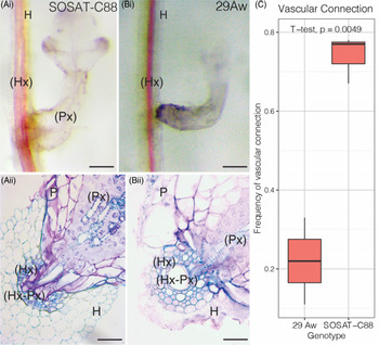
In SOSAT-C88-P10, parasite growth was vigorous, and parasite shoots were well developed within 9 DAI (Figure 3Ai). In contrast, slow parasitic growth with less vegetative tissue in the resistant 29Aw was observed (Figure 3Bi). Histological analysis showed that at 9 DAI, Striga parenchymal cells and vessel elements had successfully penetrated the host vascular system. Dense blue tissues in the hyaline body in SOSAT-C88-P10 clearly indicated successfully established vascular continuity (Figure 3Bii). In contrast, 29Aw blocked the parasite’s development at the endodermal layer (Figure 3Bii).
Additionally, notable differences were observed in the number of vascular connections made by Striga in the two pearl millet genotypes. A high frequency (74%, n = 9) of successful vascular connections were measured in SOSAT-C88-P10 compared with fewer (22%, n = 9] connections in 29Aw (Figure 3C). Results for the frequency of vascular connections in pearl millet resemble those recorded in maize. Amusan et al. (Reference Amusan, Rich, Menkir, Housley and Ejeta2008) reported frequencies of between 12% and 88% vascular connections in maize infected with S. hermonthica. Failure to form a physical parasite–host bridge with 29Aw was attributed to resistance by the host at the endodermal and cortical levels.
Mechanisms of post-attachment resistance of pearl millet against Striga hermonthica evaluated microscopically at 9 d after infection (DAI). (Ai) Close-up of successful Striga parasitism and vascular connection on susceptible SOSAT-C88-P10. (Aii) Transverse section of SOSAT-C88-P10 showing successful parasitism indicated by xylem–xylem connections between host and parasite. (Bi) Close-up of resistant 29Aw showing failure of Striga parasitism and vascular connection establishment. (Bii) Transverse section of 29Aw showing failure of the parasite to penetrate host tissue beyond the endodermis. H, host; HX, host xylem; P, parasite; PX, parasite xylem; PX-HX, parasite and host xylem connection. Scale bar: top, 2 mm; bottom, 0.1 mm. (C) Analysis of vascular connections between SOSAT-C88-P10 and 29Aw.

Striga non-host/host incompatibility and resistance post-attachment has been well described (Yoshida and Shirasu Reference Yoshida and Shirasu2009). In brief, there are four layers of incompatibility: after vascular connection (layer I), endodermis and pericylce (layer II), cortex (layer III), and failure of attachment (layer IV). The results from this study suggest that post-attachment resistance in 29Aw occur at layers II (endodermis) and III (cortex). Resistance at level II is supported by data on low frequency of vascular connection with the host. This inefficient vascular connection is reminiscent of the Striga-resistant rice variety Nipponbare reported by Gurney et al. (Reference Gurney, Slate, Press and Scholes2006), in which Striga failed to enter the host’s endodermis. Evidence for resistance at the cortical level is drawn from the deep safranin and toluidine staining observed in 29Aw (Figure 3Bi and Bii). This kind of resistance has been attributed to phenolic compounds such as suberins, lignin, accumulation of H2O2, or cell wall crosslinking and increase in peroxidase activity, respectively reported in the resistant S. leiocladum ‘WSE-1’ against S. hermonthica (Mbuvi et al. Reference Mbuvi, Masiga, Kuria, Masanga, Wamalwa, Mohamed, Odeny, Hamza, Timko and Runo2017); resistant sunflower (Helianthus annuus L.) and Orobanche cumana Wallr. (Echevarria-Zomeno et al. Reference Echevarria-Zomeno, Perez-de-Luque, Jorrin and Maldonado2006); resistant sorghum landrace Framida against S. hermonthica (Arnaud et al. Reference Arnaud, Véronési and Thalouarn1999); yellow rattle (Rhinanthus minor L.)/non-host oxeye daisy (Leucanthemum vulgare Lam.) interactions (Cameron et al. Reference Cameron, Coats and Seel2006); resistant pea (Pisum vulgare L.) against bean broomrape (Orobanche crenata Forssk.) (Echevarria-Zomeno et al. Reference Echevarria-Zomeno, Perez-de-Luque, Jorrin and Maldonado2006); and in vetch (Vicia sp.) plants and Egyptian broomrape (Orobanche aegyptiaca Pers.) (Goldwasser et al. Reference Goldwasser, Hershenhorn, Plakhine, Kleifeld and Rubin1999). Although the exact physiological and biochemical mechanisms underpinning the resistance will need further elucidation, these observations support the existence of post-attachment resistance mechanisms in 29Aw.
In summary, this study showed that the pearl millet line 29Aw possesses two levels of resistance against S. hermonthica. This finding is unexpected, because most previously investigated cereals exhibited either a pre-attachment or a post-attachment resistance to Striga. This study revealed that 29Aw is likely to exude low levels of potent Striga germination stimulants, resulting in a low germination of the parasite in the first place. The existence of post-attachment resistance to Striga in the wild pearl millet line 29Aw was also shown. This resistance is possibly due to the deposition of secondary plant metabolite elements such as phenolic compounds (Yoshida and Shirasu Reference Yoshida and Shirasu2009). As a consequence, the endodermis of 29Aw formed a nearly unbreachable physical barrier to Striga attack. The existence of the combination of pre- and post-germination resistance to Striga will increase the durability of host defense.
These findings have far-reaching implications for Striga management in pearl millet. Subsequent research using genetic and genomic tools will allow for identification of genetic loci leading to these resistance mechanisms. The identified markers can then be used in gene modification, editing, or marker-assisted breeding of pearl millet.
Acknowledgments
The authors are grateful to Kenyatta University for supporting this study. OD is a fellow of the Africa scholarship program of the German Academic Exchange Service (DAAD), which is gratefully acknowledged. We acknowledge the International Crops Research Institute for the Semi-Arid Tropics (ICRISAT) and its pearl millet breeding program for West and Central Africa (WCA) for providing us with pearl millet seeds and documentation. The authors have no conflict of interest to declare.