Introduction
Ophiothela belongs to the family Ophiotrichidae, which is considered a problem for taxonomists due to its poor morphological delineation with many overlapping features (Clark, Reference Clark1966; Hendler, Reference Hendler2005). The genus Ophiothela presently includes six species: O. danae, O. gracilis, O. mirabilis, O. tigris, O. vincula and O. venusta (Stöhr et al., Reference Stöhr, O'Hara and Thuy2019). Two of these are fissiparous (Mladenov, Reference Mladenov1996): O. mirabilis Verrill, Reference Verrill1867 and O. danae Verrill, Reference Verrill1869.
The genus Ophiothela was restricted to Pacific waters (Clark, Reference Clark1976), but recently introduced populations have been described in the Atlantic (Hendler et al., Reference Hendler, Migotto, Ventura and Wilk2012; Mantelatto et al., Reference Mantelatto, Vidon, Silveira, Menegola, Rocha and Creed2016; Araújo et al., Reference Araújo, Soares, Matthews-Cascon and Monteiro2018; Lawley et al., Reference Lawley, Fonseca, Júnior and Lindner2018). Since then, there has been no consensus in the identification of Ophiothela species on the Brazilian coast. It has been identified alternatively as O. mirabilis (Hendler et al., Reference Hendler, Migotto, Ventura and Wilk2012; Mantelatto et al., Reference Mantelatto, Vidon, Silveira, Menegola, Rocha and Creed2016), O. cf. mirabilis (Alitto et al., Reference Alitto, Bueno, Di Domenico and Borges2016; Lawley et al., Reference Lawley, Fonseca, Júnior and Lindner2018), O. danae (Viana, Reference Viana2010) and Ophiothela sp. (Alitto et al., Reference Alitto, Bueno, Guilherme, Di Domenico, Christensen and Borges2018). These disagreements are due to its fissiparous nature, which increases the difficulty in delimiting taxa based only on external morphology.
The accurate identification of Ophiothela species, particularly from Brazil, is an imperative requirement as its expansion has changed the appearance of coral reefs, sponges, algae and other echinoderms (Mantelatto et al., Reference Mantelatto, Vidon, Silveira, Menegola, Rocha and Creed2016; Araújo et al., Reference Araújo, Soares, Matthews-Cascon and Monteiro2018). The implications of these associations are still unclear. However, the identification and description of the Ophiothela species in Brazil is the first essential step in proceeding with ecological studies and environmental monitoring. In order to fill these gaps, our aim was to determine if specimens from Brazil are Ophiothela mirabilis and/or Ophiothela danae.
Materials and methods
Abbreviations: dd, disc diameter; BR, Brazil; FG, French Guyana; MA, Madagascar; MP, Mexico Pacific; NZ, New Zealand; PE, Peru; SEM, scanning electron microscope; spm, specimen(s); sq, sequence.
Delimitation of the candidate species inferred from morphological characters
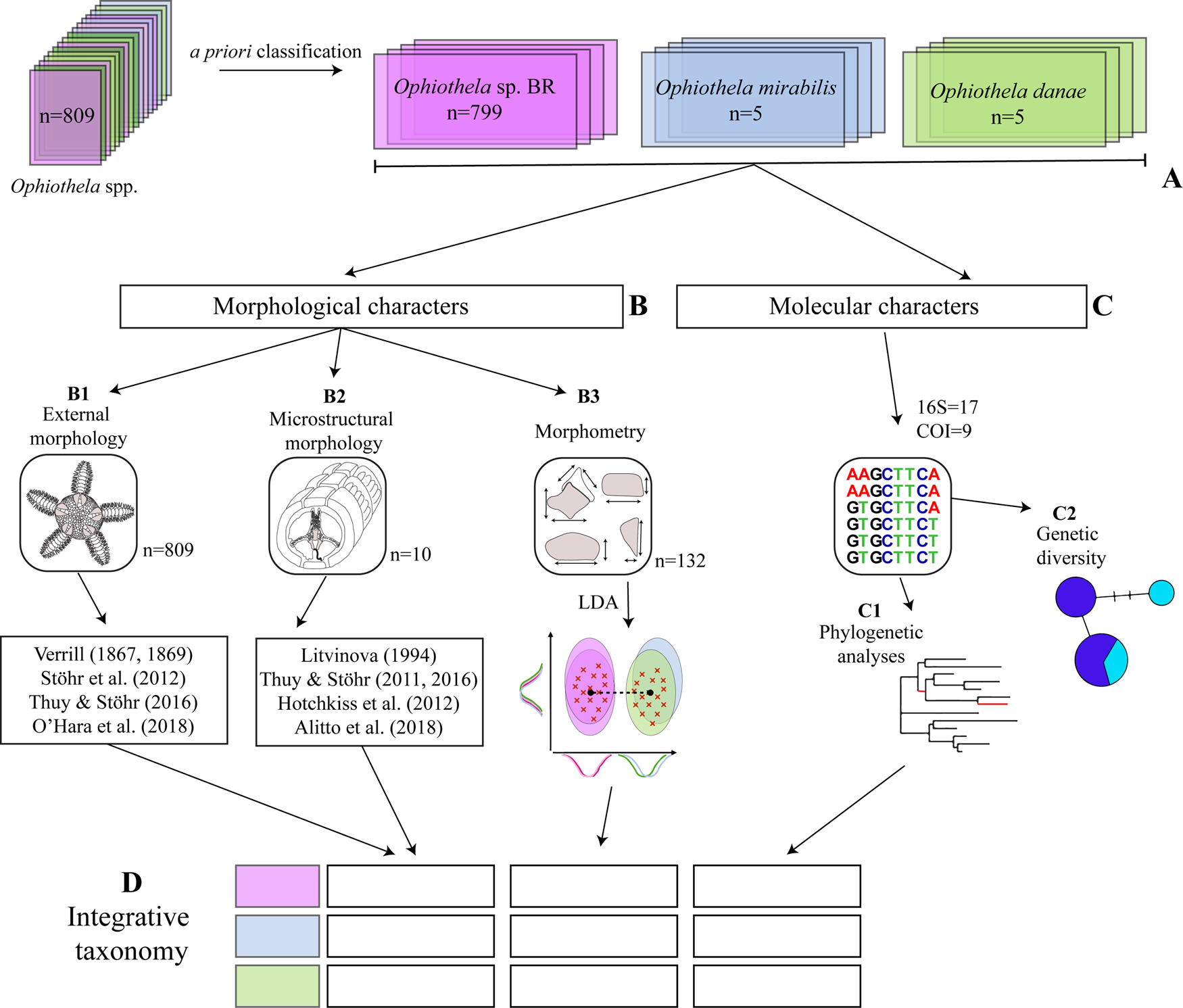
As a starting point, we performed morphological comparisons of the specimens of Ophiothela sp. from Brazil (BR) with the six Ophiothela species listed by Stöhr et al. (Reference Stöhr, O'Hara and Thuy2019): O. danae, O. gracilis, O. mirabilis, O. tigris, O. venusta and O. vincula. This comparison was based on characters typically reported as diagnostic in morphological overviews and keys (Verrill, Reference Verrill1867; Reference Verrill1869; Alitto et al., Reference Alitto, Bueno, Guilherme, Di Domenico, Christensen and Borges2018). The species most similar to the Brazilian specimens are Ophiothela mirabilis Verrill, Reference Verrill1867, and O. danae Verrill, Reference Verrill1869, as they have six arms and five arm spines (step A, Figure 1A).
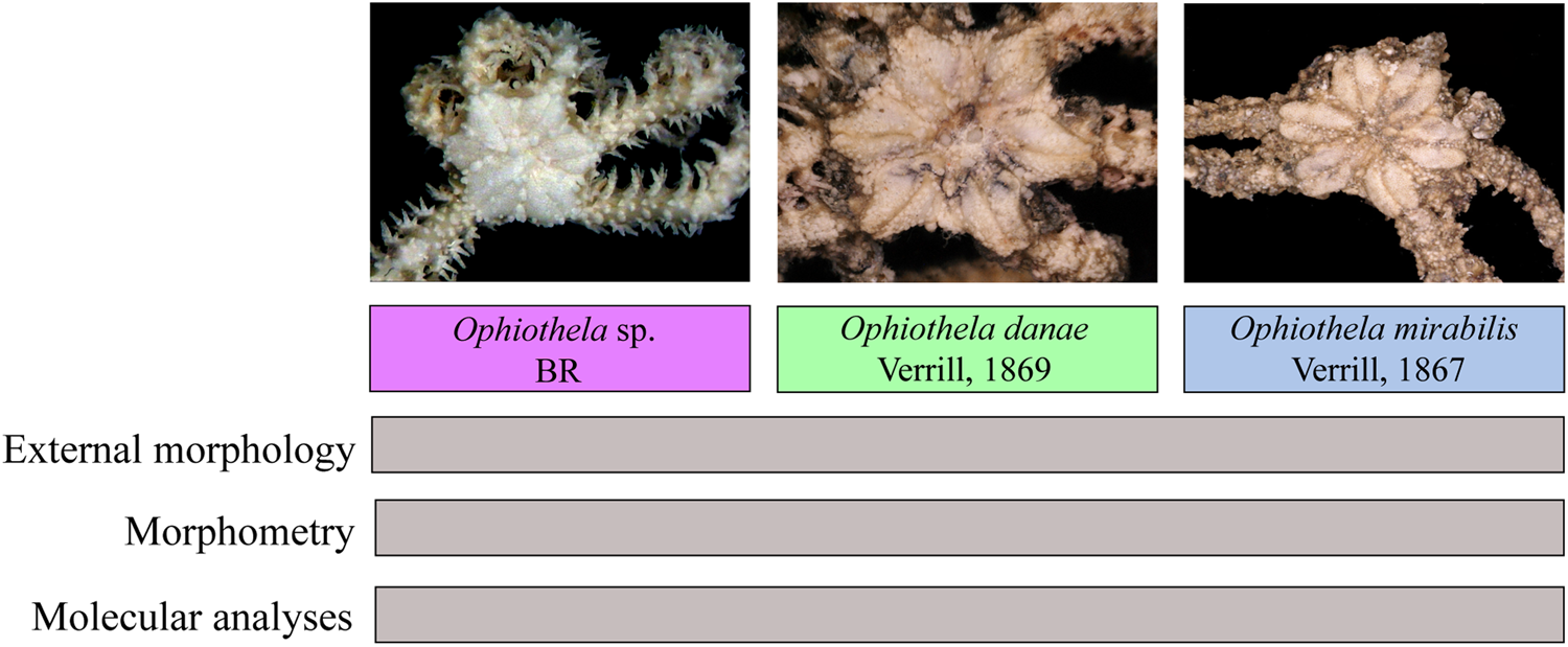
Schematic diagram of the integrative taxonomy applied to Ophiothela spp.: (A) a priori classification inferred from morphological characters based on literature; (B) morphological characters: B1, external morphology; B2, microstructural morphology; B3, morphometry; (C) molecular characters: C1, phylogenetic analyses (Maximum parsimony and Bayesian inference); C2, genetic diversity (genetic distances, AMOVA, haplotype network); and (D) congruence framework of integrative taxonomy – filled rectangles represent the species a priori and empty represent the datasets.

Institutional abbreviations: MCZ, Museum of Comparative Zoology; USNM, United States National Museum, Smithsonian Institution; ZUEC OPH, Ophiuroidea Scientific Collection from Museum of Zoology of the University of Campinas.
Morphological characters
Data collection
A total of 799 spms were studied from four sites in Brazil: (i) Todos os Santos Bay, Bahia; (ii) Araçá Bay, São Paulo; (iii) Praia Preta, São Paulo; and (iv) Estuarine Complex of Paranaguá, Paraná. These samples were then compared to those from scientific collections:
(1) MCZ: (i) Ophiothela mirabilis from Panama (1 dried syntype, OPH-2495); (ii) Ophiothela danae from Fiji Islands (1 dried syntype, OPH-2492; 1 syntype in ethanol, OPH-2631) and from South Africa Cape Colony (1 dried syntype, OPH-2493).
(2) USNM: Ophiothela mirabilis from Panama (4 dried spms, E27240 and E31768) and Ophiothela danae from the Philippines (2 dried spms, 41274 and 41275).
The geographic coordinates, substrate type and main references for each specimen studied are shown in Supplementary Table S1.
External morphology
Comparisons of external characters of Ophiothela sp. BR were made against original descriptions of Ophiothela mirabilis and O. danae (Verrill, Reference Verrill1867, Reference Verrill1869) and against museum specimens from MCZ (syntypes) and USNM (Figure 1B, Step B1). The terms applied are based on the literature (Stöhr et al., Reference Stöhr, O'Hara and Thuy2012; Thuy & Stöhr, Reference Thuy and Stöhr2016; O'Hara et al., Reference O'Hara, Stöhr, Hugall, Thuy and Martynov2018).
Microstructural morphology
The best-preserved specimens of Ophiothela sp. BR were selected to study the arm ossicles, which were extracted from a small portion of the proximal arm, between the fifth and the tenth segments. The arm portion was immersed in regular household bleach (NaClO) until the soft tissues were removed (Stöhr et al., Reference Stöhr, Conand and Boissin2008). The ossicles were then washed with distilled water, air-dried, and prepared for examination with SEM model JEOL JSM5800LV. The terms applied to the microstructures are based on the literature (Hotchkiss et al., Reference Hotchkiss, Prokop and Petr2007; Martynov, Reference Martynov2010; Thuy & Stöhr, Reference Thuy and Stöhr2011, Reference Thuy and Stöhr2016; Hotchkiss & Glass, Reference Hotchkiss, Glass, Kroh and Reich2012; Stöhr et al., Reference Stöhr, O'Hara and Thuy2012; O'Hara et al., Reference O'Hara, Stöhr, Hugall, Thuy and Martynov2018). The arm ossicles are illustrated in Alitto et al. (Reference Alitto, Bueno, Guilherme, Di Domenico, Christensen and Borges2018). Arm vertebra joints were classified into different types as proposed by Litvinova (Reference Litvinova, David, Guille and Feral1994) (Figure 1B, Step B2).
Morphometry
Measurements were taken using an ocular micrometer and through the AxioVision VS program 40.4.8.20 (Carl Zeiss Microscopy, Germany) attached to a ZEISS Discovery V20 stereomicroscope or by using digital measurement (Adobe Photoshop®). A detailed list of measurements is presented in Supplementary Table S2.
The linear discriminant analysis (LDA) was applied using the R environment (R Development Core Team, 2018). To avoid multicollinearity among morphological characters, a correlation matrix was constructed and the variables that were significantly correlated were removed with a threshold value of 0.9. To investigate the differences in morphological characters, the lda function (package MASS) (Venables & Ripley, Reference Venables and Ripley2002; R Development Core Team, 2018) was used to distinguish the Ophiothela spp. The classification rate of each Ophiothela species was assessed by the ‘lda’ function. Visualization was performed using the package ggplot2 (Wickham, Reference Wickham2009) (Figure 1B, Step B3).
Molecular characters
DNA extraction, amplification and sequencing
DNA sequences used in phylogenetic analyses were obtained as follows. Samples of tube feet from the arms were soaked in two changes of Tris-EDTA (TE) buffer for 30 min each. The samples were then macerated in 500 μl of TE buffer with a pestle. A volume of 300 μl of a 10% Chelex 100 solution (BioRad) was added. The samples were then incubated at 55°C for 60 min, boiled for 8 min, cooled to room temperature, vortexed for 20 s, followed by 60 s of centrifugation at 14,000 × g. The supernatant was decanted and kept refrigerated until use in polymerase chain reactions (PCR).
A fragment of the mitochondrial 16S gene was PCR-amplified using the forward primer 16S Sofi F (5′-CAG TAC TCT GAC TGT GCA A-3′) and the reverse primers 16S Sofi R (5′-GGA AAC TAT GAT CCA ACA TC-3′) (Pérez-Portela et al., Reference Pérez-Portela, Almada and Turon2013) and 16S Sbr-H (5′-CCG GTC TGA ACT CAG ATC ACG T-3′) (Palumbi et al., Reference Palumbi, Martin, Romano, McMillan, Stice and Grabowski1991). A fragment of the mitochondrial cytochrome oxidase subunit 1 (COI) gene was amplified using the forward primer COIceF (5′-ACT GCC CAC GCC CTA GTA ATG ATA TTT TTT ATG GTN ATG CC-3′) and the reverse primer COIceR (5′- TCG TGT GTC TAC GTC CAT TCC TAC TGT RAA CAT RTG-3′) (Hoareau & Boissin, Reference Hoareau and Boissin2010).
PCR reactions were performed using PuReTaq Ready-To-Go™ PCR Beads for 25 μl reactions (GE Healthcare), complemented with 0.5 to 1 μl of 25 mM MgCl2. Alternatively, PCR reactions were composed of 10 mM Tris-HCl, pH 8/1 mM KCl buffer, 1.5 mM MgCl2 and 200 μM each dNTP (Invitrogen). In both cases, 0.4 μM of each primer was used and template DNA concentration (ranging from 2.7 to 11 ng μl−1) was tested.
Thermal cycling consisted of a single step at 94°C for 4 min, which was followed by 40 cycles (denaturation at 94°C for 60 s, annealing at 45.7 to 55.7°C for 70 s, and extension at 72°C for 80 s) and a final extension at 72°C for 5 min on a thermal cycler (Eppendorf Mastercycler®).
PCR products were separated from excess primers and dNTPs using a purification kit (Promega). Purified product was then used as a template for DNA sequencing reactions using BigDye Terminator (Applied Biosystems) and sequenced by Serviço de Sequenciamento de DNA – SSDNA IQUSP with the same primers used in the amplification reaction. A 320 bp fragment from 17 individuals was obtained for 16S and a 704 bp fragment from five individuals for COI. All the sequences are deposited in GenBank (www.ncbi.nlm.nih.gov/Genbank) (Supplementary Table S3). The nucleotide sequences were edited using BIOEDIT Sequence Alignment Editor v. 7.0.1 (Hall, Reference Hall1999) and GeneStudio 2.2.0.0 (Genestudio, Suwanee, GA, USA).
Phylogenetic analyses
Two mitochondrial datasets were considered: (i) 16S and (ii) COI. All sequences were aligned with GeneStudio 2.2.0.0 via Clustal W 1.83 (GeneStudio, Suwanee, GA, USA) providing a unified matrix. Additional sequences from other Ophiotrichidae (16S, COI) available in GenBank were added to the matrix for comparison. Amphipholis squamata (Amphiuridae) was used as an outgroup. All samples used are listed in Supplementary Table S3.
Phylogenetic reconstructions were made on the basis of two different optimality criteria, Maximum parsimony (MP) and Bayesian inference (BI), to assess whether there were any differences in the trees recovered with regard to the method used (Figure 1C, Step C1).
The MP was performed with the TNT 1.5 program (Goloboff & Catalano, Reference Goloboff and Catalano2016). The most parsimonious trees were obtained by a heuristic search (best length was hit 100 times), using the new technology search option, which included sectorial searches, ratchet, tree drifting and tree fusing. The gaps were considered as a fifth state. The cladograms had their nodes evaluated by the Bootstrap resampling test (Felsenstein, Reference Felsenstein1985), based on 1000 pseudoreplicates using the Traditional Search.
The BI was performed with MrBayes version 3.2 (Ronquist et al., Reference Ronquist, Teslenko, van der Mark, Ayres, Darling, Höhna, Larget, Liu, Suchard and Huelsenbeck2012). The best-fitting model of molecular evolution for 16S and COI datasets were SYM + G, chosen based on the Akaike Information Criterion (AIC), and General Time Reversible (GTR), chosen based on the Hierarchical Likelihood Ratio Tests (hLRTs), according to the estimation by MR.MODELTEST v.2.3 (Nylander, Reference Nylander2004). The BI analyses used one cold and three incrementally heated Monte Carlo Markov chains (MCMC) on two simultaneous runs. The standard deviation of the split frequencies between the two runs reached a value lower than c. 0.001 at one million generations, with one tree sampled every 100th generation, each using a random tree as a starting point and a temperature parameter value of 0.2 (the default in MrBayes). The first 25% of the total sampled trees were discarded as burn-in to achieve the MCMC log-likelihoods that had become stationary and converged. The analyses resulted in similar likelihood scores, with ESS >200, as verified using TRACER. The phylogenetic trees were visualized and edited in FIGTREE v1.4.3 (http://tree.bio.ed.ac.uk/software/figtree/).
Analyses of genetic diversity
The genetic distances between and within the different groups were estimated by p-distance using MEGA v. 7.0 (Kumar et al., Reference Kumar, Stecher and Tamura2016), ignoring the alignment gaps in pairwise comparisons. Following Hart & Podolsky (Reference Hart and Podolsky2005) and Pérez-Portela et al. (Reference Pérez-Portela, Almada and Turon2013), pairwise genetic p-distances of >3% for 16S and >15% for COI were considered interspecific variations as most of the species already described have been differentiated by this minimum (Figure 1C, Step C2).
The COI matrix was submitted to the AMOVA (Analysis of Molecular Variance) test (Excoffier et al., Reference Excoffier, Smouse and Quattro1992) to evaluate the genetic difference between and among the studied groups formed by the phylogenetic analyses. Haplotype networks were constructed using the TCS method in PopART v1.7 (Leigh & Bryant, Reference Leigh and Bryant2015) (Figure 1C, Step C2).
Integrative taxonomy
Specimens of Ophiothela sp. BR were evaluated using three independent character sets in order to test: (i) external morphology, (ii) morphometry and (iii) molecular data (16S and COI fragments). The species were classified according to the cumulative framework described in Padial et al. (Reference Padial, Miralles, De la Riva and Vences2010) when there was a concordance of at least two datasets analysed (Figure 1D). Therefore, we highlight that molecular data were not weighted more heavily than morphological features.
Results
Morphological characters
External morphology
A detailed comparison between Ophiothela sp. (BR), O. danae and O. mirabilis is presented according to the main characteristics observed in their original descriptions, syntypes from MCZ and additional material from USNM. The possibility that our specimens belonged to some species was rejected as follows: O. tigris Lyman, Reference Lyman1871, has five arms and dorsal arm plates represented by a double row of irregular elongated warts, which only at the base of the arm are increased in number; O. venusta (de Loriol, Reference de Loriol1900) has five arms and triangular ventral arm plates; and O. gracilis Nielsen, Reference Nielsen1932, and O. vincula Mortensen, Reference Mortensen1913, both have five arms.
Dorsal disc
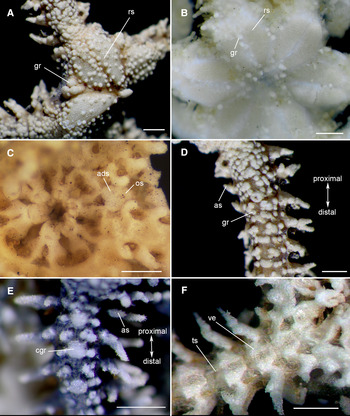
Verrill (Reference Verrill1867, Reference Verrill1869) described Ophiothela danae and O. mirabilis with rounded granules scattered on dorsal disc as we observed in our BR specimens and in the syntypes from MCZ. However, some of the specimens examined had more granules than others, which may be related to their size (Figure 2A, C, E). One specimen of O. danae had three larger and rounded granules in the interradial recesses (USNM 41275) (Figure 2A). Although uncommon, some specimens of O. danae have been described without granules on dorsal surface (Matsumoto, Reference Matsumoto1917), but this did not occur in our specimens.
Ophiothela spp. variations. (A) Granules on the radial shields and three larger and rounded granules in the interradial recesses in Ophiothela danae USNM 41275; (B) Granules only around the radial shields in Ophiothela mirabilis USNM E27240; (C) Oral and adoral shields in Ophiothela danae syntype MCZ OPH-2631 (available from http://mczbase.mcz.harvard.edu/guid/MCZ:IZ:OPH-2631); (D) A higher concentration of granules in Ophiothela danae USNM 41275; (E) A central large granule on the arm in Ophiothela mirabilis USNM E31768; (F) Thin skin highlighting the vertebrae of Ophiothela mirabilis ZUEC OPH 1020. ads, adoral shield; as, arm spine; cgr, central granule; gr, granule; os, oral shield; rs, radial shield; ts, thickened skin; ve, vertebra. Stereomicroscope photos, scale bars: 0.5 mm.

Clusters of sharp conical spines in the interradial recesses and in the centre of the disc were described by Verrill (Reference Verrill1867) for Ophiothela mirabilis. These spines were not observed in any specimen examined in this study.
Dorsal disc with central area depressed
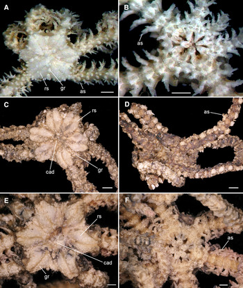
Verrill (Reference Verrill1869) described the central area depressed only for Ophiothela danae. We observed this feature in the majority of our BR specimens and in both the syntypes from MCZ (Figure 3C, E). We do not consider this character applicable for a species diagnosis since it may be related to the preservation method or possibly to food items ingested.
Comparisons of Ophiothela sp. BR with Ophiothela mirabilis and Ophiothela danae. (A, B) Ophiothela sp. BR ZUEC OPH 1020; (C, D) Syntype of O. mirabilis MCZ OPH-2495 (available from http://mczbase.mcz.harvard.edu/guid/MCZ:IZ:OPH-2495); (E, F) Syntype of O. danae MCZ OPH-2492 (available from http://mczbase.mcz.harvard.edu/guid/MCZ:IZ:OPH-2492). as, arm spine; cad, central area depressed; gr, granule; rs, radial shield. Stereomicroscope photos, scale bars: 0.5 mm.

Radial shields
Verrill (Reference Verrill1867, Reference Verrill1869) described prominent radial shields occupying almost the entire disc, covered by rounded granules in Ophiothela mirabilis and O. danae. We also observed this feature in all our BR specimens and in all syntypes from MCZ. The number of granules on the radial shields was variable, even in the same individual, and may be possibly related to their size or regeneration state. One specimen of O. mirabilis had granules only around the radial shields, but not covering them (USNM E27240) (Figure 2B).
Mouth
Plates surrounding the mouth forming a continuous ring were described for the genus Ophiothela as a diagnostic character (Verrill, Reference Verrill1867). Some of our BR specimens did not have thickened skin covering the ventral disc surface, exposing the plates. This corroborates the original description and contradicts Alitto et al. (Reference Alitto, Bueno, Guilherme, Di Domenico, Christensen and Borges2018). According to (Verrill, Reference Verrill1867), the oral and adoral shields are closely united to the surrounding parts, features also observed in some of our BR specimens. However, these plates were not observed in the syntypes of O. mirabilis (MCZ OPH-2495) and O. danae (MCZ OPH-2631) due to the poor condition of the ventral side.
Dorsal arm plates
Verrill (Reference Verrill1867) described the dorsal arm plates (DAP) as covered by rounded granules for Ophiothela mirabilis, but Verrill (Reference Verrill1869) did not describe the DAP for O. danae. A higher concentration of granules was observed proximal to the disc in both species, suggesting that older segments have more granules (Figure 2D). A lower number of granules was observed on regenerating arms of both BR and syntypes specimens. A central series of large granules (CGR) on DAP was described only for O. danae (Verrill, Reference Verrill1869), while numerous and unequal granules on DAP were described for O. mirabilis (Verrill, Reference Verrill1867). The CGR was not observed on syntypes specimens due to their poor condition. However, we observed that some specimens of O. mirabilis (USNM E31768) had a central large granule for each arm segment on the more developed arms (Figure 2E). We assume that the DAP is absent.
Ventral arm plates
Verrill (Reference Verrill1867) described the VAP in Ophiothela mirabilis approximately as long as wide, widest outwardly, outer edge convex, and sides converging to the rounded inner angle. We agree with Nielsen (Reference Nielsen1932) that Verrill's description appears to be the combination of the VAP and the vertebra as seen through the thickened skin (Figure 2F). The naked skin was observed in most of BR specimens, obscuring or making it difficult to see the ventral arm plates (VAP). The VAP is very tiny and was not observed under the stereomicroscope, but using SEM it is three times as long as wide, distal region wider than the proximal, and with a notch in the distal edge.
Arm spines
Ophiothela mirabilis was described with five to six arm spines in specimens with dd 4 to 5.5 mm (Verrill, Reference Verrill1867), while O. danae was described with five arm spines in specimens with dd 4.5 to 5 mm (Verrill, Reference Verrill1869). The number of arm spines ranged from three to five, with the ventralmost being the shortest and bearing wide serrations in all our specimens. Due to the poor condition of the arms of the syntype of O. mirabilis (MCZ OPH-2495), the arm spines were not observed. Four arm spines were observed in O. danae (MCZ OPH-2492), with wide serrations in all specimens.
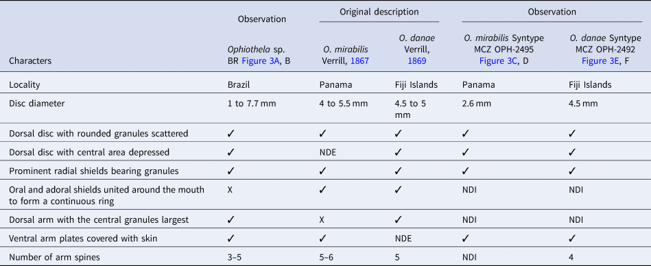
Ophiothela sp. BR and the syntypes of Ophiothela mirabilis and O. danae are represented in Figure 3. Table 1 summarizes the main characteristics observed between them.
Main morphological characters of Ophiothela sp. from Brazil compared with Ophiothela danae and Ophiothela mirabilis from their original descriptions and observations on syntypes from MCZ

NDI, not discernible; NDE, not described.
✓ present; X absent.
The following features were considered variations: higher or lower quantity of granules on the dorsal disc and on the radial shields; dorsal disc with or without the central area depressed; presence or absence of rounded granules in the interradial recesses; granules only around the radial shields, not on them; and presence or absence of a central series of large granules on dorsal arm plates.
We could not distinguish Ophiothela mirabilis from O. danae based on the descriptions of morphological characters by Verrill (Reference Verrill1867, Reference Verrill1869) and on our observations of syntypes from MCZ and specimens from USNM. This indicates that they cannot be separated morphologically.
Morphometry
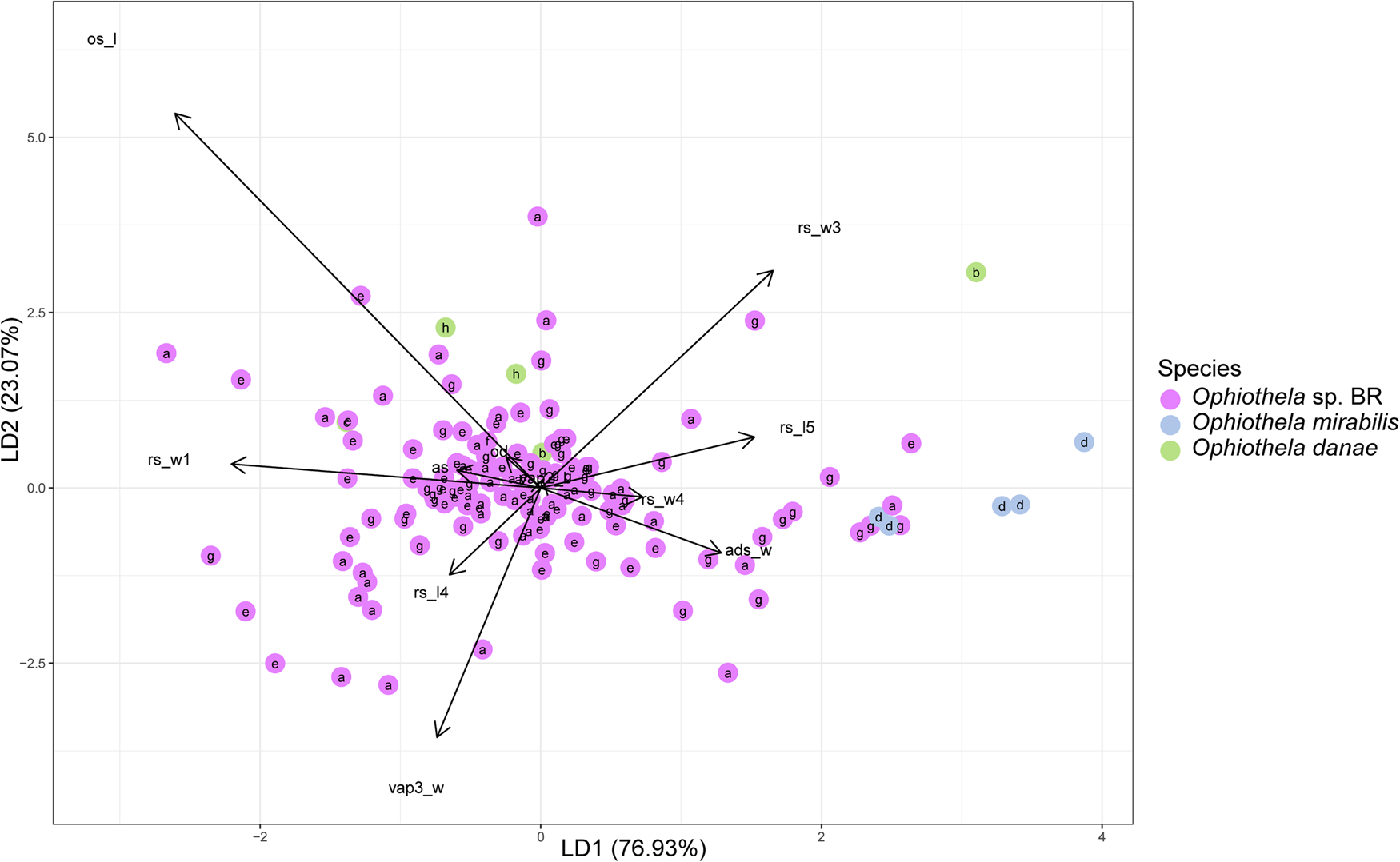
The LDA performed shows that specimens identified as Ophiothela sp. BR overlapped with specimens identified as O. danae, particularly from the Philippines. One specimen of O. danae from Fiji and all specimens of O. mirabilis were isolated from other specimens (Figure 4). The first and second linear discriminant axes described 76.93% and 23.07% of the among species variation in morphological characters, respectively. The classification success of LDA among the three species was 92%, showing Ophiothela sp. BR and O. mirabilis as the species classified correctly most often (96% and 60%, respectively), while O. danae was the most misclassified (0%).
Axes 1 and 2 from Linear Discriminant Analysis (LDA) based on 13 brittle stars characters. See Supplementary Table S2 for definitions of the morphological characters used in the morphometric analysis of Ophiothela spp. a: Araçá Bay, Brazil; b: Fiji Islands; c: South Africa Cape Colony; d: Panama; e: Estuarine Complex of Paranaguá, Brazil; f: Praia Preta, Brazil; g: Todos os Santos Bay, Brazil; h: the Philippines.

The width of the third radial shield (rs_w3) and the length of the fifth radial shield (rs_l5) were the morphological characters with the highest positive coefficients in the first discriminant vector (LD1). The highest negative coefficients were width of the first radial shield (rs_w1) and length of oral shield (os_l). Alternatively, the second discriminant vector (LD2) has length of oral shield (os_l) and width of the third radial shield (rs_w3) as positive coefficients and width of the third ventral arm plate (vap3_w) and length of the fourth radial shield (rs_l4) as negative coefficients.
In summary, the LDA results indicate that the species analysed are not morphometrically distinct. Ophiothela mirabilis, O. danae and Ophiothela sp. BR cannot be distinguished from each other based on morphological measurements.
Molecular characters
Phylogenetic analyses
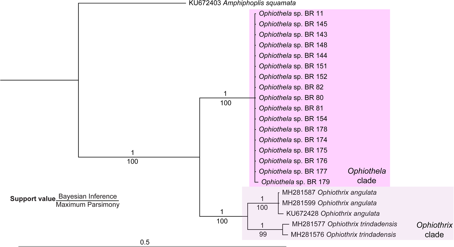
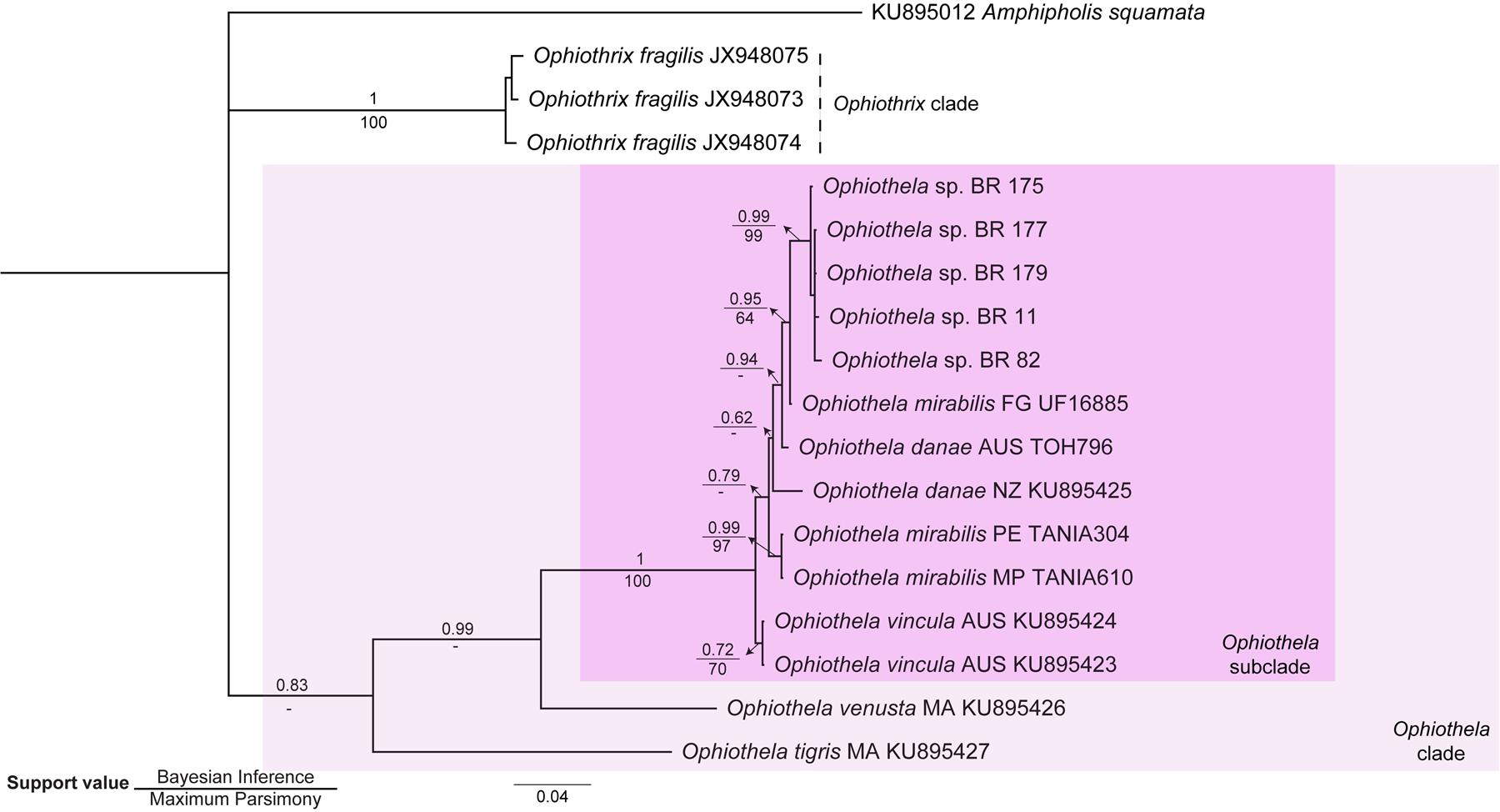
The phylogenetic cladograms obtained either in the Bayesian (BI) analyses or in the Maximum parsimony (MP) analyses of the two datasets (16S, Figure 5; COI, Figure 6) show a clade containing all the Ophiothela sp. BR with high support values (Figures 5 & 6).
Phylogenetic tree inferred from the Bayesian analysis of 16S sequences. The numbers above the branches represent posterior probabilities. For the clades also inferred in the MP analysis, bootstrap values (%) are provided (below the branches). Branches are identified by individual codes (Supplementary Table S3) and their localities: BR, Brazil.

Phylogenetic tree inferred from the Bayesian analysis of COI sequences. The numbers above the branches represent posterior probabilities. For the clades also inferred in the MP analysis, bootstrap values (%) are provided (below the branches). Branches are identified by individual codes (Supplementary Table S3) and their localities: AUS, Australia; BR, Brazil; FG, French Guyana; MA, Madagascar; MP, Mexico Pacific; NZ, New Zealand; PE, Peru. The dash (-) indicates that the branch was not recovered in MP analysis.

Using the 16S dataset, two clades were noted in BI and MP with high support values (1, 100%, respectively) (Figure 5): (i) an Ophiothela clade comprised of all Ophiothela sp. BR; and (ii) an Ophiothrix clade which contains all the Ophiothrix samples.
An Ophiothela subclade was noted in both BI and MP using the COI dataset (Figure 6) with high support values (1, 100%, respectively). The Ophiothela subclade was formed by Ophiothela sp. BR, O. mirabilis, O. danae and O. vincula. Two main clades were also noted: (i) an Ophiothela clade which contains all the Ophiothela samples, but only in BI with a low support value (0.83); and (ii) an Ophiothrix clade which contains all the Ophiothrix samples in BI and MP with high support values (1, 100%, respectively).
Analyses of the genetic diversity
Genetic distances
The genetic distances observed in 16S and COI within the Ophiothela sp. BR were low (from 0.3–0.7% for 16S and from 0.1–0.2% for COI).
Utilizing the COI dataset, the Ophiothela subclade, which is formed by Ophiothela sp. BR, O. mirabilis, O. danae and O. vincula has a range of genetic distance of 1.4–2.7% within it. The lowest genetic distances were between Ophiothela mirabilis and O. vincula (1.4%), O. mirabilis and O. danae (1.7%) and O. vincula and O. danae (1.8%). The highest genetic distances were between O. tigris and O. danae (20.6%), and O. tigris and Ophiothela sp. BR (20.3%) (Supplementary Table S4).
AMOVA
The AMOVA test revealed that 44.36% of the total genetic variability in COI sequences occurred among the Ophiothela subclade (inferred in the phylogenetics analysis) and the group formed by Ophiothela venusta and O. tigris (Supplementary Table S5). On the other hand, 34.48% of the total genetic diversity refers to variability within these groups. These results indicate that the two groups are genetically distinct and well structured, with low or null gene flow between them.
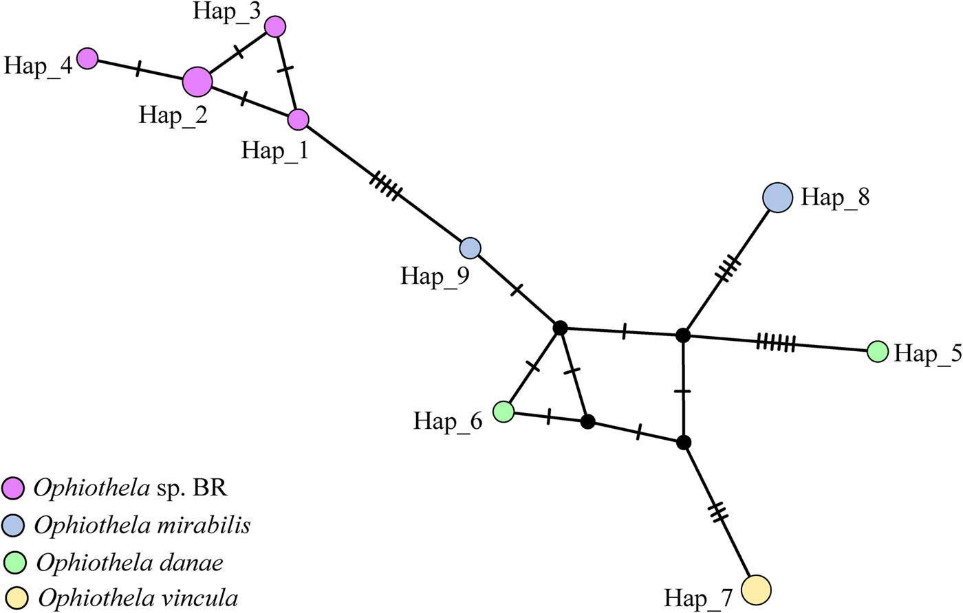
Haplotype network
The haplotype network indicated the existence of two divergent lineages (1: Ophiothela sp. BR, and 2: O. mirabilis, O. danae and O. vincula) with intermediate haplotypes. The divergence among lineages was caused by five mutations. The first lineage (Figure 7, left) is composed of low-frequency (mostly private) haplotypes, separated by a few mutational steps. The second lineage (Figure 7, right) is composed of five haplotypes sharply separated, but connected by the intermediate haplotypes (Figure 7, black circles).
Median-joining haplotype network based on COI gene sequences. Each circle represents one haplotype and their size is proportional to the number of individuals possessing it. The black circles represent possible intermediate haplotypes.

Our studies based on molecular characters had two main results. First, the analyses with 16S and COI sequences indicate that only one lineage is present in Brazil. Second, the comparisons based on COI sequences revealed that Ophiothela sp. BR, O. vincula, O. mirabilis and O. danae form a group sharing similarities, while O. tigris and O. venusta are the most different from the first group.
Integrative taxonomy
To integrate the results of the different operational criteria, a cumulative framework was followed (Padial et al., Reference Padial, Miralles, De la Riva and Vences2010). Concordance was found between the analyses based on external morphology, morphometry and molecular data (16S, genetic distances, haplotype network) indicating that Ophiothela sp. BR, O. mirabilis and O. danae are closely related (Figure 8).
Synthetic representation of the integrative taxonomy approach within the Ophiothela spp. Small rectangles would be used to represent a dataset delimiting a species while larger rectangles represent the inability of the dataset to delimit the species included. The large rectangles for all three datasets illustrate the close relationship of Ophiothela sp. BR, O. mirabilis and O. danae.

In the following, we present the description of Ophiothela sp. BR as Ophiothela mirabilis. The description contains the amended diagnosis, taxonomic comments, remarks and distribution. The classification is in accordance with O'Hara et al. (Reference O'Hara, Hugall, Thuy, Stöhr and Martynov2017, Reference O'Hara, Stöhr, Hugall, Thuy and Martynov2018).
Morphological description
Order AMPHILEPIDIDA (O'Hara et al., Reference O'Hara, Hugall, Thuy, Stöhr and Martynov2017)
Family OPHIOTRICHIDAE Ljungman, 1867
Genus Ophiothela Verrill, Reference Verrill1867
Type species. Ophiothela mirabilis Verrill, Reference Verrill1867
Amended diagnosis. Dorsal disc with scattered rounded granules. Radial shields very large, covering most of the disc. Ventral side of arms and disc covered by thickened skin, usually obscuring the arm plates and scales. Dorsal arm plates are not distinguishable. Arm spines mostly turned downward, armed with thorns or hooks. Transspondylous vertebrae and keeled. Dorsal groove of vertebrae lined by smooth knobs (Verrill, Reference Verrill1867; Alitto et al., Reference Alitto, Bueno, Guilherme, Di Domenico, Christensen and Borges2018).
Ophiothela mirabilis
Figures 9–11
Ophiothela mirabilis Verrill, Reference Verrill1867: 268
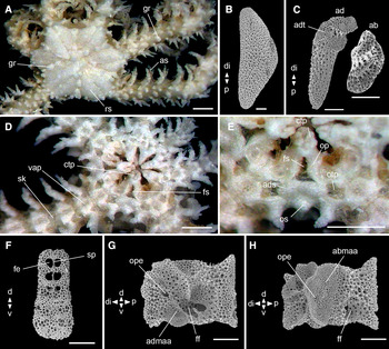
External morphology, genital and oral plates of Ophiothela mirabilis BR ZUEC OPH 1020 (2.5 mm dd). (A) dorsal view; (B) radial shield; (C) genital plate; (D) ventral view; (E) detail of the oral view; (F) dental plate; (G) oral plate in adradial view; (H) oral plate in abradial view. ab, abradial; ad, adradial; ads, adoral shield; abmaa, abradial muscle attachment area; adt, adradial-distal tip; admaa, adradial muscle attachment area; as, arm spine; cd, central depression; ctp, cluster of tooth papillae; d, dorsal; di, distal; fe, fenestration; ff, first foot; fs, fossae; gr, granule; op, oral plate; ope, opening; os, oral shield; otp, oral tentacular pore; p, proximal; rs, radial shield; v, ventral; vap, ventral arm plate; sp, septum; sk, skin. Stereomicroscope photos: (A, D, E), scale bars: 0.5 mm. SEM photos: (B, C, F–H), scale bars: 100 μm.

Ophiothela caerulea H.L. Clark 1915: 283; A. M. Clark & Rowe 1971: 116, 130
Ophiothela dividua von Martens, 1879: 127–130; H.L. Clark 1923: 343; Balinsky 1957: 22; A.M. Clark 1974: 470
Ophiothela hadra H.L. Clark, 1915: 284–285
Ophiothela involuta Koehler, 1898b: 88–90; Matsumoto, Reference Matsumoto1917: 230–232
Ophiothela isidicola Lütken, 1872: 92; de Loriol 1893: 52–53; Koehler, Reference Koehler1905: 117–118; H.L. Clark 1915: 285; Matsumoto, Reference Matsumoto1917: 230–232
Ophiothela verrilli Duncan, 1879: 477–479, Matsumoto, Reference Matsumoto1917: 230–232
Maximum size. dd up to 7 mm.
Material examined. 132 specimens (dd 0.7–7.7 mm). See Supplementary Table S1.
Amended diagnosis. Dorsal disc with scattered rounded granules. Radial shields covering ~2/3 of dd. Ventral side of arms and disc covered by thickened skin. Dorsal arm plates absent. Three to six arm spines. Transspondylous vertebrae and keeled. Dorsal groove of the vertebrae lined by a series of four smooth knobs to either side. Typically, hexamerous, occasionally pentamerous. Arms and radial shields may be of unequal lengths due to fission events.
Description. Disc: (dd: 2.5 mm). Star-shaped, with interradial recesses, rounded granules scattered over the entire dorsal surface, larger between radial shields (Figure 9A). Radial shields elevated, triangular (Figure 9B), abradial edge bowed and adradial straight, ~2/3 of dd, united distally and separated proximally by one scale. Radial shield pairs separated by a narrow row of plates bearing granules (Figure 9A). Abradial genital plate shorter than half the adradial length; adradio-distal tip shape concave (Figure 9C). Ventral side of arms and disc covered by thickened skin (Figure 9D). Mouth plating. Oral shields slightly broadened proximally. Adoral shields broadened distally and united proximally. Fossa between two oral plates. A cluster of tooth papillae on the dental plate. Oral tentacle pore large and opening at the mouth angle (Figure 9D, E). Dental plate entire; ventral half widest; dorsal half with fenestrations and a septum (Figure 9F). Oral plates longer than wide (~1.5 times) (Figure 9G, H); abradial and adradial surface with an opening smaller than the opening of the first tube foot; abradial muscle attachment area with common stereom (Figure 9H); adradial muscle attachment area in ventral position of the articulation area (Figure 9G).
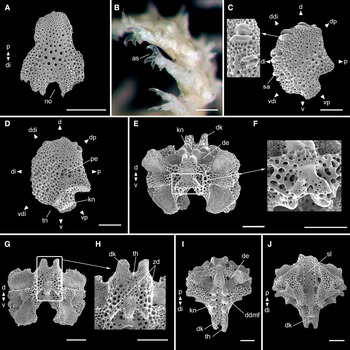
Arms. Six arms, dorsoventrally coiled and covered with scattered granules, dorsal arm plates absent (Figure 9A). Ventral arm plates three times as long as wide, proximal region is convex and distal straight, distal region wider than the proximal, notch in distal edge, lateral edges with clear incisions for tentacle openings, the first three plates contiguous, the others separated by lateral arm plates (Figure 10A). Three to five arm spines, ventral shortest and bearing sharp hyaline hooks (Figure 10B).
Vertabrae, ventral and lateral arm plates of Ophiothela mirabilis BR ZUEC OPH 1020 (2.5 mm dd). (A) ventral arm plate; (B) detail of a hooked arm spine; (C) lateral arm plate – external view; (D) lateral arm plate – internal view; (E, F) vertebra – proximal face; (G, H) vertebra – distal face; (I) vertebra – dorsal face; (J) vertebra – ventral face. as, arm spine; d, dorsal; ddi, dorso-distal; ddmf, dorsodistal muscular fossae; de, depression; di, distal; dk, distal keel; dp, dorso-proximal; kn, knob; no, notch; p, proximal; pe, perforation; sa, spine articulation; sl, suture line; sp, spine; tn, tentacle notch; v, ventral; vdi, ventro-distal; vp, ventro-proximal; zd, zygocondyle. Stereomicroscope photo (B), scale bar: 0.1 mm. SEM photos (A, C–J), scale bars: 100 μm.

Lateral arm plates (Figure 10C, D). General outline: ventral portion is slightly projecting ventro-proximalwards; ventro-distal tip not projecting ventralwards; tentacle notch pointing ventralwards. Outer surface ornamentation: trabecular intersections protruding to form knobs larger than stereom pores on most of outer surface. Outer proximal edge: central part of the proximal margin is protruding. Spine articulations: on same level as remaining outer surface; dorsalwards increasing in size; distance between spine articulations equidistant. Lobes simply separated, equal-sized; lobes parallel, straight, oriented nearly horizontal; stereom massive. Ridges and knobs: with two separate knobs; without additional dorsal structure on inner side; single large perforation on inner side.
Vertebrae. Transspondylous and keeled (although it may not be well developed due to the high incidence of fission). Two large grooves on proximal side of vertebrae dorsally corresponding to distalwards projecting dorso-distal muscular fossae of distal side (Figure 10E). Transspondylous articulation (Figure 10F). Zygosphene absent; a small zygospondyline peg on the distal side; thorns on the end of the distal keel seen from the distal aspect (Figure 10G, H). Dorso-distal muscular fossae transformed distalwards clearly projecting beyond the zygocondyles (true keel) and divided into two separate end processes matching the two large dorsal depressions proximally. Dorsal groove lined by a series of three to four smooth knobs to either side (Figure 10I). Distinct suture line in ventral view (Figure 10J). Although the vertebrae were not well developed in our specimens due to the high incidence of fission, ~90% of specimens, in O. mirabilis, we conclude that its articulation is transspondylous (Hotchkiss & Glass, Reference Hotchkiss, Glass, Kroh and Reich2012) which is very similar to the saddle-shape described by Litvinova (Reference Litvinova, David, Guille and Feral1994).
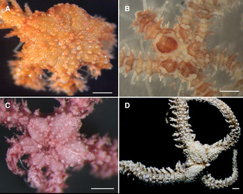
Colour patterns. The most common colour pattern in Brazil has been yellow-orange (Hendler et al., Reference Hendler, Migotto, Ventura and Wilk2012; Mantelatto et al., Reference Mantelatto, Vidon, Silveira, Menegola, Rocha and Creed2016; Lawley et al., Reference Lawley, Fonseca, Júnior and Lindner2018), but purple was also described in specimens from Araçá Bay (Alitto et al., Reference Alitto, Bueno, Guilherme, Di Domenico, Christensen and Borges2018). In the present study, the most common colouration patterns are orange and purple in living specimens and reddish purple, brown, cream, or white for those fixed in alcohol. Other patterns observed are purple with white patches, and pink with purple patches (Figure 11). While studies of O. mirabilis in the Pacific have indicated a correlation between brittle star colour and that of its host (Clark, Reference Clark1976; Granja-Fernández et al., Reference Granja-Fernández, Herrero-Pérezrul, López-Pérez, Hernández, Rodríguez-Zaragoza, Jones and Pineda-López2014), this relationship does not appear to be important in Brazilian specimens (Mantelatto et al. Reference Mantelatto, Vidon, Silveira, Menegola, Rocha and Creed2016; present study). These observations suggest that it may possess an innate predator deterrent, such as a chemical defence (Mantelatto et al., Reference Mantelatto, Vidon, Silveira, Menegola, Rocha and Creed2016), or its natural predator does not exist in Brazil.
Ophiothela mirabilis – colour patterns in living (A, B) and preserved (C, D) specimens. (A) orange ZUEC OPH 1020; (B) purple with white ZUEC OPH 2814; (C) reddish purple ZUEC OPH 2876; (D) brown USNM 41275. Stereomicroscope photos, scale bars: 0.5 mm.

Taxonomic comments. The following features were considered as variations: (i) the presence and number of granules on some structures: dorsal disc, radial shields, interradial recesses; and (ii) dorsal disc with the central area depressed. Two characters of the Ophiotrichidae diagnosis were not observed in Ophiothela mirabilis (O'Hara et al., Reference O'Hara, Stöhr, Hugall, Thuy and Martynov2018): (i) small holes at each edge on the ventral half of the dental plate, and (ii) three knobs on the inner side of the lateral arm plates (only one knob is present). An opening on both sides of the oral plate was observed. This opening is often present in self-dividing (fissiparous) species, such as Ophiocomella spp. and Ophiactis savignyi, and may suggest a relationship to the phenomenon of fissiparity (Devaney, Reference Devaney1970). Only two specimens with seven arms were observed. This is considered an unusual morphological occurrence and was described by Matsumoto (Reference Matsumoto1917) for O. danae. The series of knobs on the dorsal groove were only described for Ophiothela tigris (Mortensen, Reference Mortensen1913). Two features contradict the description of Ophiothela sp. made by Alitto et al. (Reference Alitto, Bueno, Guilherme, Di Domenico, Christensen and Borges2018): (i) one, not none, knob on the inner side of the lateral arm plate, and (ii) vertebrae are transspondylous (not zygospondylous). The vertebra is divided into two separate end processes and with thorns on the distal keel and has never been reported in the literature.
Remarks. Ophiothela mirabilis is commonly found associated with gorgonians. In the present study, it was also associated with the sponges Mycale (Zygomycale) and Callyspongia sp., and the coral Carijoa riisei. In Brazil, O. mirabilis is reported to use a total of 29 novel host taxa including sponges, corals, echinoderms, algae, bryozoans and a seahorse (Mantelatto et al., Reference Mantelatto, Vidon, Silveira, Menegola, Rocha and Creed2016).
Distribution. According to marine ecoregions (Spalding et al., Reference Spalding, Fox, Allen, Davidson, Ferdaña, Finlayson, Halpern, Jorge, Lombana and Lourie2007): Tropical Atlantic (realm), North Brazil Shelf (province): Guianan (Hendler & Brugneaux, Reference Hendler and Brugneaux2013); Tropical South-western Atlantic (province): north-eastern and eastern Brazil (Hendler et al., Reference Hendler, Migotto, Ventura and Wilk2012; Araújo et al., Reference Araújo, Soares, Matthews-Cascon and Monteiro2018); Temperate South America (realm), Warm Temperate South-western Atlantic (province): south-eastern Brazil (Hendler et al., Reference Hendler, Migotto, Ventura and Wilk2012; Bumbeer & Rocha, Reference Bumbeer and Rocha2016; Mantelatto et al., Reference Mantelatto, Vidon, Silveira, Menegola, Rocha and Creed2016).
Discussion
The combined integrative taxonomy based on external morphology, morphometry and molecular data has confirmed the identity of a single species of Ophiothela in Brazil. Ophiothela mirabilis, O. danae and Ophiothela sp. BR cannot be distinguished from each other based on the data examined here. We suggest that O. danae should be considered as a junior synonym of O. mirabilis. The species was redescribed in detail using external morphology and microstructural elements.
The genus Ophiothela was first described by Verrill (Reference Verrill1867) as a subgenus of Ophiothrix. According to the author, Ophiothela agrees well with the typical species of Ophiothrix, but differs in having the arms distinctly covered by a thickened skin with scattered granules. Due to these distinct characters, Ophiothela is currently accepted as a genus (Stöhr et al., Reference Stöhr, O'Hara and Thuy2019).
Ophiothela mirabilis was the first species described in the genus Ophiothela, followed by O. danae described by Verrill (Reference Verrill1869). Although the descriptions of these species were made by the same author and time period, he made no comparison between them. In the following years, many species were described as belonging to Ophiothela. To complicate matters, the small size (maximum dd 7 mm) and fissiparous nature, has made the morphological studies and delimitation of Ophiothela species even more difficult.
For Ophiothela, the most synonymized species is O. danae, with a total of seven synonyms. The primary argument used by the authors was the lack of differences between the species. One example is O. danae, O. isidicola and O. verrilli which all have overlapping features in their original descriptions with no differences between them (Koehler, Reference Koehler1905; Matsumoto, Reference Matsumoto1917). All three species were synonymized as O. danae.
The conclusion to synonymize Ophiothela danae Verrill, Reference Verrill1869 with Ophiothela mirabilis Verrill, Reference Verrill1867 is due to the two distinct analyses (external morphology and morphometry) applied to the Brazilian specimens and the syntypes from MCZ. As the analyses failed to separate them, they have been combined as a single species with intraspecific morphological variation, such as variable quantity of granules on the dorsal disc and number of arm spines.
The main character observed in non-regenerating arms of Ophiothela mirabilis was a large central granule for each arm segment, which may be a remnant of the dorsal arm plate (DAP). The DAP is absent in most of the specimens, particularly the ones from Brazil. Mortensen (Reference Mortensen1913) described the lateral granules on the dorsal side of arms as parts of the vertebrae for O. tigris. We assume that these granules could be the same smooth knobs observed on the dorsal face of the vertebrae in our specimens under the SEM. These knobs were observed in a series of four to either side on the vertebrae of O. mirabilis. This was not observed on the dorsal arm surface when analysed under the stereomicroscope. We concluded that the Brazilian specimens have granules on the dorsal arm and smooth knobs on the dorsal face of its vertebrae.
A true vertebral keel was observed in Ophiothela mirabilis. This keel is different from the keel described for Ophiothrix angulata (Alitto et al., Reference Alitto, Bueno, Guilherme, Di Domenico, Christensen and Borges2018), which also belongs to the family Ophiotrichidae. In O. mirabilis the keel is divided into two separate end processes matching the two large dorsal grooves proximally, while in O. angulata the keel is not divided, and its end process matches a single dorsal groove proximally. An ancestral state reconstruction of the evolutionary history of ophiuroid vertebral morphologies was recently proposed by Clark et al. (Reference Clark, Hutchinson, Darroch, Mongiardino Koch, Brady, Smith and Briggs2018). The authors suggested that the non-keeled vertebral morphology was present in the most recent common ancestors of Ophiuroidea and the keeled morphology appears to have evolved convergently at least twice within the Amphilepidida, to which Ophiotrichidae belongs.
Ophiothela mirabilis has a vertebral articulation similar to the saddle-shaped articulation which provides the arm the most flexibility (Litvinova, Reference Litvinova, David, Guille and Feral1994). This description marks the first report of this joint in Ophiotrichidae. Since the arm vertebrae have been subject to ecological adaptation (O'Hara et al., Reference O'Hara, Stöhr, Hugall, Thuy and Martynov2018), the need for further studies, such as behavioural observation and mechanical testing, are necessary to elucidate the function of the different joints present in ophiuroids.
Another feature of interest is the opening on both sides of the oral plate of Ophiothela mirabilis. We agree with Devaney (Reference Devaney1970) that this feature may suggest a relationship with the phenomenon of fissiparity, since it has only been described in self-dividing (fissiparous) species, such as Ophiocomella spp. and Ophiactis savignyi. This opening is thought to lead to the water vascular system (Devaney, Reference Devaney1970).
We provided a detailed description of the dental and oral plates of Ophiothela mirabilis, which could integrate future analyses of comparative morphology. Recently, important studies have highlighted the relevance of oral and dental plates as taxonomic, phylogenetic and ontogenetic resources (Thuy & Stöhr, Reference Thuy and Stöhr2016; Hendler, Reference Hendler2018). Moreover, through the description of these plates, it may be possible to understand some habits, such as feeding behaviour, and the diet of species in various families (Hendler, Reference Hendler2018).
The morphometric analysis of Ophiothela mirabilis from BR, O. mirabilis and O. danae from the type series shows that these species did not totally overlap on the LDA. Morphometry has been used as a powerful tool for delimiting species, particularly when applied with an integrative approach (Arribas et al., Reference Arribas, Andújar, Sánchez-Fernández, Abellán and Millán2013; Marinho et al., Reference Marinho, Cônsoli, Penteado-Dias and Zucchi2017; Di Camillo et al., Reference Di Camillo, Gravulli, De Vito, Pica, Piraino, Puce and Cerrano2018; Alitto et al., Reference Alitto, Amaral, Oliveira, Serrano, Seger, Guilherme, Di Domenico, Christensen, Lourenço, Tavares and Borges2019). In the case of O. mirabilis, the morphometry was relevant in detecting patterns related to its morphological variation. We highlight the use of morphometry in several areas of biology, such as delimiting species and ontogenetic series in order to detect patterns undetected by the unaided eye.
Attention should be given to Ophiothela vincula due to its morphological similarity and concurrent molecular divergence with Ophiothela danae. Ophiothela vincula was described from a few specimens sampled from the Philippine Islands, the same locality as the material of the O. danae we examined from USNM. The only morphological difference observed from the description of Mortensen (Reference Mortensen1913) is that O. vincula has five arms while O. danae has six arms. On the other hand, the level of genetic divergence between some five-armed specimens from NW Australia identified as O. vincula and six-armed O. danae from the eastern Pacific was 0.3–0.7% and 1.4–2.7% for 16S and COI, respectively. These COI divergence rates are higher than those found within Ophiothrix trindadensis (~1.5%) (Alitto et al., Reference Alitto, Amaral, Oliveira, Serrano, Seger, Guilherme, Di Domenico, Christensen, Lourenço, Tavares and Borges2019). Therefore, future studies should investigate if O. vincula should be recognized as a distinct species, or just a five-armed variant of O. danae.
Due to the large number of similar species, low levels of genetic distance, and the high degree of fissiparity, Ophiothela may represent a facultative asexual complex, possibly from hybrid origins similar to the Ophiocomella sexradia complex (O'Hara et al., Reference O'Hara, Hugall, Cisternas, Boissin, Bribiesca-Contreras, Sellanes, Paulay and Byrne2019). Hybridization has been evidenced in ophiuroids (Taboada & Pérez-Portela, Reference Taboada and Pérez-Portela2016; Weber et al., Reference Weber, Stöhr and Chenuil2019) and we consider that the improvement of new technologies, such as next generation sequencing hybridization-based capture, will provide new possibilities for testing for hybridization in Ophiothela spp.
Our findings have important taxonomic implications, particularly at the species level. But we also emphasize the necessity for an integrative approach at a family level since considerable morphological differences were found in Ophiothela. Further studies should include nuclear genes, such as 28S, in order to understand the phylogenetic relationships of the Ophiotrichidae family and its relatives.
Ophiothela has a widespread distribution that is continuing to expand. Recently, Ophiothela mirabilis was observed associated with octocorals at Dania Beach, Florida, USA (Glynn et al., Reference Glynn, Coffman, Primov, Renegar, Gross, Blackwelder, Martinez, Dominguez, Vanderwoude and Riegl2019). To date, Ophiothela has not been reported from the northern- and southern-most regions of the Pacific coast of the Americas. This places Ophiothela in the same group as the fissiparous and widespread brittle stars, Ophiactis lymani and Ophiactis savignyi, which have circum-tropical and circum-subtropical distribution. We propose the use of Ophiothela mirabilis as a model organism to: (i) explain the importance of asexual species in marine realms; (ii) for testing theories concerning the evolutionary advantages of fissiparity; and (iii) for testing its regeneration ability in applied biology and gene expression.
Supplementary material
The supplementary material for this article can be found at https://doi.org/10.1017/S002531542000034X.
Acknowledgements
We are grateful to Professor Cecília Amaral (IB/UNICAMP) for coordinating Project BIOTA-Araçá and to Marine Biology Center in University of São Paulo (CEBIMar-USP). We are grateful to Camila Silva, Guilherme Corte, Helio Checon, Nathalia Padovanni, Angélica Godoy, Renato Luchetti, Fabrizio Marcondes and Ariane Campos for helping collect and sieve all the Araçá Bay samples; Maristela Bueno for collecting the Paranaguá specimens; Alisson Santana for collecting the Salvador specimens and Pablo D.B. Guilherme for providing the LDA script and Figure 1. We are in debit to Adriane Sprogis and Stella de Ferraz for their great help with the scanning electron microscopy at UNICAMP and Karin R. Seger and Luciana B. Lourenço for all support provided with the genetic analysis at UNICAMP. Many thanks to the reviewers André Rinaldo Senna Garraffoni, Luciana Martins, Leonardo Yokoyama, Fabrizio Marcondes and Fosca Pedini whose valuable comments were greatly appreciated. We also thank the Smithsonian United States National Museum for loan of specimens and Museum of Comparative Zoology, particularly Adam Baldinger, for the excellent photos provided. Finally, many thanks to the reviewers whose valuable comments were greatly appreciated.
Financial support
This study was financed in part by the Coordenação de Aperfeiçoamento de Pessoal de Nível Superior – Brasil (CAPES) – Finance Code 001 and Proc. No. PDSE −88881.131940/2016-01. Grant 2011/50317-5, São Paulo Research Foundation (FAPESP), grant 2018/10313-0 (FAPESP) and 2017/09987-3 (FAPESP).
Ethical standards
Fieldwork was authorized by the Chico Mendes Institute for Biodiversity Conservation–ICMBio, under the licence number: 19887–1 to Cecília Amaral and 38463–3 to Maristela Bueno. Genetics data were registered at the National System for the Management of Genetic Heritage and Associated Traditional Knowledge–SisGen, according to Brazilian legislation Law number 13.123/2015 and Decree 8772/2016. Approval ID for this study were AF4BF9C, AB7038A and AACEF44.