INTRODUCTION
This review describes our current knowledge of glial cells that express the NG2 chondroitin sulfate proteoglycan (NG2 glia) in the human central nervous system (CNS). These cells are distinct from other well-characterized glia, such as astrocytes, oligodendrocytes and microglia. The history and scope of our knowledge of these cells based upon rodent studies has been recently reviewed (Nishiyama et al., Reference Nishiyama, Komitova, Suzuki and Zhu2009). The best documented function for these cells is an oligodendrocyte progenitor (OPC). In rodents, there is nearly complete overlap in expression of NG2 and the platelet-derived growth factor receptor alpha (PDGFRA) and oligodendrocytes are the major progeny of cells in transgenic mice expressing fluorescent proteins under the control of these promoters (Rivers et al., Reference Rivers, Young, Rizzi, Jamen, Psachoulia and Wade2008; Zhu et al., Reference Zhu, Bergles and Nishiyama2008). NG2 glial cell processes also contact nodes of Ranvier and neurons at the synapse (Butt et al., Reference Butt, Kiff, Hubbard and Berry2002), and cortical NG2 glial cells can receive synaptic input from neurons (Bergles et al., Reference Bergles, Roberts, Somogyi and Jahr2000; Lin and Bergles, Reference Lin and Bergles2004; Lin et al., Reference Lin, Huck, Roberts, Macklin, Somogyi and Bergles2005; Wigley et al., Reference Wigley, Hamilton, Nishiyama, Kirchhoff and Butt2007; Nishiyama et al., Reference Nishiyama, Komitova, Suzuki and Zhu2009). The name ‘syntanocyte’ has been proposed for the adult NG2 glial cell to reflect the contacts these cells make with neurons. ‘Polydendrocyte’ has been proposed to reflect the distinctive morphology of the majority of NG2 glia, while being inclusive of the functional heterogeneity (e.g. OPC, syntanocyte) observed in NG2-expressing cells (Butt and Nishiyama, Reference Butt and Nishiyama2002). Because there is no consensus for nomenclature at this time, we will use the term NG2 glia to refer to the entire population of glial cells that express NG2 and/or PDGFRA. This review will be limited to our knowledge of NG2 glia as they exist in intact human tissues; a detailed review on isolated cells used in culture or transplantation will not be included.
Coincident with the discovery and characterization of the NG2-positive glial cells in rodents, a monoclonal antibody to the human NG2 antigen was independently identified in a search for melanoma-specific antigens (Morgan et al., Reference Morgan, Galloway and Reisfeld1981; Bumol and Reisfeld, Reference Bumol and Reisfeld1982). The cDNA encoding this antigen was cloned in 1996 and originally designated the human melanoma-associated chondroitin sulfate proteoglycan (Pluschke et al., Reference Pluschke, Vanek, Evans, Dittmar, Schmid and Itin1996). Sequence analysis confirmed that this was the human homolog of rat NG2. The official full name for this molecule in all species is chondroitin sulfate proteoglycan 4 (CSPG4, designated by the HUGO Gene Nomenclature Committee). The gene is located on human chromosome 15q24.2.
DETECTION OF NG2 GLIA – GENE EXPRESSION AND TECHNICAL CONSIDERATIONS
Advances in our understanding of NG2 glia in the human CNS have been impeded by the fragility of the antigens that define them and the availability of robust reagents for their detection. NG2 is a 300 kDa chondroitin sulfate proteoglycan. The majority of the molecule is extracellular, and there are two proteolytic cleavage sites that can lead to cell surface removal of epitopes recognized by most antibodies (Nishiyama et al., Reference Nishiyama, Lin and Stallcup1995). A number of antibodies to NG2 are commercially available. The most sensitive and specific one in our hands is a mouse monoclonal IgG2a antibody prepared to a melanoma cell extract (Morgan et al., Reference Morgan, Galloway and Reisfeld1981; Bumol and Reisfeld, Reference Bumol and Reisfeld1982), clone 9.2.27 currently marketed by Pharmingen (catalog number 554275). The NG2 antigen is highly sensitive to fixation. It is best preserved by brief fixation in 4% paraformaldehyde. Experiments in rat tissues showed that no immunoreactivity was obtained in specimens fixed for 7 days (Reynolds et al., Reference Reynolds, Dawson, Papadopoulos, Polito, DiBello and Pham-Dinh2002). In our hands, immunoreactivity is best when 1 cm thick human brain slices are fixed for no longer than 48 h and cryoprotected for frozen sections. In their rat experiments, Reynolds et al. (Reference Reynolds, Dawson, Papadopoulos, Polito, DiBello and Pham-Dinh2002) also showed that NG2 glia staining was reduced at 12 h post-mortem and eliminated at 24 h. We have not studied the effect of post-mortem interval systematically. In general, tissue with shorter post-mortem times (<12 h) do yield more robust NG2 immunostaining results, but equivalent results have been obtained in tissues with post-mortem intervals of 28 h. Tyramide substrate amplification can be used to enhance sensitivity of NG2 immunostains (Chang et al., Reference Chang, Nishiyama, Peterson, Prineas and Trapp2000). Microwave epitope retrieval techniques should not be used on paraformaldehyde-fixed frozen sections as this treatment results in non-specific staining of myelin.
NG2 is also expressed by vascular mural cells (pericytes) in multiple organs and may play a role in vasculogenesis and angiogenesis (Ozerdem et al., Reference Ozerdem, Grako, Dahlin-Huppe, Monosov and Stallcup2001). In the CNS, immunoreactivity of these cells is usually easily distinguished from the parenchymal NG2-positive glial cells. The possibility that some perivascular NG2-positive cells represent a CNS progenitor cell residing in a perivascular niche has not been systematically examined.
Data from rodent studies demonstrate that all OPCs co-express NG2 and PDGFRA (Rivers et al., Reference Rivers, Young, Rizzi, Jamen, Psachoulia and Wade2008; Zhu et al., Reference Zhu, Bergles and Nishiyama2008; Nishiyama et al., Reference Nishiyama, Komitova, Suzuki and Zhu2009). In some studies of human tissues (Scolding et al., Reference Scolding, Franklin, Stevens, Heldin, Compston and Newcombe1998; Maeda et al., Reference Maeda, Solanky, Menonna, Chapin, Li and Dowling2001; Jakovcevski and Zecevic, Reference Jakovcevski and Zecevic2005; Wilson et al., Reference Wilson, Scolding and Raine2006), antibodies to PDGFRA are used in preference to NG2. In most of these reports, tissue is fixed with paraformaldehyde, and length of fixation is in the range of 12–16 h, indicating that detection of PDGFRA by immunohistochemistry suffers similar or greater limitations than NG2. In our hands, available PDGFRA antibodies do not work reliably (Chang et al., Reference Chang, Nishiyama, Peterson, Prineas and Trapp2000). In addition, comparison of the morphology of NG2 glia identified by NG2 versus PDGFRA immunostaining shows that PDGFRA labels fewer processes and immunoreactivity is closer to the cell body, whereas the NG2 staining extends to most distal processes. For this reason, labeling with NG2 antibodies has an advantage in terms of identifying interactions with other NG2 glia and other cell types.
A third antibody used to identify NG2 glia in vitro is A2B5, a monoclonal antibody that recognizes a group of sialogangliosides highly expressed on the surface of OPCs. A2B5-positive cells isolated from adult human white matter show increased levels of mRNA for NG2, PDGFRA and GD3 synthase (A2B5 antibody recognizes its products) compared to total white matter-derived cells (Sim et al., Reference Sim, Lang, Waldau, Roy, Schwartz and Pilcher2006). We have shown that in cultures of developing brain NG2-positive and A2B5-positive cells arise from overlapping cell populations (Baracskay et al., Reference Baracskay, Kidd, Miller and Trapp2007). NG2-positive cells appear prior to the expression of A2B5-positive cells and generate A2B5-positive cells. An OPC function for A2B5-positive, polysialic acid-neural cell adhesion molecule-negative, cells isolated from human brain has been demonstrated by production of myelin following transplantation of these cells into the dysmyelinating shiverer mutant (Windrem et al., Reference Windrem, Nunes, Rashbaum, Schwartz, Goodman and McKhann2004, Reference Windrem, Schanz, Guo, Tian, Washco and Stanwood2008). While this antibody has great utility for the isolation of OPCs from cellular suspensions, it does not yield OPC-specific staining in tissue sections because it labels the cytoplasm of multiple cell types (Miller and Raff, Reference Miller and Raff1984; Miller et al., Reference Miller, David, Patel, Abney and Raff1985).
To date, there have been no significant advances in identifying other antigens specific for NG2 glia; however, the recent availability of transgenic mice expressing fluorescent proteins under the control of the NG2 and PDGFRA promoters (Rivers et al., Reference Rivers, Young, Rizzi, Jamen, Psachoulia and Wade2008; Zhu et al., Reference Zhu, Bergles and Nishiyama2008) should enable bulk isolation of large numbers of purified NG2 glia prior to in vitro culture. We anticipate that such preparations will identify additional specific and more robust markers of NG2 glia.
In situ hybridization has been used as a surrogate or adjunct technique for detection of NG2- and PDGFRA-expressing cells (Gogate et al., Reference Gogate, Verma, Zhou, Milward, Rusten and O'Connor1994; Wilson et al., Reference Wilson, Scolding and Raine2006). This technique can be useful in settings where tissue is overfixed or paraffin embedded. On the other hand, this type of detection does not provide information on the morphology of the expressing cell, so it may not always be possible to differentiate between NG2-positive glia and NG2-positive perivascular cells. Whenever possible, validation of the cell types identified by in situ hybridization and immunohistochemistry should be performed prior to analysis and interpretation of in situ hybridization data for previously unstudied disease states. In addition, several studies have revealed that gene expression of oligodendrocyte lineage genes may be under post-transcriptional control. For example, expression of the enhanced green fluorescent protein driven by the mouse proteolipid protein promoter is seen in a significant subpopulation of NG2 glia, but there is no evidence of proteolipid protein or DM20 in these cells (Mallon et al., Reference Mallon, Shick, Kidd and Macklin2002). Similar results have been observed for mice expressing green fluorescent protein under control of the promoter for the oligodendrocyte protein 2′,3′-cyclic nucleotide 3′-phosphodiesterase (Aguirre and Gallo, Reference Aguirre and Gallo2004).
NG2 GLIA IN THE NON-DISEASED ADULT HUMAN CNS
Expression of NG2 or PDGFRA by glial cells in non-diseased human control tissue has been reported by a number of investigators. In most cases, these data were presented as a frame of reference to results on multiple sclerosis (MS) lesions. PDGFRA was first detected by in situ hybridization (Gogate et al., Reference Gogate, Verma, Zhou, Milward, Rusten and O'Connor1994) and later by immunofluorescence (Scolding et al., Reference Scolding, Franklin, Stevens, Heldin, Compston and Newcombe1998). Immunoperoxidase images of NG2 labeling were first shown by Nishiyama et al. (Reference Nishiyama, Chang and Trapp1999). Additional more detailed studies followed and are described below.
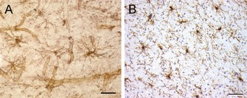
NG2 glia are widespread in all regions of the human CNS but have been most extensively studied in the cerebral cortex and white matter of adults. Like their counterparts in the rodent brain, in non-diseased human brain NG2 glia are stellate-shaped process-bearing cells which cover the CNS parenchyma. In cerebral cortex, the processes are radially oriented and symmetrical (Fig. 1), whereas in white matter processes often orient parallel to axons (data not shown). The processes of neighboring NG2 glia rarely overlap, suggesting that they may be contact inhibited. The diameter of NG2 glia in cerebral cortex can range from 50 to 150 µm. Therefore, staining of thicker sections (for example, 30 µm free-floating sections) provides a better opportunity to analyze process morphology and interactions. Comparison of Figs 1A and 2 illustrates that projections of multiple confocal microscopic images from immunofluorescence preparations provide a more detailed photographic representation of NG2 glia process morphology than immunoperoxidase preparations. Thinner sections (for example, 10 µm cryostat sections) are more appropriate for studies that require application of routine histologic stains, such as hematoxylin and eosin, to parallel sections for diagnosis of disease or where nuclear counter stain of brightfield images assists in interpretation of results (Fig. 1B). In addition, thin cryostat sections may be most appropriate for retrospective analyses where only unfixed flash frozen tissue is available for study.
NG2 glia are widespread in the human CNS. (A) Image of NG2-positive glia in cerebral cortex demonstrated by immunoperoxidase stain of a 30 µm free-floating section. NG2 expression by vascular mural cells (pericytes) is also seen. (B) NG2-positive glia in a 10 µm cryostat section. Nuclear counterstain shows that they represent 10–15% of glial cells. The overall cell density is slightly greater in panel B. This could be due to tissue shrinkage during cryoprotection followed by attachment of cryostat sections to slides or possible tissue swelling of free-floating sections. Scale bars = 50 µm.

Qualitative inspection of NG2-positive glia reveals a relatively uniform density and distribution of cells in all areas of the cerebral cortex and subcortical white matter. Detailed regional quantitative analyses have not been performed. Density (cells/mm2) of NG2 glia provides an accurate measurement of this population of cells in settings where the population of other glial cells may be affected by disease. The density of NG2-positive glial cells has been reported to be 140–150 cells/mm2 in 30 µm free-floating sections of normal appearing MS white matter (Chang et al., Reference Chang, Nishiyama, Peterson, Prineas and Trapp2000) and 155–171 cells/mm2 in 10 µm cryostat sections of control or normal appearing MS white matter (Wilson et al., Reference Wilson, Scolding and Raine2006). We have calculated a similar density in cryostat sections. By analysis of sections with nuclear counterstain (Fig. 1B), we estimate that this represents between 10 and 15% of glial nuclei and indicates that NG2 glia represent a significant proportion of CNS glial cells. This estimate is much higher than the 1–3% in tissue reported by Scolding et al. (Reference Scolding, Franklin, Stevens, Heldin, Compston and Newcombe1998) and similar yields of NG2 glia obtained by preparation of cellular suspensions and isolation using antibodies to A2B5 (Roy et al., Reference Roy, Wang, Harrison-Restelli, Benraiss, Fraser and Gravel1999; Nunes et al., Reference Nunes, Roy, Keyoung, Goodman, McKhann and Jiang2003; Ruffini et al., Reference Ruffini, Arbour, Blain, Olivier and Antel2004; Sim et al., Reference Sim, Lang, Waldau, Roy, Schwartz and Pilcher2006). Reasons for the low yield from cellular suspensions have not been systematically investigated. Possibilities include an increased sensitivity of NG2 glia to cell isolation procedures than other CNS cell types and the possibility that A2B5 is present only on a subpopulation of human NG2 glia.
There is one report comparing the density of NG2 and PDGFRA immunoreactivity in adult tissues (Wilson et al., Reference Wilson, Scolding and Raine2006). Mean PDGFRA-positive cell density was 59–64 cells/mm2 compared to 155–171 cells/mm2 for NG2-positive cells. The authors suggest that NG2-positive, PDGFRA-negative cells may represent a more mature stage, and their hypothesis is supported by studies on the developing brain (see below). It is unclear why expression of NG2 and PDGFRA in NG2 glia in human tissues would diverge so greatly from the expression pattern in rodents where co-localization is reported to be 100% (Rivers et al., Reference Rivers, Young, Rizzi, Jamen, Psachoulia and Wade2008; Zhu et al., Reference Zhu, Bergles and Nishiyama2008; Nishiyama et al., Reference Nishiyama, Komitova, Suzuki and Zhu2009). Another possibility is that available PDGFRA antibodies are not sufficiently sensitive or reliable for use in human tissues (see Technical Considerations above). Due to the number of variables that affect both NG2 and PDGFRA staining, rigorous quantitative comparisons of cell density in different blocks of tissue from different CNS regions may not be valid. Quantitative analysis of specimens that have a disease and reference control region on the same slide represents an approach that avoids many of the technical variables cited.
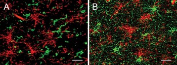
The morphology of NG2 glia is similar to, but distinct from, both microglia (Fig. 2A) and astrocytes (Fig. 2B). The published data on double labeling for NG2 and microglial antigens are mixed. Chang et al. (Reference Chang, Nishiyama, Peterson, Prineas and Trapp2000) showed no overlap between NG2-positive glia and MCH Class II-positive microglia. Wilson et al. (Reference Wilson, Scolding and Raine2006) showed no overlap between NG2-positive glia and CD68-positive microglia, but Pouly et al. (Reference Pouly, Becher, Blain and Antel1999) reported complete overlap. Double labeling of NG2 with tomato lectin also showed partial overlap (Filipovic et al., Reference Filipovic, Rakic and Zecevic2002). Data from rodent models have shown that in the setting of injury and macrophage recruitment, some macrophages display NG2 immunoreactivity (Bu et al., Reference Bu, Akhtar and Nishiyama2001; McTigue et al., Reference McTigue, Wei and Stokes2001). These macrophages are most likely of hematogenous origin (Bu et al., Reference Bu, Akhtar and Nishiyama2001). This could be a source of double labeling reported by some groups; however, we have never observed NG2 immunoreactivity in cells with the morphology of phagocytic macrophages in the many white matter lesions of MS that we have examined. Double labeling of NG2 and GFAP in human tissues has not been reported (Chang et al., Reference Chang, Nishiyama, Peterson, Prineas and Trapp2000), despite the recent observation that transgenic mice expressing the NG2 promoter can differentiate into astrocytes (Zhu et al., Reference Zhu, Bergles and Nishiyama2008).
NG2 glia in the human CNS are a distinct population of cells. (A,B) Double immunofluorescence and confocal microscopy show no overlap between NG2 glia (red) and LCA-positive microglia (A, green) or GFAP-positive astrocytes (B, green). Scale bars = 30 µm. Reproduced with permission from Journal of Neuroscience (Chang et al., Reference Chang, Nishiyama, Peterson, Prineas and Trapp2000, fig. 1E,F).

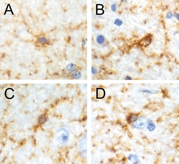
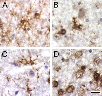
Because of the difficulties in visualizing NG2 glia in routinely processed surgical and autopsy tissues, these cells have also been under-recognized by neuropathologists. They were described in the seventh edition of Greenfield's Neuropathology (Graham and Lantos, Reference Graham and Lantos2002), the definitive text used by neuropathologists, but this was not carried into the most recent eighth edition (Love et al., Reference Love, Louis and Ellison2008). The nuclear morphology of NG2 glia is variable, and the nuclei in routine hematoxylin and eosin stained sections mimic the appearance of oligodendrocytes and astrocytes. For example, Fig. 3 shows high magnification images of NG2 glia with hematoxylin nuclear counterstain. Panel A shows a cell with a small dark round nucleus, a morphology commonly attributed to oligodendrocytes, whereas panel B shows a cell with the large pale oval nucleus that is characteristic of an astrocyte. The cells in Panels C and D have intermediate nuclear morphologies, and the one in D is a neuronal satellite cell.
The nuclear morphology of NG2 glia ranges from small round and dark (A) to large oval and pale (B). Intermediate morphologies are also seen within the parenchyma (C) and as neuronal satellite cells (D).

NG2 GLIA ARE PRESENT IN THE SECOND TRIMESTER HUMAN TELENCEPHALON
NG2 glia originate from the subventricular zone (SVZ) and migrate as unipolar and bipolar cells to colonize the entire brain. NG2 glia have been investigated in the midgestation human fetus (gestational ages from 17 to 26 weeks), and both NG2-positive and PDGFRA-positive cells could be detected (Back et al., Reference Back, Luo, Borenstein, Levine, Volpe and Kinney2001; Rakic and Zecevic, Reference Rakic and Zecevic2003; Jakovcevski and Zecevic, Reference Jakovcevski and Zecevic2005). NG2-positive cells displayed a range of morphologies from simple unipolar and bipolar to simple and complex multipolar forms (Back et al., Reference Back, Luo, Borenstein, Levine, Volpe and Kinney2001; Jakovcevski and Zecevic, Reference Jakovcevski and Zecevic2005). Both of these studies also showed double labeling with antibodies to the premyelinating oligodendrocyte antigen O4 in a subpopulation of cells. These results support the notion that NG2-positive cells in the developing human nervous system, like their rodent counterparts, represent an OPC. Jakovcevski and Zecevic (Reference Jakovcevski and Zecevic2005) also quantified the percentage of cells double labeled with NG2 and PDGFRA in different brain regions. There was nearly 100% double labeling in the SVZ. The number of double-labeled cells decreased in white matter with increasing distance from the SVZ, and in the cortical plate only 50% of the NG2-positive cells were reported to be PDGFRA-positive. Rakic and Zecevic (Reference Rakic and Zecevic2003) also reported double labeling of NG2 or PDGFRA with Dlx2 in the midgestation human cerebrum. In rodents, Dlx2 is expressed by cells of the ganglionic eminence that can produce either interneurons or oligodendrocytes (He et al., Reference He, Ingraham, Rising, Goderie and Temple2001; Marshall and Goldman, Reference Marshall and Goldman2002).
NG2 GLIA IN THE DISEASED HUMAN CNS
Multiple sclerosis
NG2 glia can be detected in a variety of human CNS diseases. The most extensively studied disease is MS. NG2 glia have been identified in active, chronic active and chronic inactive lesions in subcortical white matter (Scolding et al., Reference Scolding, Franklin, Stevens, Heldin, Compston and Newcombe1998; Nishiyama et al., Reference Nishiyama, Chang and Trapp1999; Chang et al., Reference Chang, Nishiyama, Peterson, Prineas and Trapp2000; Maeda et al., Reference Maeda, Solanky, Menonna, Chapin, Li and Dowling2001; Filipovic et al., Reference Filipovic, Rakic and Zecevic2002; Reynolds et al., Reference Reynolds, Dawson, Papadopoulos, Polito, DiBello and Pham-Dinh2002; Wolswijk, Reference Wolswijk2002; Wilson et al., Reference Wilson, Scolding and Raine2006) and in cerebral cortical lesions (Chang et al., Reference Chang, Nishiyama, Peterson, Prineas and Trapp2000; Reynolds et al., Reference Reynolds, Dawson, Papadopoulos, Polito, DiBello and Pham-Dinh2002). The relative number of NG2-positive cells in different lesion types reported by different authors varies. NG2-positive glia have been reported to be decreased (Reynolds et al., Reference Reynolds, Dawson, Papadopoulos, Polito, DiBello and Pham-Dinh2002) or unchanged (Wilson et al., Reference Wilson, Scolding and Raine2006) in active lesions and decreased (Wilson et al., Reference Wilson, Scolding and Raine2006) or variable (Reynolds et al., Reference Reynolds, Dawson, Papadopoulos, Polito, DiBello and Pham-Dinh2002) in chronic inactive lesions. NG2-positive glia have been reported to be increased (Filipovic et al., Reference Filipovic, Rakic and Zecevic2002) or unchanged (Wilson et al., Reference Wilson, Scolding and Raine2006) at the edge of lesions.
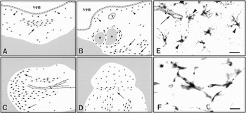
Our analyses showed a wide variation in NG2 glia density within individual chronic lesions (Chang et al., Reference Chang, Nishiyama, Peterson, Prineas and Trapp2000). NG2 glia are commonly present adjacent to the SVZ (Fig. 4A–D), supporting generation from primitive cells in the SVZ. There is no obvious decrease in NG2 glia in cortical MS lesions (Chang et al., Reference Chang, Nishiyama, Peterson, Prineas and Trapp2000; Chang, Trapp, unpublished data). These results suggest that the environment of demyelination in white matter is destructive either to the NG2 antigen or to the cell. The latter interpretation is favored by the observation of two morphologies of NG2-positive cells in chronic MS lesions. The most common morphology of NG2-positive cells was stellate (Fig. 4E), similar to that seen in non-lesion white matter, but we also observed cells with an elongated morphology that often formed interconnected networks (Fig. 4F). Bipolar cells have also been reported by others (Filipovic et al., Reference Filipovic, Rakic and Zecevic2002; Reynolds et al., Reference Reynolds, Dawson, Papadopoulos, Polito, DiBello and Pham-Dinh2002). Such morphologies are correlated with immature migrating cells (Jakovcevski and Zecevic, Reference Jakovcevski and Zecevic2005) and could represent repopulation of the lesion by OPCs capable of differentiation and remyelination. A subpopulation of these cells is also labeled with antibodies to the p75 neurotrophin receptor. This receptor can function to promote death or survival, depending upon which Trk receptor is co-expressed. We favor the latter interpretation because we were unable to identify any p75-positive, NG2-positive cells that were labeled by TUNEL (Chang et al., Reference Chang, Nishiyama, Peterson, Prineas and Trapp2000). In many chronic MS lesions, we also detected newly generated premyelinating oligodendrocytes (Chang et al., Reference Chang, Tourtellotte, Rudick and Trapp2002). This supports the notion that NG2-positive glial cells can function as OPCs in chronic MS lesions.
NG2 glia in MS lesions. (A–D) Artistic renditions of the distribution of NG2 cells in four MS lesions. The white areas are demyelinated periventricular (A,B) or subcortical (C,D) white matter, and the gray areas are myelinated white matter. NG2 cells (black cells) are distributed unevenly in the MS lesions (A–D). They have stellate (arrowheads) or elongated (arrows) shapes. ven, ventricle; V, vessels; *, myelinated areas within a periventricular lesion. (E,F) NG2-stained sections of MS lesions that contain stellate (E, arrowheads) or elongated (F) cells. Endothelial cells (E, arrow) are stained by NG2 antibodies also. Scale bars (E,F) = 30 µm. Reproduced with permission from Journal of Neuroscience (Chang et al., Reference Chang, Nishiyama, Peterson, Prineas and Trapp2000, fig. 2A–F).

Other non-neoplastic human diseases
Publications depicting NG2 glia in tissue from other human diseases are rare. Pouly et al. (Reference Pouly, Prat, Blain, Olivier and Antel2001) present an image from a temporal lobe epilepsy specimen showing immunoreactive cells with the typical stellate morphology. A second paper, focused on astrocytes in tuberous sclerosis, shows one figure that includes NG2 and PDGFRA immunolabeling to demonstrate no obvious morphological alterations in NG2 glia in this disease (Sosunov et al., Reference Sosunov, Wu, Weiner, Mikell, Goodman and Crino2008). We have examined the morphology and distribution in several types of human specimens commonly encountered in surgical and autopsy pathology. Analysis of human tissues was performed with approval of the Cleveland Clinic Institutional Review Board, which waived the requirement for informed consent. All specimens were fixed overnight in 4% paraformaldehyde, cryoprotected in sucrose, and 10 µm cryostat sections were cut and stained using the 9.2.27 monoclonal antibody.
Tissue was available from 20 patients with surgical resections for epilepsy, six with resection of metastasis and 12 patients at autopsy. Patient age ranged from 6 months to 91 years. Pathologic diagnoses for the epilepsy tissues were subpial gliosis (n = 3), focal perivascular lymphocytes (n = 3), chronic encephalitis (n = 1), non-neoplastic mass lesion (n = 3) and malformation of cortical development (n = 10). Causes of death and neuropathologic diagnoses for the autopsy tissues were: acute death (fatal cardiopulmonary arrest, no known premortem CNS disease), with no neuropathology (n = 3) or with diffuse senile plaques insufficient for diagnosis of Alzheimer's disease (n = 2), premortem sepsis and/or multiorgan failure, with no neuropathology (n = 2), with remote infarcts (n = 2), with metabolic gliosis (Alzheimer Type 2 astrocytes) (n = 1), global cerebral ischemia and coma × 1 week (n = 1), cerebral hemorrhage and coma × 1 week (n = 1).
NG2 glia could be detected in all cases examined, but the signal intensity varied with the degree of pathology seen by routine hematoxylin and eosin stain. NG2-immunoreactivity was readily identifiable in specimens that contained any kind of discreet structural abnormality or evidence of significant inflammation or gliosis. They were not readily identified in three epilepsy resection specimens that showed minimal pathology (subpial gliosis or mild focal perivascular infiltrates); however, application of the Tyramide Substrate Amplification procedure (Invitrogen, Carlsbad, CA) increased sensitivity and enabled detection of NG2 glia.
The morphology of NG2 glia also changes with degree of pathology by routine stain. For example, Fig. 5 shows the morphology of NG2-positive cells within a region of subacute infarction and in the adjacent penumbra. NG2 glia in the penumbra have multiple long thin processes (Fig. 5A), whereas the processes are shorter and thicker in the infarcted area (Fig. 5B). The morphological changes seen in NG2-positive cells correlate with changes in other reactive cell populations, such as microglia and astrocytes. Figure 5C shows ramified microglia in the penumbra and ameboid microglia and phagocytic macrophages in the infarct (Fig. 5D).
Morphology of NG2 glia and MHC Class II-positive cells in subacute infarction. NG2- and MHC Class II-positive cells are present in the penumbra and the region of infarction, but their morphologies differ. The NG2-positive cells in the penumbra display multiple delicate processes characteristic of NG2 glia in regions without pathology (A, cf. Fig. 1), but they have fewer, shorter and thicker, processes in the infarcted region (B). The MHC Class II-positive cells have the morphology of ramified microglia in the penumbra (C) and phagocytic macrophages in the infarct (D). Scale bar = 20 µm.

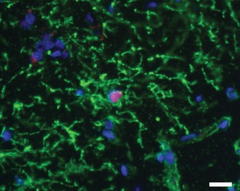
The reactive changes seen in NG2 glia, as well as the evidence from animal models that NG2 glia are a cycling OPC population, led us to investigate whether there is evidence for their proliferation in the human brain. We performed double labeling for NG2 and Ki-67 on 13 specimens where the NG2 immunolabeling was sufficiently intense by simple indirect immunofluorescence. Ki-67 is a nuclear antigen that is highly expressed in cycling cells and rapidly down-regulated in quiescent cells (Bullwinkel et al., Reference Bullwinkel, Baron-Luhr, Ludemann, Wohlenberg, Gerdes and Scholzen2006). It is currently the most commonly used proliferation marker in human diagnostic pathology. We were able to identify double-labeled cells in 6/9 epilepsy specimens, but not in tissue from each of two autopsy and metastasis cases (Fig. 6, Table 1). A proliferation index was not calculated for NG2 glia specifically. In most of our specimens, fewer than 0.1% of total nuclei are Ki-67-positive.
NG2 glia (green) can express the nuclear proliferation antigen Ki-67 (red). Blue = DAPI nuclear marker. Scale bar = 20 µm.

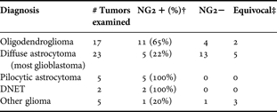
Glial and glioneuronal neoplasms examined for NG2 immunoreactivity*.
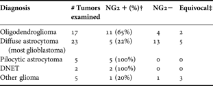
* Only tissue blocks with histologically recognizable neoplastic mass lesions were assessed for NG2 immunoreactivity. Any specimens showing only infiltrating edge of neoplasm were excluded.
† The majority of cells recognizable as neoplastic labeled with NG2 antibodies.
‡ Neoplasms were assessed as equivocal for NG2 immunoreactivity if only rare cells were positive or if immunoreactivity was a diffuse cytoplasmic, rather than a plasma membrane, distribution.
These data provide evidence that NG2 glia have the capacity to proliferate in the adult human brain.
Gliomas
Gliomas are a diverse group of primary CNS neoplasms that display a wide variety of histopathology and biological behavior. Gliomas are classified by the mature cells they resemble, e.g. astrocytoma and oligodendroglioma, but the cell or cells from which any specific glioma arises is unknown. Since the first histologic description of gliomas (Bailey and Cushing, Reference Bailey and Cushing1926), and particularly in the past decade (Shoshan et al., Reference Shoshan, Nishiyama, Chang, Mork, Barnett and Cowell1999; Sanai et al., Reference Sanai, Alvarez-Buylla and Berger2005; Canoll and Goldman, Reference Canoll and Goldman2008), there has been growing speculation that gliomas are more likely to arise from an immature or cycling cell capable of acquiring and propagating genetic alterations in growth-regulating genes than from a mature non-cycling cell. Among glial cells, OPCs are the best characterized and most studied. Both NG2 and PDGFRA have been detected on glioma cells in tissue specimens, but as described in other parts of this report, there are technical obstacles to their application to routinely processed diagnostic specimens. The first report on NG2 immunoreactivity in human glioma tissue was by Schrappe et al. (Reference Schrappe, Klier, Spiro, Waltz, Reisfeld and Gladson1991), who examined glioblastomas. The most prominent NG2-positive cell type was endothelial cells, but weak focal immunoreactivity on neoplastic cells was reported. We subsequently reported NG2 and PDGFRA immunoreactivity, with confirmation by Western blot, on 16 gliomas (Shoshan et al., Reference Shoshan, Nishiyama, Chang, Mork, Barnett and Cowell1999). Either NG2, PDGFRA, or both were expressed by 7/7 oligodendrogliomas, 1/5 glioblastomas and 3/3 pilocytic astrocytomas. One grade II diffuse astrocytoma was negative. Follow-up analysis of 52 additional independent neoplasms (Table 1) confirmed that NG2 immunoreactivity was more common in oligodendrogliomas than diffuse astrocytomas and is commonly encountered in WHO Grade I neoplasms characterized as ‘astrocytic’ (pilocytic astrocytomas) or ‘glioneuronal’ (dysembryoplastic neuroepithelial tumors, DNET). Our data on pilocytic astrocytomas are supported by an independent report of PDGFRA immunoreactivity in 37/64 (58%) of these neoplasms (Takei et al., Reference Takei, Yogeswaren, Wong, Mehta, Chintagumpala and Dauser2008).
The expression of NG2 and PDGFRA in glial and glial neuronal neoplasms can be interpreted as evidence for gliomas arising from an immature cycling cell from the oligodendrocyte lineage. This is supported by the frequent observation of Olig2 expression in a wide variety of gliomas (Ligon et al., Reference Ligon, Alberta, Kho, Weiss, Kwaan and Nutt2004). Whether or not such expression has prognostic or therapeutic significance is debatable because it is observed in neoplasms from all WHO Grades. There is increasing evidence that genetic and epigenetic alterations within different primary CNS neoplasms play an important role in their biological behavior. Nevertheless, the biological effect of any molecular alteration is manifested in the context of the gene expression profile of the cell in which the alteration exists, so there remains the possibility that expression of NG2 glia antigens in neoplasms may have clinical significance.
DETECTION OF NG2 GLIA – TECHNICAL ADVANCES
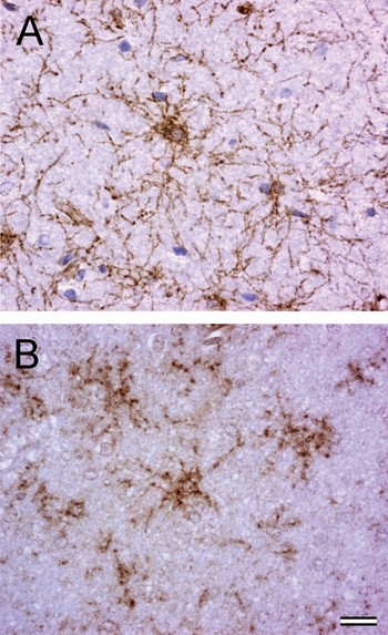
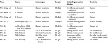
Our understanding of NG2 glia in humans can best be advanced by identification of reagents that can reliably identify these cells in specimens fixed for routine pathology examination, i.e. formalin-fixed paraffin-embedded (FFPE) tissue. To this end, we assembled a panel of antibodies (Table 2) known to react with the NG2 antigen and screened them on control tissue (skin containing NG2-positive vascular cells) that was formalin-fixed overnight and paraffin embedded. Only one antibody, a rabbit polyclonal prepared to a recombinant protein representing the extracellular domain of NG2 (NG2 EC, Table 2), showed valid reactivity. Both heat-induced epitope retrieval and enzymatic tyramide substrate amplification techniques were required to detect NG2 with this antibody in these tissues. Prolonged formalin fixation (greater than 24 h) severely impaired immunoreactivity. We then applied this antibody to 15 specimens for which comparable paraformaldehyde-fixed cryoprotected frozen tissue and FFPE tissue was available. The NG2 immunoreactivity in FFPE tissue was judged as a good representation of the PFA fixed counterpart in 10/15 comparisons (Fig. 7). We were able to achieve immunoreactivity similar to that seen in paraformaldehyde-fixed tissue in paraffin sections, but only after using tyramide substrate amplification. The specificity of immunolabeling was confirmed by elimination of staining of parallel sections with primary antibody that was pre-incubated with B49 cells that express rat NG2. Even in these cases, staining in FFPE tissue was often non-uniform across the specimen, and appreciation of the complex process morphology was limited by the relative thinness of paraffin sections. The observation that the only antibody that showed utility in FFPE tissues was a polyclonal antibody generated to the extracellular domain indicates that recognition of multiple epitopes in the context of molecular secondary structure is essential for detection of NG2 in routinely processed tissues. Hopefully, the observations reported here will stimulate investigators or industry to develop additional antibody reagents of this type.
Comparison of morphology of NG2 glia in paraformaldehyde-fixed 10 µm cryostat sections (A) and FFPE 6 µm sections (B). The appearance is similar; however, the complex process morphology is better demonstrated in the paraformaldehyde-fixed sections. Detection of NG2 in FFPE sections required tyramide substrate amplification. Scale bar = 20 µm.

Sources of NG2 antibodies evaluated for reactivity in FFPE tissues.

These antibodies were initially screened on cryostat sections of human skin and rodent brain fixed in 4% paraformaldehyde by immunohistochemistry using Vectastain ABC reagent and DAB detection. Antibodies reactive with vascular cells in human skin were confirmed for reactivity with NG2 glia in human brain and then tested on FFPE human skin.
CONCLUSIONS AND FUTURE PERSPECTIVES
NG2 glia represent a numerically significant population of cells in the human CNS, and their morphological changes in the setting of disease suggest that they may play a role in pathogenesis and/or repair. Active investigation of NG2 glia function in animal models is critical for our increased understanding of the role that these cells can play in human disease and for the development of more robust markers for their detection in routinely processed human specimens.
ACKNOWLEDGEMENTS
We thank Drs. Michael Obiwoyi and Ansi Chang for immunohistochemistry and photography of the figures presented in this report, and Drs. William Stallcup, Burnham Institute for Medical Research, and Soldano Ferrone, University of Pittsburgh, for antibodies. This work was supported by NIH R01 NS 29818 (Trapp).
Statement of interest
None.