Implications
Adipose tissue is an organ of energy storage, but also an endocrine organ regulating body homeostasis. The mass of adipose tissue is largely determined by the number and size of adipocytes. The increase in cell number results from recruitment of multipotent stem cells/progenitors resident in tissues. A better knowledge of the cellular and molecular basis of adipose tissue development should help us to develop new strategies to optimize the management of the lean-to-fat ratio in farm animals and thus to improve production efficiency.
Introduction
The developmental biology of adipose tissue is a topic of great interest in the field of animal production sciences. Indeed, the surplus or extra energy of feed after digestion processes is mainly converted into fat stored in different parts of the body. Because animals are intermittent feeders, short and long-term managements of energy resources are important elements for survival, adaptation and robustness. Body fat also acts as a barrier against cold, participates to immunity and influences reproductive ability such as fertility, pregnancy outcomes and lactation (Norgan, 1997). Lastly, the control of body fat content and distribution is of the upmost importance for different values of meat producing animals (Sillence, 2004). Because fat has a high-energy cost of deposition, any lipid depots in excess are energetically unfavorable and reduce production efficiency. The lean to fat ratio also determines carcass grading in different species, and greater body fatness decreases the payments granted to producers and increases the costs of carcass trimming in slaughtering and packing plants. Moreover, excessive consumption of animal products in some social classes and countries has been suspected of leading to excessive intake of fat detrimental to human health. In pork (Fernandez et al., 1999) and beef (Hausman et al., 2009), flavor intensity and consumers’ acceptability of meat are however positively related to intramuscular (IM) lipid content, so that the IM fat often remains a desired component for taste. Altogether, there is a need to identify the key elements that control adipose tissue deposition and distribution in different locations to optimize the volume of meat produced while maintaining high quality standards of meat products.
Adipose tissue development is basically triggered by variations in the number and size of adipose cells. These cells are recruited from various populations of multipotent stem cells/progenitors. In the past two decades, there has been an explosion of research dealing with the biology of stem cells derived from adipose tissue, showing that a diverse subset of multipotent stem cells can contribute to adipose tissue homeostasis (Lafontan, Reference Lafontan2012; Dodson et al., Reference Dodson, Allen, Du, Bergen, Velleman, Poulos, Fernyhough-Culver, Wheeler, Duckett, Young, Voy, Jiang and Hausman2015). The diversity of stem cell populations, the biological fates of each of these populations depending on the tissues where they are issued, and the flexibility in abundance and properties of these cell populations are thus bottlenecks to consider for a better understanding of the mechanisms governing adipose tissue development. However, one important point to consider is that available data are based mainly on the investigation of adult tissues and are dealing with cell-based therapies in human and veterinary medicine (Gimble et al., 2007; Casteilla et al., Reference Casteilla, Planat-Benard, Laharrague and Cousin2011; Arnhold and Wenisch, 2015). In contrast, farm animals are often taken to slaughter before they reach full maturity, so that dedicated studies are needed. It is thus essential to investigate the sequence of events associated with adipose tissue development during prenatal and neonatal life in domestic animals. In view of the numerous papers dealing with adipose cell biology, this review cannot be exhaustive. It is an update of cellular and molecular mechanisms associated with white and brown adipose tissue development in farm animals, with special emphasis on the impact of early nutrition on later expansion of adipose tissue mass. Data obtained in human or rodents will be also considered when data in domestic species are missing.
Overview of structure, location and functions of adipose tissues
There are two main types of adipose tissue with differences in morphology, location and functions in mammals: white (WAT) and brown (BAT) adipose tissues. As further described below, WAT and BAT contain mainly white and brown adipocytes, respectively. Recently, another type of brown adipocyte-like cells, so called beige/brite cells, has been also described in different species (Giralt and Villarroya, Reference Giralt and Villarroya2013).
White adipose tissue appears as a number of individual depots throughout the body, some in the abdominal cavity (generally surrounding viscera such as mesenteric and perirenal fat depots, around epididymis, etc.), some under the skin (subcutaneous depots) and some within the musculature (inter- and intramuscular depots), these depots having large differences in size and in relative quantitative importance depending on age and species (Komolka et al., Reference Komolka, Albrecht, Wimmers, Michal and Maak2014). For instance, the predominant adipose tissue depot is subcutaneous AT in growing pigs (Kouba et al., 1999; Kouba and Bonneau, 2009) whereas it is intermuscular adipose tissue in cattle (Robelin, 1981; Cianzio et al., 1985).
Brown adipose tissue has been originally described in hibernating animals and in small non hibernating mammals like rodents (Cannon and Nedergaard, 2004). It has been also found in neonates of large mammalian species and more recently, in adults (Lee et al., Reference Lee, Swarbrick and Ho2013). As observed in human, BAT depots in sheep are mainly found in the supraclavicular/neck region and in the pericardial and perirenal regions (Symonds et al., Reference Symonds, Pope and Budge2015). Perirenal, pericardial and peritoneal adipose tissues in calf neonates have been essentially recognized as brown (Casteilla et al., 1989; Martin et al., 1999; Smith et al., 2004), but recent studies revealed that fetal bovine perirenal adipose tissue may have much more in common with WAT than with BAT (Taga et al., Reference Taga, Chilliard, Meunier, Chambon, Picard, Zingaretti, Cinti and Bonnet2012a and Reference Taga, Chilliard, Picard, Zingaretti and Bonnet2012b). Two recent studies also indicate that ovine adipose tissue undergoes pronounced changes in cell composition and gene expressions between mid-gestation and 1 month of age (Pope et al., 2013; Basse et al., Reference Basse, Dixen, Yadav, Tygesen, Qvortrup, Kristiansen, Quistorff, Gupta, Wang and Hansen2015), leading to a gradual disappearance of brown adipocytes. The pig neonate with its lack of BAT represents an exception among mammals (Trayhurn et al., 1989; Jastroch and Andersson, Reference Jastroch and Andersson2015).
Both WAT and BAT are recognized to be differently involved in energy metabolism (Himms-Hagen, 1990; Ailhaud, 1992). White adipose tissue has been considered for many years as a preferential site to store energy in the form of triacylglycerols during excessive energy disposal and to restore it during fasting periods. It also forms an insulating layer with a protective function. Brown adipose tissue is a site of energy expenditure, because it metabolizes both fatty acids and glucose to produce heat; it participates to non-shivering thermogenesis. The unique thermogenic capacity of BAT results from the expression of uncoupling protein 1 (UCP1) in the mitochondrial inner membrane, which uncouples the mitochondrial proton gradient from ATP synthesis and generates heat (Cannon and Nedergaard, 2004). The UCP1 gene has been shown to be disrupted in the pig lineage (Berg et al., 2006), providing an explanation for the lack of BAT in this species.
These traditional functions of adipose tissues have also been extended after the discovery of different secretory products by white and brown adipocytes, so that adipose tissues are now recognized as playing much more important roles in whole-body physiology than was previously thought. The discovery of leptin as a hormone produced by white adipocytes in mice (Zhang et al., 1994) and other species including farm mammals (Barb et al., 2001; Chilliard et al., 2005) has initiated this process. In addition, cells other than white adipocytes also contribute to the secretory function of WAT (Fain et al., 2006). Over the past years, the identification of multiple other secretory products termed adipokines or adipocytokines, has now allowed considering the WAT as a true secretory or endocrine organ (Romacho et al., Reference Romacho, Elsen, Röhrborn and Eckel2014). It is important to note that the list of proteins secreted by WAT is still expanding including in livestock species (Komolka et al., Reference Komolka, Albrecht, Wimmers, Michal and Maak2014; Restelli et al., Reference Restelli, Codrea, Savoini, Ceciliani and Bendixen2014). These adipokines function as classic circulating hormones to communicate with other organs including brain, liver, muscle, the immune system, and adipose tissue itself. These molecules control eating behavior, peripheral insulin sensitivity and even the development of the female reproductive system. Accumulating evidence indicates that BAT similarly releases many signaling molecules that act either on brown adipocytes themselves (autocrine action) or on other cell types nearby (paracrine action). Some of these released molecules are common to WAT notably after thermogenic stimuli (e.g. IGF-I, interleukin-1/6) but the main adipokines produce by WAT (leptin, adiponectin) are poorly expressed in BAT, so that BAT has its own pattern of secreted factors to control distinctive biological actions (Villarroya et al., Reference Villarroya, Cereijo and Villarroya2013).
A detailed description of the diversity of cells in adipose tissues
When isolated from WAT or from BAT by mechanical dissection and chemical digestion (collagenase), cells can be divided into two main groups: mature adipocytes and cells from the stromal vascular fraction (SVF). Mature adipocytes with their high triglyceride content are floating in the upper phase. The SVF cells found in the pellet contain a variety of cell types, including adipose progenitors that can differentiate into adipocytes in culture (e.g. Gerfault et al., 1999 in pigs; Grant et al., 2008 in bovine; Ma et al., Reference Ma, Hou, Dahanayaka, Satterfield, Burghardt, Bazer and Wu2015 in sheep), fibroblasts, vascular cells and immune cells. The relative amounts of the various cells have been estimated in human adipose tissue even though these proportions can vary substantially according to age, anatomical location, BW and a variety of other factors (Hauner, 2005): mature adipocytes comprise the majority of the tissue volume and represent about 50% to 70% of the total cells in adipose tissue, while progenitors represent 20% to 40% of the total cells.
Features of mature adipocytes
As previously mentioned, mature adipocytes are subdivided into white and brown adipocytes with drastically different phenotypes (Table 1). White adipocytes are spherical cells with a wide range of diameters (10 to 250 µm; Hood and Allen, 1973) and are characterized by the presence of a large lipid droplet/vacuole. Adipocytes from different WAT are not strictly identical in size, metabolic and secretory properties. Intramuscular adipocytes display the largest differences compared with subcutaneous and perirenal adipocytes in pigs (Gardan et al., 2006 and 2007; Gondret et al., 2008) and bovine (Schoonmaker et al., 2004; Bonnet et al., 2007). Their lower diameters are associated with a lower lipogenic, lipolytic, fatty acid oxidative, fatty acid transport and(or) energy transfer capacities compared with larger adipocytes isolated from subcutaneous or perirenal adipose tissue in growing pigs and steers. The IM adipocytes also distinguish themselves by the expression profiles of genes encoding for leptin, adiponectin and various hormonal receptors. Interesting characteristics may be also the lower expression of the IGF1 gene but the greater expression of the IGF2 gene in IM adipocytes as compared with adipocytes isolated from subcutaneous and perivisceral adipose tissues (Gardan et al., 2006). These data support the view that triggering adipogenesis rather than cell metabolism per se might be a valuable strategy to control lipid deposition in skeletal muscles.
White adipocyte features compared with brown and beige adipocytes

Brown adipocytes are smaller and more elongated than white adipocytes and contain multiple and small lipid droplets (Cannon and Nedergaard, 2004). Mitochondria are large and numerous in brown adipocytes, and expression of UCP1 is generally recognized as the molecular hallmark of these adipocytes. Recently, a third type of adipocytes has emerged in WAT in response to cold and hormonal stimuli in mice and human (Sanchez-Gurmaches and Guertin, Reference Sanchez-Gurmaches and Guertin2014): the beige/brite adipocytes. These beige adipocytes, as brown adipocytes, store lipids as small droplets and have the potential to express UCP1, so that they are considered as a new thermogenic cell type. However, thermogenic genes in these cells are expressed at low levels in basal state, although they can be induced at levels quite similar to those reported in brown adipocytes under hormonal stimuli. The presence of beige adipocytes has been also suggested in white adipose depots of fattening cattle (Asano et al., Reference Asano, Yamada, Hashimoto, Umemoto, Sato, Ohwatari, Kanamori, Terachi, Funaba and Matsui2013) and sheep (Pope et al., Reference Pope, Budge and Symonds2014).
Adipose tissue-derived stromal vascular fraction: a heterogeneous population of cells
The SVF obtained from adipose tissue contains a heterogeneous population of progenitors. Besides pre-adipocytes which are defined as immature cells already engaged in the adipogenic lineage, it also includes adult mesenchymal stem cells (MSCs) also known as adipose tissue-derived stromal cells (ADSCs; Casteilla et al., Reference Casteilla, Planat-Benard, Laharrague and Cousin2011; Cawthorn et al., Reference Cawthorn, Scheller and Macdougald2012). A common characteristic of all stem cells is that they can self-renew (make copies of themselves) and differentiate into various cell types when cultured in defined media (Figure 1). Contrary to embryonic stem cells which are pluripotent cells (i.e. they are able to give rise to all cell types of the body) found in the blastocyst, MSCs are multipotent (i.e. they can develop into more than one cell type, but are more specialized than pluripotent cells). They were originally isolated from bone marrow but they reside in most fetal and adult tissues of the body where they are involved in growth, expansion and repair mechanisms (Zuk et al., 2001; Kolf et al., 2007). Among other MSCs, the investigation of ADSCs is quite recent but is of considerable interest because of their quantity and accessibility compared with bone marrow cells. They share many biological similarities with bone marrow MSCs but exhibit some specificity as shown in pigs (Monaco et al., Reference Monaco, Bionaz, Rodriguez-Zas, Hurley and Wheeler2012).
An overview of terms used to categorize stem cells during development.

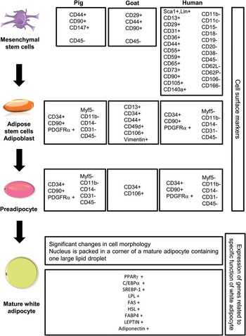
The characterization of ADSCs is based on the expression of several clusters of differentiation (CD) markers (Figure 2) using flow cytometry and fluorescence-activated cell sorting (FACS). This technology requires the use of antibodies raised against these cell surface proteins. Because there is no complete consensus on the antigen expression pattern of these cells, the use of a number of markers has been recommended for a basic characterization of ADSCs and other SVF cells in human and rodents (Planat-Benard et al., 2004; Bourin et al., Reference Bourin, Bunnell, Casteilla, Dominici, Katz, March, Redl, Rubin, Yoshimura and Gimble2013), where most of the knowledge has been acquired. The cell surface markers include CD11b and CD14 for innate immune cells, CD31 for endothelial cell, CD34 for hematopoietic stem cells, CD45 for hematopoietic cells, CD56 for satellite cells and CD90 /Thy’s-1 for mesenchymal stem cells. Data obtained in human muscle have shown that the combination of CD34 and CD56 markers allow the discrimination between adipogenic and myogenic progenitors, respectively (Pisani et al., Reference Pisani, Clement, Loubat, Plaisant, Sacconi, Kurzenne, Desnuelle, Dani and Dechesne2010). Moreover, platelet derived growth factor receptor alpha (PDGFRα) has recently been shown to distinguish adipogenic from non adipogenic cells among interstitial cells in murine muscle (Uezumi et al., Reference Uezumi, Fukada, Yamamoto, Takeda and Tsuchida2010). Because available tools have been rather limited until recently in livestock species (Rozemuller et al., Reference Rozemuller, Prins, Naaijkens, Staal, Bühring and Martens2010), few data have been obtained in pigs (Perruchot et al., Reference Perruchot, Lefaucheur, Barreau, Casteilla and Louveau2013) and ruminants (Ren et al., Reference Ren, Wu, Zhou, Wen, Jin, Cang, Guo, Wang, Liu and Ma2012; Sampaio et al., Reference Sampaio, Chiaratti, Santos, Bressan, Sangalli, Sá, Silva, Costa, Cordeiro, Santos, Ambrosio, Adona, Meirelles, Miranda and Ohashi2015). Clear advancement was allowed by the selection of antibodies against CD34, CD45, CD31, CD56, CD11b, CD14, CD90 and PDGFRα markers in porcine adipose tissue and skeletal muscle (Perruchot et al., Reference Perruchot, Lefaucheur, Barreau, Casteilla and Louveau2013). Cell populations from these tissues are negative for CD11b, CD14, CD31 and CD45 markers. As expected, adipose tissue appears to be a less heterogeneous tissue than skeletal muscle with two main populations, CD90+/CD34− and CD90+/CD56−in adipose tissue and more than five in skeletal muscle using the same markers. The relative low proportion of CD34+cells in adipose tissue of pigs agrees with other data obtained in equine adipose tissue (Ranera et al., Reference Ranera, Lyahyai, Romero, Vázquez, Remacha, Bernal, Zaragoza, Rodellar and Martín-Burriel2011) but differ from most reports in human considering adipose tissue as a large reservoir of adult stem cells (24% to 90% of SVF) identified as CD34+/CD31− cells. These discrepancies may arise from methodological aspects (freshly isolated native SVF cells v. cultured cells), and from differences in studied subjects (growing v. adult, normal-weight v. overweight and obese).
White adipocyte development from multipotent mesenchymal stem cells. Specific markers of each cell type and genes expressed in white adipocytes are described in pig, goat and human (adapted from Cawthorn et al., Reference Cawthorn, Scheller and Macdougald2012; Ren et al., Reference Ren, Wu, Zhou, Wen, Jin, Cang, Guo, Wang, Liu and Ma2012; Bourin et al., Reference Bourin, Bunnell, Casteilla, Dominici, Katz, March, Redl, Rubin, Yoshimura and Gimble2013; Perruchot et al., Reference Perruchot, Lefaucheur, Barreau, Casteilla and Louveau2013).

Origin of stem cells in adipose tissues
The origin of ADSCs and the detailed events leading to the early commitment of embryonic stem cells towards the adipocyte lineage are known only partially, with available data mainly obtained in rodents but not in farm animals. It is admitted that the types of progenitors, and even the embryonic origin of these cells, depend on adipocyte types and anatomical location of adipose tissues (Chau et al., Reference Chau, Bandiera, Serrels, Martinez-Estrada, Qing, Lee, Slight, Thornburn, Berry, McHaffie, Stimson, Walker, Chapuli, Schedl and Hastie2014; Sanchez-Gurmaches and Guertin, Reference Sanchez-Gurmaches and Guertin2014). Based on the Myf5 markers (an early marker of the myogenic lineage), MSC can be divided into two groups: the Myf5− progenitors can differentiate into white and beige adipocytes (Wu et al., Reference Wu, Boström, Sparks, Ye, Choi, Giang, Khandekar, Virtanen, Nuutila, Schaart, Huang, Tu, van Marken Lichtenbelt, Hoeks, Enerbäck, Schrauwen and Spiegelman2012), while the Myf5+progenitors can give rise to brown adipocytes and to muscle cells (Timmons et al., 2007; Crisan et al., 2008). Consistent with this, cells derived from classical BAT in primary cultures express at low levels some gene characteristics of skeletal muscle (Timmons et al., 2007). This situation is however complicated by the fact that white and brown adipocytes could also differentiate from a common progenitor at certain WAT locations (Xue et al., 2007, Seale et al., 2008; Tseng et al., 2008). Lineage tracing studies have shown that cells expressing PDGFRα can give rise to both white and brown adipocytes in adult WAT (Lee and Granneman, Reference Lee and Granneman2012). The multiple embryonic origins of white adipocytes are underlined by the fact that in mice, half of the fat cells of the retroperitoneal adipose tissue derives from progenitors expressing Myf5, a marker of the paraxial mesoderm (Sanchez-Gurmaches and Guertin, Reference Sanchez-Gurmaches and Guertin2014) but the other half derives from progenitors expressing the Wilms tumor 1 gene (Wt1), a marker of the intermediate mesoderm (Chau et al., Reference Chau, Bandiera, Serrels, Martinez-Estrada, Qing, Lee, Slight, Thornburn, Berry, McHaffie, Stimson, Walker, Chapuli, Schedl and Hastie2014). These authors indicate that Wt1 is expressed in several visceral fat depots but not in subcutaneous WAT. The neural crest of ectodermic origin can also produce cephalic white fat cells in the mouse (Billon et al., 2007). Fat white cells in the trunk that are formed near blood vessels (Hausman and Richardson, 2004) can derive from endothelial progenitors (Tang et al., 2008). Finally, the precursor(s) of white adipocytes in skeletal muscle have not been determined yet.
From multipotent stem cells to adipocytes
The process whereby pre-engaged progenitors further develop into functioning adipocytes refers to adipogenesis (Figure 2). It is characterized by changes in cell morphology. It includes proliferation (i.e. an exponential growth phase) and differentiation processes. These events are contrary to each other because the former requires cell cycle activity, whereas the latter requires cell cycle withdrawal, so that cell cycle progression and cell cycle arrest are two major cellular events for the balance between ADSCs proliferation and differentiation (Grégoire et al., 1998). Although most of the research in the field of adipogenesis has been performed with immortalized murine cell lines, some data have been also obtained in livestock species using cells isolated from adipose tissue and cultured in vitro. Some mechanisms have been also described in vivo using successive sampling of animal tissues along fetal and postnatal development. The main mechanisms seem common to all species but the kinetics of expression of key transcriptional regulators and actors may slightly vary, as shown in pigs (e.g. Ding et al., 1999; Kim et al., 2000; McNeel et al., 2000). The transcriptional program is also largely similar for the white and brown adipocytes (Koppen and Kalkhoven, Reference Koppen and Kalkhoven2010), and involves many genes that are sequentially under- or over-expressed as revealed by expression studies including high-throughtput omics technologies (see for pigs Monaco et al., Reference Monaco, Bionaz, Rodriguez-Zas, Hurley and Wheeler2012; and for cattle, Taga et al., Reference Taga, Chilliard, Meunier, Chambon, Picard, Zingaretti, Cinti and Bonnet2012a). A concerted cooperation of numerous transcription factors together with their coactivators and corepressors is also needed. Only key points of these complex events are briefly summarized here. Increased expressions in cyclin-related cell cycle inhibitors (Zhang et al., Reference Zhang, Suh, Choi, Ahn, Davis and Lee2014) but decreased expression in DLK1/Pref1 (Gondret et al., Reference Gondret, Père, Tacher, Daré, Tréfeu, Le Huërou-Luron and Louveau2013), a factor expressed in preadipocytes and known to inhibit adipogenesis, and in IGF2 (Gardan et al., 2008), a fetal mitogen highly expressed during the proliferative phase that prevents leptin induction, have been enlightened during early development of adipose tissues in pigs. Transient increased abundances of cyclin-related cell cycle partners or other proteins have been also observed in bovine fetal adipose tissue. For example, the low abundance in YWHAB, YWHAE and YWHAG proteins may favor cell cycle progression to increase adipocyte number, while their high abundances in enlarged adipocytes may balance cell cycle progression to cell cycle arrest (Taga et al., Reference Taga, Chilliard, Meunier, Chambon, Picard, Zingaretti, Cinti and Bonnet2012a). Concomitantly, a decreased abundance of PTGR2 and HSPD1, two proteins that have been rarely identified in adipose tissue so far, may sustain proliferative phase in adipose tissue development by delaying preadipocyte differentiation through inactivation of the peroxisome proliferator-activated receptor gamma (PPARG, Taga et al., Reference Taga, Chilliard, Meunier, Chambon, Picard, Zingaretti, Cinti and Bonnet2012a). In all species, PPARG is indeed considered as one of the master regulators of adipocyte differentiation; it cooperates with other transcription factor families including three members of the CCAAT/enhancer-binding protein (C/EBP) family and SREBF1 (Hausman et al., 2009). Up-regulations of PPARG-induced proteins induce lipid transport, lipid metabolism, insulin signaling and adipokine production (e.g. Samulin et al., 2009 in pigs) and result in adipocyte volume increase. Subtle differences in activity or expression of few other PPARG co-regulators such as PGC1-α (PPARGC1A) may further explain the specification between white and brown adipocytes (Koppen and Kalkhoven, Reference Koppen and Kalkhoven2010). In vitro studies have largely demonstrated that the various steps of adipocyte differentiation are under the control of hormones and growth factors (Grégoire et al., 1998; Louveau and Gondret, 2004; Hausman et al., 2009; Obregon, Reference Obregon2014).
Prenatal and postnatal growth of adipose tissues
The chronologies of appearance of WAT and BAT vary according to species and their anatomical locations. The first groups of adipocytes located in the anatomical sites of the future fat depots are detected during the fetal period in mammals of large size, while adipose tissues are hardly detectable before birth in rodents (Hausman et al., Reference Hausman, Basu, Wei, Hausman and Dodson2014). In pigs, the first groups of adipocytes appear subcutaneously between 50 and 75 days of gestation (43% to 65% of gestation; Hausman and Kauffman, 1986), then, perirenal adipose tissue develops at approximately 70 days of gestation (61% of the gestation; Hausman and Thomas, 1986). The first IM adipocytes appear during the first postnatal month (Hauser et al., 1997). Although fetal fat gain in the body accelerates from 69 days post-conception onwards, pig neonates like sheep are characterized by a small amount of total body fat at birth (1% to 2% of the live weight; Frondas-Chauty et al., Reference Frondas-Chauty, Louveau, Le Huërou-Luron, Rozé and Darmaun2012), contrasting with the situation in human neonates (~10%; Carberry et al., Reference Carberry, Colditz and Lingwood2010). At birth, the percentage of multilocular adipocytes is very high, but by day 3 postpartum, many unilocular adipocytes (one major central lipid droplet) are already observed (Hauser et al., 1997). Contrary to pigs, the first groups of adipocytes in cattle are located around the kidney (perirenal fat depot) and appear at 110 days of gestation (40% of gestation); then, adipocytes are apparent at the intermuscular or subcutaneous level at approximately 180 days of gestation (66% of gestation; Vernon, 1986). Intramuscular fat appears after three months of age in cattle, so that IM fat is generally regarded as the last developing adipose tissue in all species (Bonnet et al., Reference Bonnet, Cassar-Malek, Chilliard and Picard2010). In bovine, adipose tissues represent 4% to 7% of the live weight at birth (Robelin and Casteilla, 1990). The bovine perirenal and intermuscular adipose tissues are heterogeneous and consist of both brown and white adipocytes around birth (Taga et al., Reference Taga, Chilliard, Meunier, Chambon, Picard, Zingaretti, Cinti and Bonnet2012a and Reference Taga, Chilliard, Picard, Zingaretti and Bonnet2012b), while subcutaneous bovine adipose tissue would be white from its formation (Alexander et al., 1975; Casteilla et al., 1989).
The expansion in mass of adipose tissue is predominating after birth in pigs as well as in ruminants (Robelin, 1981; Cianzio et al., 1985; Kouba et al., 1999; Kouba and Bonneau, 2009). For instance, adipose tissues represent between 7% and 35% of the live weight in young bullocks White Belgian Blue and Japanese Black, respectively, at the age of commercial slaughter (24 months; Gotoh et al., 2009). The postnatal growth of adipose tissue results from an increase in the number (hyperplasia) and especially in the size (hypertrophy) of adipocytes.
Unlike other tissues, adipose tissue mass has considerable capacity to expand throughout life as underlined in studies comparing lean and obese subjects (Hausman et al., 2001; Bonnet et al., Reference Bonnet, Cassar-Malek, Chilliard and Picard2010). The number of adipocytes is set during adolescence in human (Spalding et al., 2008) or in 100 kg of live weights in bovine with some differences between early and late adipose tissues (Bonnet et al., Reference Bonnet, Cassar-Malek, Chilliard and Picard2010). Weight variations in the 20% range over periods of several months/years, do not modify the total number of fat cells in obese or normo-balanced individuals (Spalding et al., 2008) which suggests the presence of mechanisms preserving tissue homeostasis of an individual.
As adipocytes are terminally differentiated cells unable to divide, changes in adipose mass in adult animals are mostly due to changes in their metabolic capacities. However, adipocyte number can increase with the sustained capacities of proliferation/differentiation of ADSCs to maintain/self-renew the tissue (Grégoire et al., 1998; Hausman et al., 2009). Modulation of adipocyte number in adult animals may be thus influenced by the balance between differentiation of ADSCs or quiescent preadipocytes and the dedifferentiation or transdifferentiation of adipocytes. The recent evidence of age-related changes in the relative proportions of ADSCs in porcine adipose tissue (Perruchot et al., Reference Perruchot, Lefaucheur, Barreau, Casteilla and Louveau2013) may argue for a contribution of ADSCs to adipocyte hyperplasia and adipose tissue mass expansion.
Early adipose tissue development: importance for neonatal survival and impact on subsequent adiposity
Body fat mass and more generally body composition in early life play a key role in neonatal survival and in a variety of health outcomes in later life. First, the regulation of body temperature is of the utmost importance for successful adaptation to extra-uterine life. The insulating role of WAT at subcutaneous location to participate in the maintenance of core temperature during cold exposure is well-recognized. Moreover, the presence of BAT at birth confers a capacity for thermogenesis, although animals lacking BAT such as pigs are able to cope with cold exposure with the presence of the nest littermates and shivering (Herpin et al., 2002).
Low birth weight has been identified as a key detrimental factor for neonatal survival (Milligan et al., 2002), and huge differences in birth weights exist between littermates in polytocus species such as pigs or sheep, due to placental insufficiency leading to under-nutrition and selective intra-growth restriction in a subset of fetuses (Warshaw, 1990). Similarly to human where subcutaneous adipose tissue thickness is positively associated with birth weight (Assimakopoulos et al., 2007), piglets with low weight have lower body fat content at birth than their appropriately grown littermates (Morise et al., 2009, Frondas-Chauty et al., Reference Frondas-Chauty, Louveau, Le Huërou-Luron, Rozé and Darmaun2012). At birth, the low weight observed in response to high or low protein maternal diets during gestation was also associated with a reduction in body fat in piglets (Rehfeldt et al., Reference Rehfeldt, Lefaucheur, Block, Stabenow, Pfuhl, Otten, Metges and Kalbe2012a). In sheep, fetal adipose tissue development is also highly sensitive to nutritional changes (Symonds et al., 1998; Budge et al., 2003). Maternal undernutrition (~50% requirement of controls) had however opposite effects according to the stage of pregnancy. Maternal undernutrition during early to mid-gestation (days 28 to 80 of gestation) increased the mass of perirenal adipose tissue in near-term fetuses; undernutrition occurring during late gestation (days 115 until term) had the opposite effect. The increased adiposity induced by early maternal undernutrition was also greater in lambs born from mothers fed to requirement than to appetite in late gestation (Bonnet et al., Reference Bonnet, Cassar-Malek, Chilliard and Picard2010). On the opposite, maternal overfeeding (approx.+55% of control) during mid- to late gestation did not affect the weight of perirenal adipose tissue or the adipocyte volume of the near-term fetuses (Muhlhausler et al., 2007).
According to the Barker hypothesis or the concept of ‘metabolic programming,’ any alteration of nutrition at a critical period of development in prenatal or early postnatal life can affect the subsequent pattern of growth and development of tissues and organs and may predispose individuals to metabolic disorders in later life (Hales and Barker, 1992). This is thus an area of very active research with specific concerns on adipose tissue development (Du et al., Reference Du, Wang, Fu, Yang and Zhu2015). After birth, a catch-up fat growth has been observed in low birth weight piglets compared with their normal BW siblings (Morise et al., 2009), so that pigs having being small at birth may have an increased subcutaneous fat thickness and percentage at peripubertal age when compared with their larger littermates (Poore and Fowden, 2004; Gondret et al. 2006; Madsen and Bee, Reference Madsen and Bee2015). However, others did not find any differences in adiposity at commercial slaughter between low and normal pig littermates (Gondret et al., 2005; Bérard et al., 2008; Beaulieu et al., Reference Beaulieu, Aalhus, Williams and Patience2010). Differences in postnatal feeding regimen (nutrient composition, feeding scales, etc.) have been suggested as interfering in growth. This raises the question of whether reduction in body fat mass at birth by inadequate nutritional supplies may only delay the development of low birth weight piglets or induce a true perinatal programming of adipose tissue mass. To answer this question, different experiments have examined the relationships between nutritionally induced variations in fetal adipose tissue development and later postnatal adiposity and adipocyte features. Dietary protein intake of gilts during gestation below (50%) or above (250%) recommendations reduced body fat content in offspring at birth (Rehfeldt et al., Reference Rehfeldt, Lefaucheur, Block, Stabenow, Pfuhl, Otten, Metges and Kalbe2012a); maternal low-protein diet was also associated with greater abundance of proteins involved in glucose and lipid metabolisms in adipose tissue of 1-day-old piglets (Sarr et al., Reference Sarr, Gondret, Jamin, Le Huërou-Luron and Louveau2011), suggesting a greater capacity of these piglets to develop fat later in life. Carcasses of pigs born from low-protein fed gilts and then cross-fostered to sows fed a standard diet contained less lean but more fat than control pigs at 6 months of age, while excess dietary protein during gestation seems to have little effect on the fetal programming of postnatal adipose tissue phenotype of the progeny (Rehfeldt et al., Reference Rehfeldt, Stabenow, Pfuhl, Block, Nürnberg, Otten, Metges and Kalbe2012b). An exposure of piglets to a high protein formula during the suckling period induces a transitory reduction in adipose tissue development in pigs born with a low birth weight, with greater proportion of adipocytes with small diameters and various changes in protein abundance in WAT at weaning compared with pigs receiving formula with an adequate protein content (Sarr et al., Reference Sarr, Louveau, Le Huërou-Luron and Gondret2012). This results in modifications in subsequent fat growth trajectories at peripubertal age with enlarged adipocytes and altered rates of glucose incorporation into lipids, despite a similar body composition (Sarr et al., Reference Sarr, Gondret, Jamin, Le Huërou-Luron and Louveau2011). Even though the impact on adipocyte features suggests some changes at the metabolic levels, the underlying mechanisms remain to be investigated. Finally, a compensatory growth feeding strategy during the growing-finishing period in pigs was inadequate in overcoming the disadvantages of low birth weight on long-term adiposity (Madsen and Bee, Reference Madsen and Bee2015). Altogether, inadequate nutrition during gestation and suckling periods may promote adipocyte proliferation (thus favoring the establishment of a greater number of ADSCs), delay adipocyte differentiation, and results in catch-up fat growth during later life. Interactions with subsequent feeding regimen should determine the ultimate effects of early nutrition on body adiposity around puberty.
Similarly to pigs, the long term consequences of maternal nutrition on the adiposity of the ovine offspring are inconsistent. Maternal undernutrition during early pregnancy either increased backfat thickness, and perirenal adipocyte volume and absolute and relative visceral fat mass (Gardner et al., 2005; Daniel et al., 2007; Ford et al., 2007) in lambs of 4 to 12 months of age or had no effect at 6 or 36 months of age (Nodby et al., 1987; Gopalakrishnan et al., 2004). Differences between studies probably result from the levels and timing of nutritional challenge, the confounding and remnant effects of maternal nutrition during lactation or from the feeding level during the post-natal period (Bonnet et al., Reference Bonnet, Cassar-Malek, Chilliard and Picard2010). Whether the occasionally-observed fetal programming of the post-natal adiposity has affected the commitment, proliferation or differentiation of adipocyte progenitors remains to be unraveled. With their key role in tissue homeostasis throughout life, ADSCs may thus represent a relevant level of tissue adaptation in response to factors inducing changes in growth and body composition. Because the abundance of ADSCs declines with animal’s age and their respective proportions in adipose tissue also vary with advancing growth (Perruchot et al., Reference Perruchot, Lefaucheur, Barreau, Casteilla and Louveau2013), it is generally admitted that it is more effective to manipulate progenitor cell fate at an early developmental stage to influence further body composition. However, as mentioned above, ADSCs are resident in lean and fat tissues of animals whatever their age. Some variations in their number and properties may influence tissue composition and mass throughout the life. The number of studies in this area of research remains still limited whatever the species considered (Mihaylova et al., Reference Mihaylova, Sabatini and Yilmaz2014). Recently, it has been reported that transcriptomic profiles and properties of stem cells isolated from subcutaneous adipose tissue were different in obese patients compared with non-obese individuals (Oñate et al., Reference Oñate, Vilahur, Ferrer-Lorente, Ybarra, Díez-Caballero, Ballesta-López, Moscatiello, Herrero and Badimon2012; Reference Oñate, Vilahur, Camino-López, Díez-Caballero, Ballesta-López, Ybarra, Moscatiello, Herrero and Badimon2013). Taken together, these studies support the view that flexibility of adult stem cells in response to external changes deserves to be further investigated.
Conclusion
Even though it is recognized that the increase in cell number results from recruitment of cells from multipotent stem cells/progenitors resident in tissues, the mechanisms governing the number of adipocytes remain largely unknown whatever the species. The diversity of stem cell populations, the biological fate of each population depending on the tissue of origin, and the flexibility in abundance and properties of these stem cell populations, are bottlenecks to consider for a better understanding of the mechanisms governing body and tissue compositions. Indeed, adult stem cells/progenitors in adipose tissue may represent a relevant level of tissue adaptation to different factors.
Acknowledgments
This review is based on an invited presentation at the 65th Annual Meeting of the European Association for Animal Production held in Copenhagen, Denmark, August 2014.
Supplementary Material
To view supplementary material for this article, please visit http://dx.doi.org/10.1017/S1751731116000872