Implications
The decreasing physical contact between man and milking cow in modern production systems calls for other ways of inspecting the cow udder health and the milk quality. In-line measurements in modern milking systems open up the prospect of this surveillance. This paper compares the kinetics and fold changes of somatic cell counts, four enzymes and two acute phase proteins as indicators of bovine mastitis in proportional milk using an inflammatory model simulating mastitis caused by Gram-negative bacteria. Together with a proper modeling of variable distribution and alarm thresholds, this is relevant in direct surveillance of animal health and product quality.
Introduction
Mastitis is one of the most critical diseases in dairy production. Early detection of mastitis is of utmost importance in order to take steps toward isolation and/or treatment of the cow. Conventional milking holds the possibility of detecting mastitis in a rather advanced stage; however, robotic milking systems, with less ‘cow side inspection,’ aggravate the situation. On the other hand, in-line analyses of milk, for example, from robotic systems open up opportunities to establish the disease status at early subclinical stages and take action if proper indicators of mastitic milk are available.
Tissue injury due to inflammation in the udder epithelium causes a cascade of steps leading to a local and systemic acute phase response (APR) with release of acute phase proteins (APP). Various APP have been identified in bovine serum. However, circulating levels of acute phase reactants may arise from tissue injury or infection elsewhere than in the udder epithelium, that is, they are not specific for mastitic inflammation. Analytical developments have therefore focused on detection of APP in milk in order to increase specificity of diagnosis. Haptoglobin (Hp) and milk amyloid A (MAA) are both acute phase reactants that, for the time being, seem most promising as indicators of inflammation (Eckersall et al., Reference Eckersall, Young, McComb, Hogarth, Safi, Weber, McDonald, Nolan and Fitzpatrick2001; Pyörälä, Reference Pyörälä2003; Grönlund et al., Reference Grönlund, Hultén, Eckersall, Hogarth and Waller2003).
Earlier investigations have revealed that enzyme activities in the udder epithelium change markedly (Bogin et al., Reference Bogin, Ziv and Avidar1976; Banga et al., Reference Banga, Gupta, Ahuja, Srivastava and Roy1989) due to mastitic inflammation. Similarly, enzyme activities in blood serum/plasma or fractions of blood cells have proven to be indicative of experimentally induced mastitis (Symons and Wright, Reference Symons and Wright1974; Banga et al., Reference Banga, Gupta, Ahuja, Srivastava and Roy1989; Heyneman and Burvenich, Reference Heyneman and Burvenich1992). More practical attention has been given to detection of enzyme activity in milk, and numerous enzymes have been proposed and listed as reliable markers of bovine mastitis (Kitchen, Reference Kitchen1981; Korhonen and Kaartinen, Reference Korhonen and Kaartinen1995). Among milk enzymes, NAGase (N-acetyl-β-D-glucosaminidase, EC 3.2.1.30) has obtained the greatest attention due to its relative simplicity of analyses and its high correlation with somatic cell counts (SCC) of milk (Kitchen et al., Reference Kitchen, Middleton and Salmon1978; Kitchen et al., Reference Kitchen, Durward, Middleton, Andrews and Salmon1980). NAGase has attained the status of ‘gold standard’ for mastitis detection within the last 2 to 3 decades, but other enzymes have been suggested as valuable indicators. Recently, we have shown that lactate dehydrogenase (LDH, EC 1.1.1.27) possesses comparable qualities for mastitis detection (Chagunda et al., Reference Chagunda, Larsen, Bjerring and Ingvartsen2006a). Former analytical procedures for the LDH analyses were methodologically biased; however, the use of this parameter is now more attractive due to new analytical principles (Larsen, Reference Larsen2005).
Coliform mastitis may cause serious illness in the cow, especially in the early phase of lactation. Coliform mastitis is caused by Gram-negative bacteria of which Escherichia coli is the most prevalent. The coliform bacterial cell wall contains lipopolysaccharides (LPS) often designated as endotoxin. The endotoxins are the key molecules in the induction of inflammation and the inflammational response in the animal; furthermore, pathophysiologically, the response is dose-dependent (Sandholm and Pyörälä, Reference Sandholm and Pyörälä1995; Burvenich et al., Reference Burvenich, Bannerman, Lippolis, Peelman, Nonnecke, Kehrli and Paape2007). It is therefore possible to induce experimental coliform mastitis based on intramammary (IM) infusions of LPS instead of intact, living bacterial cells – a ‘sterile and non-infectious’ technique. The technique has been used extensively for decades because the administration of LPS induces the same local signs as observed during E. coli mastitis.
The objective of this study was to investigate the usefulness and the validity of enzyme activities in proportional milk as indicators of acute mastitis induced by Gram-negative bacteria. The activity of selected enzymes, that is, NAGase, LDH, alkaline phosphatase (AP, EC 3.1.3.1) and acid phosphatase (AcP, EC 3.1.3.2) was determined days before and after the onset of an LPS-induced mastitis and compared with the content of APP and SCC in milk samples.
Material and methods
Experimental procedure
The Ethical Committee for Animal Experiments under the Danish Ministry of Justice approved the experimental protocol in advance of this study.
Animals and feeding
Eight primiparous cows of the Danish Holstein-Friesian breed, all in mid-lactation, were enrolled in the trial. The cows were all healthy, and they had no preceding history of mastitis in their first lactation based on monthly SCC recordings and veterinary treatment registrations. The cows were selected based on a low Californian Mastitis Test (CMT range 1 to 5) and bacteriological examinations. The SCC were determined twice separated by 1 week before the selection procedure, and all cows had SCC below 100 000 on front quarters (mean ± s.d.: 35 000 ± 13 000/ml) and below 138 000 on hindquarters (mean ± s.d.: 52 000 ± 28 000/ml). The animals were tethered in a tie stall barn spaced by empty boxes.
The cows were fed a total mixed ration twice daily at 0800 and 1400 h, the composition of the ration being in accordance with Danish recommendations.
Infusions
Only front quarters were used in the trial. Two quarters per cow were infused, one with LPS solution and the other with placebo infusion (saline). Quarters were selected based on CMT scores (<2) and SCC in fore milk using the portable DeLaval cell counter (range 1 to 6000 × 103 cells/ml). The front quarter with the lowest SCC (<26 000/ml) in the selected pair was chosen for LPS infusions, whereas the other was chosen for placebo infusion (SCC < 69 000/ml).
The LPS infusion consisted of 200 μg E. coli LPS (0111:B4; Sigma-Aldrich, Denmark) dissolved in 10 ml 0.9% sterile and endotoxin-free NaCl solution. This LPS dose is considered to be a medium dose, and was chosen as it introduces a systemic and stereotypic clinical response in the LPS-treated cows (Vels et al., Reference Vels, Røntved, Bjerring and Ingvartsen2009). Placebo infusions were the same pyrogene-free physiological, sterile saline. Infusions were given only once, that is, after the morning milking at 0900 h on day 0 of the experiment (t = 0). Each teat was gently disinfected with 70% ethanol. The LPS or NaCl solutions were infused into the gland with a sterile teat cannula and the quarters were thoroughly massaged.
Milking
The cows were milked twice daily, at 0600 and 1600 h, on days −2, −1, 0, 1, 2, 3, 4 and 5 of the experiment. The quarter infused with LPS solution and the quarter infused with placebo solution were used in the experiment. A proportional sample was subsequently taken using a single quarter milking equipment developed locally. All milk samples were directly filtered through a 100-μm filter into a funnel. Immediately after sampling, 10 to 15 ml of milk was transferred to diagnostic SCC tubes preserved with 2-bromo-2-nitropropan-1,3diol (Bronopol®; Merck-Schuchardt, Hoherbrunn, Germany), resulting in a 0.01% to 0.02% concentration in milk. Milk for enzyme and APP analysis was immediately frozen and stored at −18°C. CMT was performed using commercial equipment from Kruuse (Marslev, Denmark). Samples were taken from all quarters. Determination of SCC was performed at a commercial laboratory (Eurofins, Holstebro, Denmark) using a standard Fossomatic cell counter (EN ISO 13366-3, Foss Electric Ltd, Hillerød, Denmark).
Enzymes and APP
Milk samples were pipetted, further diluted and distributed to different analyses using a Biomek 2000©, Laboratory Automation Workstation (Beckman Coulter, Fullerton, California, USA). Reagents for Hp, MAA, NAGase, LDH, AcP and AP assays were added in the robotic system as well as in the spectrophotometer/fluorometer, Fluostar© (BMG Labtechnologies, Offenburg, Germany). Analyses were performed in 96-well plates; 2 × 7 standards and 2 × 2 control samples were used for every analysis and plate.
The milk content of Hp and MAA was assayed using a solid phase enzyme-linked immunosorbent assay and ELISA assay, respectively, as prepared by Tridelta Developments Ltd (County Wicklow, Ireland). The procedure recommended by the manufacturer was followed. Hp: the intra-assay variation was 9.0 and 4.3 CV% for low (100 ng/ml) and high (167 ng/ml) control samples, respectively (n = 12). The between-plate variation was 9.5 and 8.6 CV% for low and high control samples, respectively (n = 12). MAA – The intra-assay variation was 8.8 and 8.2 CV% for low (30 ng/ml) and high (120 ng/ml) control samples, respectively (n = 12). The between-plate variation was 28.3 and 22.0 CV% for low and high control samples, respectively (n = 12). NAGase-activity, N-acetyl-β-d-glucosaminidase (EC 3.2.1.30), was determined by an endpoint fluorometric assay, in principle according to Kitchen et al. (Reference Kitchen, Middleton and Salmon1978) and Schüttel (Reference Schüttel1999). The accuracy (relative bias) was −7.2% (low) and 1.1% (high), intra-assay precision was 5.0 and 3.7 (CV%), respectively, and inter-assay precision was 6.3 and 5.4 (CV%) for low (1.11 U) and high (5.56 U) controls, respectively. LDH activity, LDH (EC 1.1.1.27), was analyzed according to Larsen (Reference Larsen2005) by a fluorometric kinetic method. The intra-assay and inter-assay precisions were 6.6 and 4.2 (CV%) and 10.0 and 11.8 (CV%) for low and high controls, respectively. AcP (EC 3.1.3.2) and AP (EC 3.1.3.1) activities were determined by kinetic, fluorometric detection, using 4-methylumbelliferone phosphate, 4-MeU-P, as substrate (Fernley and Walker, Reference Fernley and Walker1969; Acros Organics, 41504-0010, Janssen Pharmaceuticalaan 3a, Geel, Belgium). AcP was determined at pH 4.9 (citrate buffer) and AP was determined at pH 10.0 (DEA buffer). Excitation wavelength was 355 nm, and emission was detected at 460 nm. The intra-assay and inter-assay precisions were 8.3 and 7.7 (CV%) and 22.2 and 9.1 (CV%) for low (1.67 U) and high (8.33 fU) AcP controls, respectively. Corresponding figures for AP were 3.9 and 5.2 (CV%), respectively, and inter-assay precision was 19.9 and 7.7 (CV%) for low (85 U) and high (425 U) AP, respectively.
Statistics
The effect of IM LPS infusion or NaCl solution on repeated measurements of milk SCC, enzymes and APP was statistically analyzed using the MIXED procedure in SAS (SAS Institute Inc. 2004).

where: Yjjt = dependent variable for treatment i and cow j at time t; μ = intercept; αi = fixed effect of quarter treatment i {i = LPS solution, NaCl solution}; γt = fixed effect of time t from infusion of LPS/NaCl solution for milk variables: {t = −41, −27, −17,−3, 7, 21, 31, 45, 55, 69, 79, 93, 103, 117 and 127 h from infusion}; λij are the random regression coefficients for cow j {j = 1, 2, …., 8} and quarter treatment i. εijt = measurement error associated with time t. Pm(t) is the mth Legendre polynomial evaluated at time t (t has been normalized to be −1; 1). k is the order of the random regression. For random regressions 0 ⩽ m ⩽ k − 1.
All values are reported as least square means.
Results
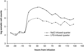
The method used for SCC was insufficient in cases where the number of cells exceeded approximately 20 million/ml. Counts are therefore missing for milk collections at 21 h after injection for the LPS milk samples. SCC in milk, both NaCl-infused quarters and LPS-infused quarters, are shown in Figure 1.
Somatic cell counts in milk (NaCl-infused and lipopolysaccharide (LPS) infused) during the experimental period.

All the milk parameters tested in this study, that is, SCC, NAGase, LDH, AP, AcP, Hp and MAA, responded significantly to LPS infusions in the quarters.
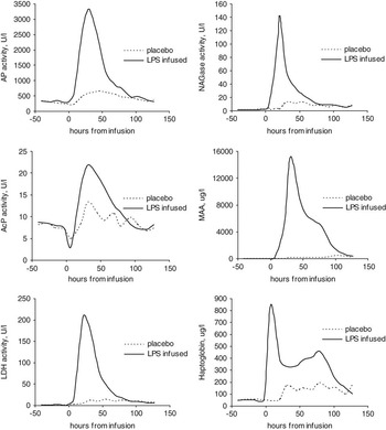
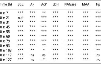
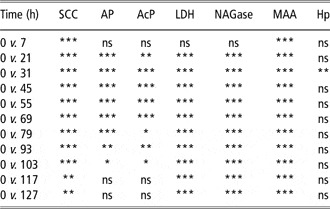
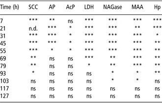
The general appearance of enzyme activity and APP in milk is given in Figure 2, where smoothed curves indicate both the level in LPS-infused and placebo-infused quarter samples over a 7-day time span. Table 1 summarizes the comparisons of the enzyme activity and acute phase levels at various time stages v. the basic level for the LPS-infused quarter milk samples, whereas Table 2 compares the same parameters for the placebo-infused samples. The comparison between the LPS-infused and the placebo-infused quarter samples is given in Table 3.
Milk from lipopolysaccharide (LPS)-infused and placebo-infused quarters was measured at fixed time intervals for enzyme activities and acute phase proteins.

Milk samples from LPS-infused quarters were analyzed for seven indicators of inflammation at fixed time intervals from infusion

LPS = lipopolysaccharides; SCC = somatic cell counts; AP = alkaline phosphatase activity; AcP = acid phosphatase activity; LDH = lactate dehydrogenase activity; NAGase = NAGase activity; MAA = milk amyloid A content; Hp = Haptoglobin content; n.d. = not determined; ns = not significantly different.
ns, *(P < 0.05), **(P < 0.01) and ***(P < 0.001) indicate the probability of being equal to the basic level before infusion (t = 0).
Milk samples from placebo-treated quarters were analyzed for seven indicators of inflammation at fixed time intervals from infusion

SCC = somatic cell counts; AP = alkaline phosphatase activity; AcP = acid phosphatase; LDH = lactate dehydrogenase activity; NAGase = NAGase activity; MAA = milk amyloid A content; Hp = Haptoglobin content; ns = not significantly different.
ns, *(P < 0.05), **(P < 0.01) and ***(P < 0.001) indicate the probability of being equal to the basic level before infusion (t = 0).
Milk samples from LPS-infused quarters and milk from sham-infused quarters (placebo) were analyzed for seven inflammation parameters at fixed time intervals after infusion

LPS = lipopolysaccharides; SCC = somatic cell counts; AP = alkaline phosphatase activity; AcP = acid phosphatase activity; LDH = lactate dehydrogenase activity; NAGase = NAGase activity; MAA = milk amyloid A content; Hp = Haptoglobin content; n.d. = not determined; ns = not significantly different.
ns, *(P < 0.05), **(P < 0.01) and ***(P < 0.001) indicate the probability of equality between milk from LPS-infused quarters and sham-infused quarters.
From Figure 2, it is evident that all parameters fluctuate within the observed interval. The times when maximum value (peak value) appeared for the separate parameters are given in Table 4. Furthermore, the measured level at peak time is compared with the initial level at t = 0; the ratio between peak and initial level is indicated by the multiplication factor in the parentheses.
The parameters measured in milk samples from LPS-infused and placebo-infused quarters peaked in intensity at different times

LPS = lipopolysaccharides; SCC = somatic cell counts; AP = alkaline phosphatase activity; AcP = acid phosphatase activity; LDH = lactate dehydrogenase activity; NAGase = N-acetyl-β-d-glucosaminidase activity; MAA = milk amyloid A content; Hp = haptoglobin content; n.d. = not determined.
This table shows the peaking time, hours after infusion, for the individual parameters, and the maximum level compared with the initial level at t = 0, that is, before infusion is given in brackets.
LPS-infused quarters
All measured parameters were markedly elevated within 7 h of LPS infusion. The significantly higher level persisted for 4 days compared with the initial level. The phosphatase enzyme activities and the Hp level seemed to return to the basic level first; SCC, LDH and NAGase activities and the MAA level stayed significantly elevated for a longer time compared with the basic level even 5 days after LPS infusion. Hp peaked early (7 h), whereas the other parameters peaked 20 to 30 h after LPS infusion (Table 4).
Placebo-infused quarters
SCC and MAA levels appeared markedly above the initial level (P < 0.001) already 7 h after placebo infusion. The other parameters, except Hp, appeared significantly higher than the basic level 21 h after infusion. Most parameters were still above the basic level 5 days after infusion; again, the phosphatase enzyme activities seemed to decline first. The Hp level seemed to be steadily increasing, but only slightly significantly from the basic level. SCC, NAGase and AcP peaked 31 h after placebo infusion, the other parameters later, and MAA peaked substantially later, that is, 103 h after infusion (Table 4).
Discussion
A very important criterion for a useful indicator of early mastitis should be a fast reaction (response) to the infection/inflammation condition, so that treatment toward the attack could be initiated as early as possible. An early and robust response would open the possibility to avert a serious and prolonged period of subclinical and clinical mastitis and the unfavorable health and economic consequences.
Another important factor in the early warning procedure is the detection power in the warning action, that is, how distinguishable is the response from the basic value in milk. An indicator that multiplies significantly because of the mastitic attack is of paramount interest.
Furthermore, the duration of the inflammation response would also be of a certain value because a prolonged period of maximum alarm would blur or even hide an assessment of time for infection start if milk samples are not analyzed and examined very regularly, for example, daily. From an objective point of view, it is furthermore important that the indicator appears in the infected quarters only, and that it has not spread to neighboring not infected quarters. Referring to the above criteria, it is essential that the intended mastitis parameter is adapted to be used at the single quarter level or composite milk level. An increased level of enzyme activity or APP in a single quarter will inevitably be diluted considerably in composite milk because milk from three healthy quarters will mix up with the infected milk. Furthermore, infected quarters are known to produce a little less milk than healthy quarters (Schaar and Funke, Reference Schaar and Funke1986). Monitoring of quarter milk will, in contrast, request less precision of the indicator because the milk from the sick quarter here will be undiluted and the indicator level is therefore higher compared to the basic level of the same quarter or the neighboring quarter.
The APP in serum and especially milk, dominated by MAA and Hp, have gained a lot of attention within the last 10 years as indicators of mastitis (e.g. Eckersall et al., Reference Eckersall, Young, McComb, Hogarth, Safi, Weber, McDonald, Nolan and Fitzpatrick2001; Grönlund et al., Reference Grönlund, Hultén, Eckersall, Hogarth and Waller2003; Winter et al., Reference Winter, Fuchs, Walshe and Colditz2003; Lehtolainen et al., Reference Lehtolainen, Røntved and Pyörälä2004; Grönlund et al., Reference Grönlund, Sandgren and Waller2005; Jacobsen et al., Reference Jacobsen, Niewold, Kornalijnslijper, Toussaint and Gruys2005; Nielsen et al., Reference Nielsen, Larsen, Bjerring and Ingvartsen2005). Hp and isoforms of serum amyloid A (SAA) are produced in the liver and udder during E. coli and LPS-induced mastitis (Rinaldi et al., Reference Rinaldi, Li, Bannerman, Daniels, Evock-Clover, Silva, Paape, Van Ryssen, Burvenich and Capuco2009; Vels et al., Reference Vels, Røntved, Bjerring and Ingvartsen2009). In the inflamed udder, Hp and MAA may originate from structural tissue, leukocytes migrating into the tissue or extravasation of APP from the systemic APR. Hp is secreted from blood and milk leukocytes (Thielen et al., Reference Thielen, Mielenz, Hiss and Sauerwein2005; Cooray et al., Reference Cooray, Waller and Venge2007) and from the mammary gland epithelium (Thielen et al., Reference Thielen, Mielenz, Hiss, Zerbe, Petzi, Schubert, Seyfert and Sauerwein2007), whereas only the mammary gland epithelium appears to secrete MAA (SAA3) in cattle (Larson et al., Reference Larson, Weber, Weber and McDonald2005).
Natural mastitis cases as well as inoculations with living bacteria (e.g. Staphylococcus aureus or Staphylococcus epidermidis; E. coli) or inoculations with LPS have been described. The general conclusion among the authors of the studies is that the milk acute phase reactants under consideration react immediately after infection/inoculation and that the APP level multiplies tremendously from a negligible level in the non-inflamed udder to a ‘subclinical level’ and subsequently to a clinical level. Many studies reveal a variable response in the acute phase level on infection (e.g. Grönlund et al., Reference Grönlund, Sandgren and Waller2005). The exact discrepancy between mild and moderate mastitis may be questionable for MAA (Eckersall et al., Reference Eckersall, Young, McComb, Hogarth, Safi, Weber, McDonald, Nolan and Fitzpatrick2001), as was the separation of artificially infected acute and chronic mastitis (Grönlund et al., Reference Grönlund, Hultén, Eckersall, Hogarth and Waller2003). Hp in milk seems to distinguish more successfully between moderate and severe cases. Other drawbacks connected with the present acute phase reactants, from a diagnostic point of view, seem to be a rather prolonged phase of decline, that is, the level of the protein, for instance, peaked more than once (Winter et al., Reference Winter, Fuchs, Walshe and Colditz2003) and the declining phase may last for days (all studies cited here). This also seems to be reflected in this study. In comparison, all enzymes had a monophasic response. MAA had a wider monophasic response with a ‘shoulder’ in the recovery period and Hp had a biphasic response with the first peak appearing simultaneously with the enzymes and the second peak appearing simultaneously with the MAA shoulder. As damaged leukocytes are major contributors of enzymes, it may be speculated that the first Hp peak also derives from these cells, and the second peak partly derives from the systemic APR (hepatic response). Nevertheless, further investigations must be conducted to enlighten how the release and turnover of inflammatory indicators in different body compartments contribute to various response curves in milk during mastitis.
The prolonged and biphasic response, together with a variable response to infection, may clearly obstruct the use of APP in modeling of the inflammation, which is a prerequisite in modern in-line detection of udder health. Another question is whether Hp and MAA analyses are suitable for in-line measurements. The chemically based method for Hp that may be used in blood plasma is based on spectrophotometry, which is not adaptable for milk analyses; the assays normally used in milk are based on ELISA-principles that may be considered disadvantageous for in-line systems (Pyörälä, Reference Pyörälä2003). In future, however, biosensors are expected to appear for practical purposes; recently, a biosensor assay for determination of Hp in bovine milk was evaluated (Åkerstedt et al., Reference Åkerstedt, Björk, Waller and Stjernesjö2006).
The four milk enzyme activities tested in this study all responded to the LPS infusion assayed in milk. Furthermore, placebo-infused quarters produced milk that only reacted slowly to the infusion and generally at a much lower level. This fact contrasts with the present and the usual SCC recordings, where cell counts may increase significantly in healthy quarters rapidly after inflammation of the neighboring quarters. In practice, this will have the implication that SCC measurements do not differentiate between healthy and inflamed quarters. However, the response to inflammation was not of equal quality among the enzyme markers if the intention was to focus on subclinical mastitis. AcP reached activities only 3 to 4 times the background level (Figure 2, Table 4), and furthermore, the milk from the placebo-infused quarters rose relatively fast to proportionally high levels resulting in only minor and less discriminate differences between non-LPS-infused quarters and LPS-infused quarters. A number of papers have been devoted to milk AcP and clinical mastitis, for example, Anderson et al. (Reference Anderson, Brooker, Andrews and Alichanidis1974), Kitchen (Reference Kitchen1976) and Andrews (Reference Andrews1991), showing more or less comparable results with this study.
LDH, NAGase and AP reacted fast to the LPS infusion in milk samples. The enzyme activity in the inflamed quarters was significantly higher than in the pre-infused quarters after only a few hours. Furthermore, the inflamed quarters were clearly distinct from the placebo-treated neighboring quarter for the first 2 to 3 days after infusion.
NAGase, LDH, AcP and AP activity are widespread in the entire organism, intracellularly as well as extracellularly. The enzymes are furthermore native constituents of healthy milk; among these, AcP and AP may even be categorized as indigenous. Basic AcP activity is believed to affect casein micellar structure (Andrews, Reference Andrews1991), whereas the elevated AcP activity seen in mastitis is connected with the appearance of isoenzymes of leukocyte origin (Anderson et al., Reference Anderson, Brooker, Andrews and Alichanidis1975; Andrews and Alichanidis, Reference Andrews and Alichanidis1975). Basic AP activity is associated with microsomal particles and is particularly enriched in the cream phase, since the microsomes are absorbed onto the surface of fat globules (Andrews, Reference Andrews1991). It seems most probable that the mammary gland itself is the source of the (basic) milk enzyme (Kitchen, Reference Kitchen1985; Andrews, Reference Andrews1991). The elevated activity seen in mastitis is also likely to originate from other sources. Neutrophils have shown to respond in vitro to LPS by upregulation of AP activity on the cell surface (Aida and Pabst, Reference Aida and Pabst1991). Heyneman and Burvenich (Reference Heyneman and Burvenich1992) studied the activity of AP in bovine blood neutrophiles in experimentally induced E. coli mastitis. The authors concluded that the leukocyte enzyme from healthy and mastitic cows displayed very similar characteristics, suggesting that the increase in activity during mastitis is most probably related to the enhanced expression of the normal leukocyte AP enzyme under direct or indirect influence of inflammatory mediators.
Early investigations indicated that elevated LDH activity in mastitic milk would not originate from blood plasma alone, but were more likely from parenchyma cells and disintegrated leukocytes as well (Bogin and Ziv, Reference Bogin and Ziv1973). Kato et al. (Reference Kato, Mori and Katoh1989) confirmed the contribution of LDH activity in mastitic milk from leukocytes and further specified the major increase in activity to originate from granulocytes and lymphocytes.
Kitchen et al. (Reference Kitchen, Kwee, Middleton and Andrews1984) stated that NAGase is an intracellular, lysosomal enzyme that is released into milk from neutrophils during phagocytosis and cell lysis, and to some degree from damaged epithelial, mammary cells. Numerous papers have described, for example, NAGase activity as an indicator of mastitis caused by various agents, and a few studies have even compared the NAGase response between pathogens (e.g. Pyörälä and Pyörälä, Reference Pyörälä and Pyörälä1997). Pyörälä (Reference Pyörälä2003) emphasized that NAGase accurately reflects the degree of inflammation so that, in mastitis caused by major pathogens, milk NAGase levels are significantly higher than in mastitis due to minor pathogens. It is therefore justifiable to assume that a basic activity of the four enzymes is present, even in healthy milk, and also that the activity in milk increases due to host immunological responses and, in certain cases, damage to the mammary epithelium.
In general, however, in this context unimportant response to saline infusions is believed to be due to the systemic APR induced by the LPS. This will increase the migration of leukocytes to other body compartments, including the saline-infused control quarter.
Based on this study, milk AP, LDH and NAGase enzyme activities appear as reliable indicators of udder inflammation in proportional milk when tested in an LPS mastitis model. In the early inflammatory phase, the three enzymes were significantly increased as the APP and SCC and reached their peak value at 21 to 31 h, simultaneously with Hp, MAA and SCC. In the late inflammatory phase, they followed the kinetic pattern of MAA and SCC.
Especially NAGase, as well as LDH activity in milk, has been advocated as reliable early indicators of acute mastitis. LDH activity may, in the in-line situation, be preferable due to analytical constraints connected with NAGase determinations. Consequently, mathematical models using LDH response in milk as predictors in in-line measurement systems have recently been developed (Chagunda et al., Reference Chagunda, Friggens, Rasmussen and Larsen2006b; Friggens et al., Reference Friggens, Chagunda, Bjerring, Ridder, Højsgaard and Larsen2007).
A logical continuation of this trial will be to investigate the same parameters in experimental and spontaneous mastitis cases caused by various pathogens including different breeds of animals and different lactation stages of cows.
Acknowledgments
Dorte Agnholt, Jens Clausen and Carsten Berthelsen are kindly acknowledged for excellent technical assistance and skilled analytical work. This study, which was part of the Biosens project, was funded by the Danish Ministry of Food, Agriculture and Fisheries and the Danish Cattle Association.