INTRODUCTION
In the absence of oxygen, most organisms convert glucose into partially oxidized organic molecules, including succinate and acetate, which are excreted to the environment. This fermentative strategy is used by organisms that live in predominantly microaerophilic (or anaerobic) conditions, like several protist and metazoan parasites. Glycolysis oxidizes glucose to two molecules of pyruvate while generating a net output of two ATP molecules, in both aerobiosis and anaerobiosis. Oxidation of glucose also produces two molecules of NADH corresponding to four reducing equivalents (step 8 in Figs. 1–4). These need to be reoxidized to NAD+ to maintain the glycolytic flux. In anaerobiosis, this can be achieved by the transfer of the reducing equivalent onto organic catabolites, thus producing lactate, succinate, formate, ethanol, glycerol, etc. Excretion of these partially oxidized metabolic intermediates disposes of reducing equivalents at the expense of energy stored in their carbon backbones. However, production of some excreted catabolites, such as acetate or propionate, is associated with production of additional energy in the form of ATP. In this review we aim to describe the pathways leading to production of acetate as an excreted end product in early branching eukaryotes such as the anaerobic amoebae, diplomonads, trichomonads and helminths, as well as the aerobic trypanosomatids. In the procyclic stage of Trypanosoma brucei, succinate production is concomitant to acetate excretion and will also be addressed here.
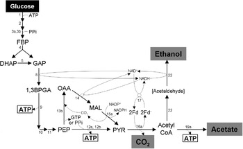
Carbohydrate metabolism in the trophozoite stage of Entamoeba histolytica (adapted from (Köhler and Voigt, Reference Köhler, Voigt and Mehlhorn1988; Coombs and Müller, Reference Coombs, Müller, Marr and Müller1995)). Excreted end products of glucose metabolism are in black characters on a grey background. The enzyme-bound metabolite is indicated by brackets (acetaldehyde). ATP molecules produced by substrate level phosphorylation are boxed. Abbreviations used in Figs 1–4: 1,3BPGA, 1,3-bisphosphoglycerate; c, cytochrome c; CIT, citrate; DHAP, dihydroxyacetone phosphate; e−, electrons; FBP, fructose 1,6-bisphosphate; G3P, glycerol-3-phosphate; GAP, glyceraldehyde-3-phosphate; Fd, ferredoxin; FUM, fumarate; GLUT, glutamate; ISC, isocitrate; KET, 2-ketoglutarate; MAL, malate; MCoA, methylmalonyl-CoA; OAA, oxaloacetate; PCoA, propionyl-CoA; PEP, phosphoenolpyruvate; PPi, inorganic pyrophosphate; PYR, pyruvate; SCoA, succinyl-CoA; SUC, succinate. Enzymes in Figs 1–4 are: 1, hexokinase: 2, glucose-6-phosphate isomerase; 3a, ATP-dependent phosphofructokinase (ATP-PFK); 3b, PPi-dependent phosphofructokinase (PPi-PFK); 4, aldolase; 5, triose-phosphate isomerase; 6, glycerol-3-phosphate dehydrogenase; 7a, glycerol-3-phosphatase; 7b, glycerol kinase; 8, glyceraldehyde-3-phosphate dehydrogenase; 9, phosphoglycerate kinase; 10, phosphoglycerate mutase; 11, enolase; 12a, pyruvate kinase (PYK); 12b, pyruvate phosphate dikinase (PPDK); 13a, phosphoenolpyruvate carboxykinase (PEPCK); 13b, phosphoenolpyruvate carboxytransphosphorylase; 14, malate dehydrogenase; 15a, malic enzyme; 15b, malate dehydrogenase (decarboxylating); 16a, pyruvate:ferredoxin oxidoreductase; 16b, pyruvate dehydrogenase complex; 17, ferredoxin:NAD oxidoreductase; 18, hydrogenase; 19a, Acetyl-CoA synthetase ‘ADP forming’ (‘ADP forming’ AceCS); 19b, acetate:succinate CoA-transferase (ASCT); 20, succinyl-CoA synthetase (SCoAS); 21, unknown enzyme; 22, bifunctional acetyl-CoA reductase/alcool dehydrogenase; 23a, lactate dehydrogenase; 23b, unknown enzyme; 24, fumarase, 25a, membrane-bound fumarate reductase (FRD); 25b, NADH-dependent fumarate reductase (NADH-FRD); 26, propionate:succinate CoA-transferase; 27, methylmalonyl-CoA mutase; 28, methylmalonyl-CoA racemase; 29, propionyl-CoA carboxylase; 30, citrate synthase; 31, aconitase; 32, citrate lyase; 33, isocitrate dehydrogenase; 34, acetyl-CoA synthetase ‘AMP forming’ (‘AMP forming’ AceCS); 35, L-proline dehydrogenase; 36, pyrroline-5 carboxylate dehydrogenase; 37, alanine aminotransferase; 38, 2-ketoglutarate dehydrogenase complex; 39, L-threonine dehydrogenase; 40, acetyl-CoA:glycine C-acetyltransferase; 41a, rotenone-sensitive NADH dehydrogenase (complex I of the respiratory chain); 41b, rotenone-insensitive NADH dehydrogenase; 42, FAD-dependent glycerol-3-phosphate dehydrogenase (FAD-GPDH); 43a, rhodoquinone, such as menaquinone; 43b, ubiquinone; 44, SHAM-sensitive alternative oxidase; 45, complex III of the respiratory chain; 46, complex IV of the respiratory chain; 47, F0F1-ATP synthase.

Carbohydrate metabolism in the adult worms of Fasciola hepatica (adapted from Köhler and Voigt, Reference Köhler, Voigt and Mehlhorn1988; van Hellemond et al. Reference Van Hellemond, Van Der Klei, Van Weelden and Tielens2003). Excreted end products of glucose metabolism are in black characters on a grey background. ATP molecules produced by substrate level phosphorylation and oxidative phosphorylation are boxed and circled, respectively. For abbreviations and enzyme names, see Fig. 1.

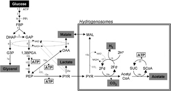
Carbohydrate metabolism in Trichomonas vaginalis (adapted from Coombs and Müller, Reference Coombs, Müller, Marr and Müller1995). Excreted end products of glucose metabolism are in black characters on a grey background. ATP molecules produced by substrate level phosphorylation are boxed. For abbreviations and enzyme names, see Fig. 1.

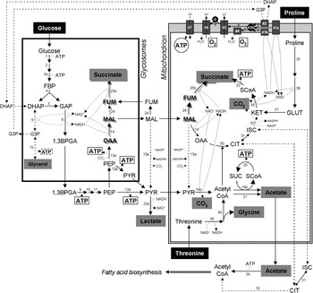
Energy metabolism in the procyclic form (insect stage) of Trypanosoma brucei grown in glucose-rich medium. Excreted end products of glucose, proline and threonine metabolism are in black characters on a grey background. Dashed arrows indicate steps which have not been shown to significantly occur or which are supposed to occur at a background level or not at all under standard growth conditions (glucose-rich medium). The glycosomal and mitochondrial succinic fermentation pathways are shadowed. ATP molecules produced by substrate level phosphorylation and oxidative phosphorylation are boxed and circled, respectively. For abbreviations and enzyme names, see Fig. 1.

PARASITES PRODUCING ACETATE
Entamoeba histolytica
E. histolytica (amoeba of the order Amoebida) infections cause amoebic dysentery, and remain an important cause of morbidity, mortality and health burden, particularly in developing countries (World Health Organization, 1997; Ximenez et al. Reference Ximenez, Moran, Rojas, Valadez and Gomez2009). Transmission is due to the ingestion of food or water contaminated by infective cysts, which excyst into metacystic trophozoites in the small intestine and then differentiate into trophozoites in the large intestine (Marshall et al. Reference Marshall, Naumovitz, Ortega and Sterling1997). The parasitic cycle of this protist is closed with the production of cysts in the colon and rectum, and their subsequent release in the faeces.
E. histolytica cells are devoid of true mitochondria and harbour relict mitochondria, named mitosomes, which do not appear to be involved in energy production or redox balance maintenance (see Box 1). As a consequence, all of the ATP is produced by substrate level phosphorylation1. E. histolytica is an obligate fermenter, using bacterial-like fermentation enzymes. It lacks enzymes of the mitochondrial electron transport chain and of the tricarboxylic acid (TCA) cycle (Loftus et al. Reference Loftus, Anderson, Davies, Alsmark, Samuelson, Amedeo, Roncaglia, Berriman, Hirt, Mann, Nozaki, Suh, Pop, Duchene, Ackers, Tannich, Leippe, Hofer, Bruchhaus, Willhoeft, Bhattacharya, Chillingworth, Churcher, Hance, Harris, Harris, Jagels, Moule, Mungall, Ormond, Squares, Whitehead, Quail, Rabbinowitsch, Norbertczak, Price, Wang, Guillen, Gilchrist, Stroup, Bhattacharya, Lohia, Foster, Sicheritz-Ponten, Weber, Singh, Mukherjee, El-Sayed, Petri, Clark, Embley, Barrell, Fraser and Hall2005). The trophozoite stage, the only E. histolytica stage analysed in vitro, has an absolute requirement for carbohydrates as an energy source, especially glucose. However, amino acids may play a larger role in energy metabolism than previously thought (Anderson and Loftus, Reference Anderson and Loftus2005). Energy metabolism of the trophozoites takes place exclusively in the cytosol, where glucose is converted anaerobically into acetate, ethanol and CO2 (Coombs and Müller, Reference Coombs, Müller, Marr and Müller1995) (Fig. 1). The classical Embden–Meyerhoff glycolytic pathway is present in E. histolytica, but this parasite expresses glycolytic enzymes relying on inorganic pyrophosphate (PPi) instead of ATP for catalysis (Mertens, Reference Mertens1993) (see Box 2). In the trophozoite stage, most of the glycolytic flux goes through the PPi-dependent PFK (step 3b), although the ATP-dependent enzyme is also present (step 3a) (Chi et al. Reference Chi, Deng, Albach and Kemp2001). Similarly, this parasite expresses two different enzymes for pyruvate production from phosphoenolpyruvate (PEP), pyruvate kinase (PYK, step 12a) that produces ATP from ADP and pyruvate phosphate dikinase (PPDK, a PPi-dependent enzyme, step 12b) that produces ATP from AMP with the contribution of PPi. However, the contribution of PYK seems to be minor in E. histolytica (Saavedra-Lira et al. Reference Saavedra-Lira, Ramirez-Silva and Perez-Montfort1998; Moreno-Sanchez et al. Reference Moreno-Sanchez, Saavedra, Rodriguez-Enriquez and Olin-Sandoval2008). PEP carboxytransphosphorylase, another PPi-dependent enzyme (step 13b), is also expressed instead of the canonical PEP carboxykinase (step 13a) (Mertens, Reference Mertens1993). This amitochondrial protist has an extended glycolytic pathway in which pyruvate is converted in the cytosol to acetyl-coA by pyruvate:ferredoxin oxidoreductase (PFOR, step 16a). In this reaction, ferredoxin accepts electrons generated by PFOR, which are transferred to NAD+ by ferredoxin:NAD oxidoreductase (step 17). Then, acetyl-CoA can either be converted to acetate by the ‘ADP-forming’ acetyl-CoA synthetase (AceCS), with ATP generation (step 19a) or reduced to ethanol with regeneration of NAD+ (step 22) (Reeves, Reference Reeves1984).
Box 1: Mitochondria-Like Organelles
Mitochondria are endosymbiont-derived organelles, thus possess a double membrane and retain a small DNA genome. They supply cellular ATP by oxidative phosphorylation2 as well as substrate level phosphorylation1. They are present in most eukaryotic cells, however, a number of eukaryotes that lack mitochondria contain other double membrane-enclosed mitochondria-like organelles involved or not in energy production, such as hydrogenosomes and mitosomes. Hydrogenosomes were the first characterized mitochondria-like organelles (Lindmark and Muller, Reference Lindmark and Muller1973) initially identified in trichomonads and then also discovered in a broad range of phylogenetically distant protists and fungi, all of them sharing the anaerobic life style (Hackstein et al. Reference Hackstein, Akhmanova, Boxma, Harhangi and Voncken1999; Stechmann et al. Reference Stechmann, Hamblin, Perez-Brocal, Gaston, Richmond, Van Der Giezen, Clark and Roger2008). Like mitochondria, hydrogenosomes are involved in energy production and maintenance of the redox balance. However, oxygen is not used as an electron acceptor, rather, they use a ferredoxin-mediated electron transport to reduce protons to molecular hydrogen (see Fig. 3). Mitochondria and hydrogenosomes were believed to share a common ancestor but, until recently, reconstructing the evolutionary origin of hydrogenosomes has been hampered by the unavailability of organellar DNA (Dyall and Johnson, Reference Dyall and Johnson2000). In 2005, Boxma et al. sequenced the DNA genome contained in hydrogenosomes of the anaerobic ciliate Nycototherus ovalis and identified genes encoding components of a mitochondrial electron transport chain. Consequently, hydrogenosomes are, without any doubt, mitochondria in the evolutionary sense, even though they differ from typical mitochondria in various biochemical properties. A mitochondrion-like organelle recently characterized in Blastocystis hominis shows biochemical steps encountered in traditional mitochondria and/or hydrogenosomes, however it lacks hydrogenase activity (Lantsman et al. Reference Lantsman, Tan, Morada and Yarlett2008). Other relict mitochondria, named mitosomes, have been identified more recently in E. histolytica, Giardia and in the parasitic fungus Microsporidium (Tovar et al. Reference Tovar, Fischer and Clark1999; Williams et al. Reference Williams, Hirt, Lucocq and Embley2002; Dolezal et al. Reference Dolezal, Smid, Rada, Zubacova, Bursac, Sutak, Nebesarova, Lithgow and Tachezy2005). These tiny mitochondrial remnants lack genomic DNA, produce neither hydrogen nor ATP and no function in energy metabolism has been attributed to this organelle (Tovar et al. Reference Tovar, Fischer and Clark1999). To date, the only metabolic function assigned to mitosomes is the biosynthesis of Fe-S clusters that are essential for cytosolic Fe–S proteins (Goldberg et al. Reference Goldberg, Molik, Tsaousis, Neumann, Kuhnke, Delbac, Vivares, Hirt, Lill and Embley2008).
Box 2: ATP-Dependent and PPi-Dependent Glycolytic Enzymes
The classical Embden–Meyerhoff glycolytic pathway requires ATP as phosphoryl group donor to phosphorylate glucose to fructose 1,6-bisphosphate. This is achieved by the canonical hexokinase (step 1, Figs. 1–4) or glucokinase and phosphofructokinase (PFK) (step 3a). ATP consumed by the first two kinases is then regenerated by phosphoglycerate kinase (step 9). Finally, pyruvate is produced by the last glycolytic step catalysed by pyruvate kinase (PYK) with a net production of ATP (step 12a). One significant variation of the standard glycolytic pathway that has been described in some anaerobic protists is pyrophosphate-dependent glycolysis, which uses pyrophosphate (PPi) instead of ATP as phosphate donor (Mertens, Reference Mertens1993). Indeed, a PPi-dependent PFK (PPi-PFK) is found instead of or together with an ATP-dependent PFK (ATP-PFK) in many prokaryotes and plants as well as in different protists, including E. histolytica, Giardia, Trichomonas, Toxoplasma gondii and Euglena gracilis (Li and Phillips, Reference Li and Phillips1997; Mertens et al. Reference Mertens, Ladror, Lee, Miretsky, Morris, Rozario, Kemp and Muller1998; Chi et al. Reference Chi, Deng, Albach and Kemp2001). The other glycolytic enzyme using PPi rather than adenine nucleotides as phosphoryl donor is pyruvate phosphate dikinase (PPDK), which replaces or coexists with PYK in plants, many prokaryotes and some protists, including E. histolytica, Giardia, oxymonads and trypanosomatids (Bruderer et al. Reference Bruderer, Wehrli and Kohler1996; Saavedra-Lira et al. Reference Saavedra-Lira, Ramirez-Silva and Perez-Montfort1998; Bringaud et al. Reference Bringaud, Baltz and Baltz1998; Slamovits and Keeling, Reference Slamovits and Keeling2006). The irreversible and reversible reactions catalysed by these ATP- and PPi-dependent enzymes, respectively, are shown here: Anaerobic organisms, including the parasites mentioned above, rely almost exclusively on glycolysis and fermentation for ATP production, so optimizing the energy output from glycolysis may be subject to strong selective pressure. Thus, the rationale for the use of PPi-dependent glycolysis is certainly linked to the improvement of energy efficiency (Mertens, Reference Mertens1993). Inorganic pyrophosphatase activity regarded as the principal enzyme responsible for maintaining a low intracellular PPi concentration in most cells is missing in these anaerobic parasites (Searle and Muller, Reference Searle and Muller1991). As a consequence, the energy of most of PPi generated during anabolic processes remains conserved, thus yielding a total of upto five ATP molecules per glucose molecule consumed instead of two produced by conventional glycolysis. Of the PPi-dependent glycolytic enzymes, PFK has been studied most extensively. PPi-PFK and ATP-PFK share a common ancestry, but phylogenies show a very complex evolutionary pattern (Bapteste et al. Reference Bapteste, Moreira and Philippe2003). For instance, the T. brucei and Plasmodium falciparum ATP-PFK are homologous to PPi-PFK, but belong to different PFK classes (McNae et al. Reference McNae, Martinez-Oyanedel, Keillor, Michels, Fothergill-Gilmore and Walkinshaw2009; Mony et al. Reference Mony, Mehta, Jarori and Sharma2009). To complicate the phylogenetic reconstruction, a single point mutation can induce a change in the preference of the phosphoryl donor and many phosphoryl donor changes and horizontal gene transfers seem to have occurred in the evolution of PFKs (Chi and Kemp, Reference Chi and Kemp2000; Bapteste et al. Reference Bapteste, Moreira and Philippe2003; McNae et al. Reference McNae, Martinez-Oyanedel, Keillor, Michels, Fothergill-Gilmore and Walkinshaw2009). Whatever, the presence of a latent nucleotide binding site in the active site of PPi-PFK E. histolytica enzyme, suggested that the PFK originally evolved as an enzyme that used ATP as a phosphoryl donor, and the utilization of PPi was a late evolving adaptation (Chi and Kemp, Reference Chi and Kemp2000).
Giardia lamblia
The intestinal protist Giardia lamblia (synonymous with Giardia intestinalis and Giardia duodenalis), which belongs to the order Diplomonadida, is a major cause of diarrhoeal diseases throughout the world (Adam, Reference Adam2001). The life cycle is simple and consists of two major stages (Bogitsh et al. Reference Bogitsh, Carter and Oeltmann2005). After infection of the host by ingestion of cysts from contaminated water or food, the cysts pass through the stomach to the small intestine where they excyst and differentiate into trophozoites. The non-infective vegetative trophozoites pass through the digestive tract and usually encyst in the colon. It is in the small intestine that the trophozoites replicate and cause symptoms of the disease to its host (malabsorption and diarrhoea).
Giardia species and other diplomonads contain mitosomes instead of mitochondria and are lacking cytochrome-mediated oxidative phosphorylation2. The microaerotolerant trophozoite stage of the parasite relies on fermentative metabolism for energy generation, even in the presence of oxygen. Glucose is the major source of energy and is metabolised in the cytosol to yield acetate, ethanol, alanine, and CO2, using a metabolic network similar to that of E. histolytica (Coombs and Müller, Reference Coombs, Müller, Marr and Müller1995; Brown et al. Reference Brown, Upcroft, Edwards and Upcroft1998) (see Fig. 1). Some differences exist: under strictly anaerobic conditions this organism produces equimolar ethanol and alanine as well as CO2 and some acetate. However, in the presence of oxygen the production of both alanine and ethanol are inhibited and more acetate and CO2 are formed (Paget et al. Reference Paget, Raynor, Shipp and Lloyd1990).
Fasciola hepatica
The helminth Fasciola hepatica, the common liver fluke, is a metazoan parasitic flatworm that infects a large variety of herbivorous animals and has humans as an accidental host. Recognizing that food-borne diseases are an important cause of illness and death worldwide, the World Health Assembly in 2000 identified prevention and control of food-borne diseases as a public health priority (World Human Organization, 2006). Not only is fascioliasis a human health burden, but the infection of livestock causes an economic burden resulting in the loss of meat and milk products (McManus and Dalton, Reference McManus and Dalton2006; Keiser and Utzinger, Reference Keiser and Utzinger2009).
The life cycle of F. hepatica starts with release of eggs into the host's alimentary tract that eventually reach the environment with faeces. The miracidium develops and hatches in fresh water, where amphibious snails serve as intermediate hosts to the developing miracidium (Keiser and Utzinger, Reference Keiser and Utzinger2009). Eventually, free-swimming cercariae emerge from the intermediate hosts, encysting, and are taken up by feeding animals. The encysted metacercariae excyst and produce adult worms that perform extensive destruction of liver tissue and bile ducts of their mammalian hosts (Bogitsh et al. Reference Bogitsh, Carter and Oeltmann2005; Keiser and Utzinger, Reference Keiser and Utzinger2009).
Adult worms of F. hepatica are the parasitic stage considered here. For energy production they primarily use carbohydrates, like glucose, which are metabolised in the cytosol into lactate and ethanol and in the mitochondria to produce acetate, propionate and CO2 (Fig. 2). Glucose is first degraded by glycolysis to PEP, part of which is converted to oxaloacetate and consequently malate within the cytosol. Within the mitochondrion, malate is processed via two distinct and complementary branches – an oxidative branch (steps 24–29) and a reductive branch (steps 15a–20), a pathway named ‘malate dismutation’ (Van Hellemond et al. Reference Van Hellemond, Van Der Klei, Van Weelden and Tielens2003). Oxidation results in the production of reducing equivalents (steps 15a, 16b), which are oxidized by the electron transport chain (steps 41a, 43a, 25a) using, as final electron acceptor, fumarate produced by the reductive branch (step 24). Both oxidative and reductive arms of malate dismutation yield ATP by substrate level phosphorylation in the acetate (steps 19b, 20) and propionate (steps 26–29) producing reactions, respectively. The mitochondrial transmembrane proton gradient generated by complex I of the respiratory chain (step 41a) can also be used to produce ATP by oxidative phosphorylation (step 47). Thus F. hepatica depends on an anaerobic mitochondrial metabolism where malate is converted to generate the electrons for oxidative phosphorylation (Tielens et al. Reference Tielens, Van Den Heuvel and Van Den Bergh1984). In contrast, the free-living stages of F. hepatica undergo aerobic metabolism, using a conventional TCA cycle.
Trichomonas vaginalis
Trichomoniasis, a common cause of vaginitis and the most common sexually transmitted parasitic infection, is readily diagnosed and treatable (Johnston and Mabey, Reference Johnston and Mabey2008). Control of trichomoniasis has received very little emphasis from public health control programmes (Schwebke and Burgess, Reference Schwebke and Burgess2004). However, trichomoniasis in women and its link to adverse outcomes during pregnancy as well as an increased risk for HIV infection has given the disease more importance (Laga et al. Reference Laga, Manoka, Kivuvu, Malele, Tuliza, Nzila, Goeman, Behets, Batter and Alary1993; Cotch et al. Reference Cotch, Pastorek, Nugent, Hillier, Gibbs, Martin, Eschenbach, Edelman, Carey, Regan, Krohn, Klebanoff, Rao and Rhoads1997). The protist parasite T. vaginalis populates lumen and mucosal surfaces of the vagina in the female and the urethra, epididymis and prostate gland in the male (Bogitsh et al. Reference Bogitsh, Carter and Oeltmann2005). There is no known cyst stage.
Trichomonads are anaerobic organisms, satisfying their energy requirements by incomplete degradation of simple sugars with consequent production of catabolites including lactate, glycerol, malate and acetate, as well as CO2 and molecular hydrogen (H2) (Muller, Reference Muller1992; Coombs and Müller, Reference Coombs, Müller, Marr and Müller1995) (Fig. 3). They contain mitochondria-like organelles, called hydrogenosomes, lacking the TCA cycle and electron transport chain (see Box 1). Pyruvate is produced in the cytosol from glycolysis, where it is partly converted to lactate or it enters the hydrogenosomes to be converted into acetate, malate, CO2 and H2. Energy in the form of ATP is produced in the cytosol and in the hydrogenosomes by substrate level phosphorylation. The main function of the hydrogenosomes is oxidative decarboxylation of pyruvate by a pathway coupled to ATP production and linked to a ferredoxin-mediated electron transport resulting in the production of H2 and acetate (Steinbuchel and Muller, Reference Steinbuchel and Muller1986; Williams et al. Reference Williams, Lowe and Leadlay1987; Muller, Reference Muller1993). Upon decarboxylation of pyruvate into acetyl-CoA by PFOR, ferredoxin is used as an electron acceptor (step 16a). Electrons are then either transferred to hydrogen ions forming H2 (hydrogenase, step 18) or to NAD+ (ferredoxin:NAD oxidoreductase, step 17) produced by conversion of pyruvate into malate in the organelle (step 15b). Acetyl-CoA is finally converted into acetate with a net production of ATP (steps 19b, 20).
Trypanosoma brucei
Devastating epidemics of Human African Trypanosomiasis (HAT), or sleeping sickness, re-emerged in many sub-Saharan countries in the 1990s (Stich et al. Reference Stich, Barrett and Krishna2003). The invariably fatal disease puts at risk 60 million of the 400 million people inhabiting 36 sub-Saharan African countries (Barrett et al. Reference Barrett, Burchmore, Stich, Lazzari, Frasch, Cazzulo and Krishna2003). Of those at risk from the disease only a mere 4 million are under surveillance (Etchegorry et al. Reference Etchegorry, Helenport, Pecoul, Jannin and Legros2001). The annually reported cases are therefore an underestimation of the real problem.
Trypanosoma brucei, the causative agent, has a complicated life cycle in the mammalian and Glossina (tsetse) insect hosts. The infective stage of this protist parasite enters the human host with the bite of the infected tsetse vector. Metacyclic trypanosomes injected by the fly differentiate to the proliferating long slender that eventually develop into the non-dividing short stumpy stages (Vickerman, Reference Vickerman1985). The short stumpy parasites are competent to further differentiate to the dividing procyclic stage in the midgut of the insect. Then the parasites undergo additional stage differentiations upon migrating and attaching to the salivary glands, from where infective metacyclic forms are shed to the saliva (Bogitsh et al. Reference Bogitsh, Carter and Oeltmann2005). The bloodstream stages and the procyclic stage of T. brucei are amenable to in vitro culture and consequently their metabolism has been extensively analysed.
The distinct environments to which the parasite has to adapt throughout its life cycle require it to remodel its metabolism upon differentiation (Besteiro et al. Reference Besteiro, Barrett, Riviere and Bringaud2005). The mammalian bloodstream stage of T. brucei relies solely on the glucose present in the host fluids to produce energy, by converting glucose into pyruvate, the only end product excreted to the environment (Michels et al. Reference Michels, Bringaud, Herman and Hannaert2006). The first seven steps of glycolysis are compartmentalised in a peroxisome-like organelle, called the glycosome (Opperdoes and Borst, Reference Opperdoes and Borst1977). In the midgut of the insect, the procyclic stage of T. brucei is thought to encounter an amino acid-rich environment. However, in vitro this stage has been traditionally cultivated in glucose-rich media. Under these growth conditions, the procyclic trypanosomes convert glucose, threonine and proline, the main carbon sources (Cross et al. Reference Cross, Klein and Linstead1975; Lamour et al. Reference Lamour, Riviere, Coustou, Coombs, Barrett and Bringaud2005), into partially oxidized end products by aerobic fermentation (Cazzulo, Reference Cazzulo1992). The major end products of glucose metabolism, which have attracted most attention, are succinate, acetate, lactate and CO2 (Fig. 4). While most of glycolysis takes place in the glycosomes (steps 1–5, 8), PEP is produced in the cytosol, where it is located at a branching point. It can re-enter the glycosomes to be converted to succinate in either the glycosomes or the mitochondrion (steps, 13a, 14, 24, 25b) (Besteiro et al. Reference Besteiro, Biran, Biteau, Coustou, Baltz, Canioni and Bringaud2002; Coustou et al. Reference Coustou, Besteiro, Riviere, Biran, Biteau, Franconi, Boshart, Baltz and Bringaud2005, Reference Coustou, Biran, Besteiro, Riviere, Baltz, Franconi and Bringaud2006). Alternatively, pyruvate produced in the cytosol from part of the PEP enters the mitochondrion and is converted to acetate via acetyl-CoA (steps 12a, 16b, 19a–21) (Van Hellemond et al. Reference Van Hellemond and Tielens1998; Riviere et al. Reference Riviere, Van Weelden, Glass, Vegh, Coustou, Biran, Van Hellemond, Bringaud, Tielens and Boshart2004). It is noteworthy that acetyl-CoA does not feed the TCA cycle, despite all the TCA cycle enzymes being expressed (Durieux et al. Reference Durieux, Schutz, Brun and Kohler1991; Van Weelden et al. Reference Van Hellemond, Van Der Klei, Van Weelden and Tielens2003). Threonine is converted into equimolar amounts of glycine and acetate (steps 39, 40, 19b–21), and is considered to be the main source of acetyl-CoA for lipid biosynthesis (Cross et al. Reference Cross, Klein and Linstead1975; Klein and Linstead, Reference Klein and Linstead1976). In the presence of glucose, the rate of proline consumption is relatively low and succinate is the main metabolic end product from proline metabolism (steps 35–38, 20) (Lamour et al. Reference Lamour, Riviere, Coustou, Coombs, Barrett and Bringaud2005; Coustou et al. Reference Coustou, Biran, Breton, Guegan, Riviere, Plazolles, Nolan, Barrett, Franconi and Bringaud2008). The procyclic trypanosomes have also been adapted to culture in glucose-depleted media to meet more physiological conditions (Furuya et al. Reference Furuya, Kessler, Jardim, Schnaufer, Crudder and Parsons2002; Morris et al. Reference Morris, Wang, Drew and Englund2002; van Weelden et al. Reference Van Weelden, Van Hellemond, Opperdoes and Tielens2005). This leads to a switch from the metabolism of glucose to high consumption of proline, which is converted into alanine instead of succinate (Lamour et al. Reference Lamour, Riviere, Coustou, Coombs, Barrett and Bringaud2005; Coustou et al. Reference Coustou, Biran, Breton, Guegan, Riviere, Plazolles, Nolan, Barrett, Franconi and Bringaud2008; Ebikeme et al. Reference Ebikeme, Peacock, Coustou, Riviere, Bringaud, Gibson and Barrett2008). In glucose-depleted conditions, the parasite depends on oxidative phosphorylation for ATP production (Coustou et al. Reference Coustou, Biran, Breton, Guegan, Riviere, Plazolles, Nolan, Barrett, Franconi and Bringaud2008). However, oxidative phosphorylation becomes dispensable for growth in glucose-rich medium and most of ATP is produced by substrate-level phosphorylation (Bochud-Allemann and Schneider, Reference Bochud-Allemann and Schneider2002; Coustou et al. Reference Coustou, Besteiro, Biran, Diolez, Bouchaud, Voisin, Michels, Canioni, Baltz and Bringaud2003, Reference Coustou, Biran, Breton, Guegan, Riviere, Plazolles, Nolan, Barrett, Franconi and Bringaud2008). This implies that the generation of the proton gradient by the respiratory chain, which is essential to maintain many mitochondrial activities, is not required for ATP synthesis. It is noteworthy that strain differences have been recently reported regarding requirement of the mitochondrial F0F1-ATP synthase for ATP production in glucose-rich conditions (Zikova et al. Reference Zikova, Schnaufer, Dalley, Panigrahi and Stuart2009).
CATABOLIC PATHWAYS PRODUCING ACETATE
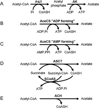
Formation of acetate from acetyl-CoA as an end product of energy metabolism is observed in many prokaryotes and in a few eukaryotic species, all predominantly living in anaerobic or hypoxic environments, with the exception of the aerobic trypanosomatids (see below). Five distinct reactions have been described so far for the conversion of acetyl-CoA into acetate (Fig. 5), two of which are used by parasites. As mentioned above, the ‘ADP-forming’ AceCS is present in E. histolytica and Giardia (step 19a, Fig. 1), where it is essential for ATP production (Sanchez and Muller, Reference Sanchez and Muller1996). The other enzyme used by T. vaginalis (Steinbuchel and Muller, Reference Steinbuchel and Muller1986), trypanosomatids (Van Hellemond et al. Reference Van Hellemond, Opperdoes and Tielens1998) and the flatworm F. hepatica (Saz et al. Reference Saz, Debruyn and De Mata1996) to produce acetate is the acetate:succinate CoA-transferase (ASCT, step 19b, Figs. 2–4), which transfers the CoA group of acetyl-CoA to succinate to produce acetate and succinyl-CoA. ASCT activity has also been detected in the anaerobic fungus Neocallimastix and the anaerobic unicellular stramenopile Blastocystis hominis (Marvin-Sikkema et al. Reference Marvin-Sikkema, Pedro Gomes, Grivet, Gottschal and Prins1993; Lantsman et al. Reference Lantsman, Tan, Morada and Yarlett2008). The ASCT reaction in this phylogenetically diverse group of organisms is directly linked to ATP production through succinyl-CoA synthetase (SCoAS, step 20) by conversion of succinyl-CoA back into succinate (Fig. 5). Biochemical evidence for this ASCT/SCoAS cycle has been obtained for T. vaginalis, F. hepatica and several trypanosomatid species (Steinbuchel and Muller, Reference Steinbuchel and Muller1986; Saz et al. Reference Saz, Debruyn and De Mata1996; Van Hellemond et al. Reference Van Hellemond and Tielens1998). ASCT belongs to family I of the CoA-transferase superfamily. Family I enzymes share conserved protein domains (CoA-trans in protein families database, accession PF01144), have a broad range of substrate specificity, and include CoA-transferases for 3-oxoacids, short-chain fatty acids and gluconate (Heider, Reference Heider2001). Most of them operate with acetyl-CoA or succinyl-CoA as possible CoA donors, although some of them can accept several different CoA donors or acceptors. This is exemplified by the recent characterization of the F. hepatica ASCT enzyme, which is probably involved in both acetate and propionate production (van Grinsven et al. Reference Van Grinsven, Van Hellemond and Tielens2009). As mentioned above, F. hepatica also produces propionate from a CoA-transferase, which transfers the CoA group from propionyl-CoA to succinate (step 26, Fig. 2). Succinyl-CoA produced by this reaction is converted via a three-step mechanism to propionyl-CoA, thus, forming a four-step cycle producing ATP (steps 26–29). Van Grinsven et al. (Reference Van Grinsven, Van Hellemond and Tielens2009) showed that the F. hepatica ASCT accepts both acetyl-CoA and propionyl-CoA as CoA donors and thus appears to be a multifunctional protein involved in both acetate and propionate production in this parasitic flat worm. The genes encoding the ASCT in F. hepatica, T. vaginalis and B. hominis are similar and closely related to the yeast ACH1 gene (Lantsman et al. Reference Lantsman, Tan, Morada and Yarlett2008; van Grinsven et al. Reference Van Grinsven, Rosnowsky, Van Weelden, Putz, Van Der Giezen, Martin, Van Hellemond, Tielens and Henze2008, Reference Van Grinsven, Rosnowsky, Van Weelden, Putz, Van Der Giezen, Martin, Van Hellemond, Tielens and Henze2009). ACH1 was originally identified as an acetyl-CoA hydrolase (this enzyme catalyses the hydrolysis of acetyl-CoA with no associated ATP production, see Fig. 5) and recently re-characterized as a bona fide CoA-transferase, which transfers CoA groups from succinyl-CoA to acetate (reverse reaction catalysed by ASCT) (Fleck and Brock, Reference Fleck and Brock2009). It is noteworthy that acetyl-CoA transferases (family I of CoA transferases) and acetyl-CoA hydrolases belong to the same acetyl-CoA hydrolase/transferase superfamily. The proportion of CoA transferase enzymes, including acetyl-CoA transferases, in this family will certainly increase as exemplified by the re-characterization of the yeast ACH1. The first identified ASCT gene was that of T. brucei (Riviere et al. Reference Riviere, Van Weelden, Glass, Vegh, Coustou, Biran, Van Hellemond, Bringaud, Tielens and Boshart2004), yet this gene is more distantly related to the yeast ACH1 group. Trypanosomatid ASCT shows high sequence similarity to succinyl-CoA:3-oxoacid CoA-transferases, which are part of ketone body and sterol biosynthetic pathways (Orii et al. Reference Orii, Fukao, Song, Mitchell and Kondo2008). Apparently, different genes have been recruited by trichomonads/helminths and trypanosomatids for an enzymatic function shared by a diverse, mostly anaerobically living, group of organisms. This is suggestive of convergent evolution. It will be interesting to identify new ASCT genes, such as the one responsible for the ASCT activity detected in the anaerobic fungus Neocallimastix (Marvin-Sikkema et al. Reference Marvin-Sikkema, Pedro Gomes, Grivet, Gottschal and Prins1993). It may belong to a distinct subfamily of CoA-transferases or acetyl-CoA hydrolases.
Acetate production from acetyl-CoA. All the known catalytic reactions leading to acetate production from acetyl-CoA are presented here. (A) In many eubacteria, the phosphate acetyltransferase (PAT) and acetate kinase (AK) produce acetate via a two-step mechanism in which acetyl phosphate is an intermediate (Brown et al. Reference Brown, Jones-Mortimer and Kornberg1977; Wolfe, Reference Wolfe2005). (B and C) Acetate can also be produced by two types of acetyl-CoA synthetases (AceCS), the ‘ADP-forming’ enzyme produces ATP from ADP and inorganic phosphate, while the ‘AMP-forming’ enzyme uses AMP and pyrophosphate instead of ADP and inorganic phosphate. The ubiquitous ‘AMP-forming’ AceCS performs the reverse reaction (acetyl-CoA production), except in Aspergillus nidulans, where the enzyme produces acetate and is essential for ATP generation (Takasaki et al. Reference Takasaki, Shoun, Yamaguchi, Takeo, Nakamura, Hoshino and Takaya2004). In contrast, the ‘ADP-forming’ enzyme is a true acetate producer primarily present in archeabacteria (Glasemacher et al. Reference Glasemacher, Bock, Schmid and Schonheit1997) as well as in E. histolytica and Giardia (Sanchez and Muller, Reference Sanchez and Muller1996). (D) The acetate:succinate CoA-transferase (ASCT) transfers the CoA group of acetyl-CoA to succinate to produce acetate and succinyl-CoA. The ASCT reaction is directly linked to ATP production through the succinyl-CoA synthetase (SCoAS) by converting succinyl-CoA back into succinate (Steinbuchel and Muller, Reference Steinbuchel and Muller1986; Van Hellemond et al. Reference Van Hellemond, Opperdoes and Tielens1998). (E) Another reaction corresponds to the hydrolysis of acetyl-CoA by acetyl-CoA hydrolase (ACH), as observed in mammals (Suematsu et al. Reference Suematsu, Okamoto, Shibata, Nakanishi and Isohashi2001). In this reaction, the high-energy thioester bound is not conserved to form ATP.

ROLE OF ACETATE PRODUCTION
All the acetate-producing reactions or cycles, except the reaction catalysed by acetyl-CoA hydrolase, have in common the net production of one molecule of ATP per molecule of acetate produced. Clearly, acetate production is coupled to ATP generation and may be essential for the organisms described here. The essential role of ATP production by substrate level phosphorylation has been genetically demonstrated for the procyclic trypanosomes (Bochud-Allemann and Schneider, Reference Bochud-Allemann and Schneider2002; Coustou et al. Reference Coustou, Besteiro, Biran, Diolez, Bouchaud, Voisin, Michels, Canioni, Baltz and Bringaud2003, Reference Coustou, Biran, Breton, Guegan, Riviere, Plazolles, Nolan, Barrett, Franconi and Bringaud2008). This includes the SCoAS step of the ASCT/SCoAS cycle (Bochud-Allemann and Schneider, Reference Bochud-Allemann and Schneider2002). The strong growth phenotype observed in the ASCT knock-out procyclic cell line (approximately 3-fold increase of the doubling time), while this mutant still produces some acetate from glucose metabolism, strongly supports the view that the ASCT/SCoAS cycle is essential due to its associated ATP production (Riviere et al. Reference Riviere, Van Weelden, Glass, Vegh, Coustou, Biran, Van Hellemond, Bringaud, Tielens and Boshart2004). The alternative source of acetate in this mutant is unknown yet.
Recently, we showed that acetate production in the procyclic trypanosomes is also essential for de novo biosynthesis of fatty acids (Riviere et al. Reference Riviere, Moreau, Allmann, Hahn, Biran, Plazolles, Franconi, Boshart and Bringaud2009). In non-photosynthetic eukaryotes, nearly all the acetyl-CoA used in fatty acid synthesis is formed in mitochondria from pyruvate oxidation and from the catabolism of amino acids. The citrate/malate shuttle is the ubiquitous route used by all eukaryotes to transfer acetyl group equivalents from the mitochondrial matrix to the cytosol across the acetyl-CoA-impermeable mitochondrial inner membrane (see Fig. 4, steps 30, 32). In this shuttle, intramitochondrial acetyl-CoA first reacts with oxaloacetate to form citrate in the TCA cycle reaction catalyzed by citrate synthase. Citrate passes through the mitochondrial inner membrane on a citrate transporter or a citrate/malate exchanger, and is converted back to acetyl-CoA and oxaloacetate by the cytosolic citrate lyase. In contrast to other eukaryotes, trypanosomes do not use a citrate/malate shuttle for lipid synthesis since the enzymatic activity of one of the key players, the cytosolic citrate lyase, is missing (Riviere et al. Reference Riviere, Moreau, Allmann, Hahn, Biran, Plazolles, Franconi, Boshart and Bringaud2009), at least in the procyclic stage. A putative citrate lyase gene was reported in trypanosomatids on the basis of weak sequence similarity (Van Weelden et al. Reference Van Weelden, Van Hellemond, Opperdoes and Tielens2005), but the function and subcellular location of its encoded protein is debatable (Riviere et al. Reference Riviere, Moreau, Allmann, Hahn, Biran, Plazolles, Franconi, Boshart and Bringaud2009). Alternatively, this parasite uses an ‘acetate shuttle’. Acetate produced in the mitochondrion from acetyl-CoA reaches the cytosol where the ‘AMP-forming’ AceCS converts it back to acetyl-CoA at the expense of ATP (step 34). Lipid biosynthesis is not an issue in T. vaginalis and F. hepatica, since these biosynthetic pathways are missing in these parasites, which are dependent on uptake of lipids from their hosts (Das et al. Reference Das, Stevens, Castillo, Villasenor, Arredondo and Reddy2002). In contrast, B. hominis has the capacity to synthesize de novo most cellular lipids, including fatty acids, from acetyl-CoA produced in its mitochondrial-like organelle (see Box 1) (Keenan and Zierdt, Reference Keenan and Zierdt1994). The absence of a detectable citrate synthase activity in B. hominis extracts suggests that again the citrate/malate shuttle is not used for this purpose in this parasitic protist (Lantsman et al. Reference Lantsman, Tan, Morada and Yarlett2008). Thus, acetate production in B. hominis is possibly involved in biosynthesis of fatty acids. The completion of the B. hominis ongoing genome project will reveal the eventual presence of a AceCS-related gene (http://tbestdb.bcm.umontreal.ca/searches/organism.php).
The production of the metabolic end product acetate in the mitochondria of helminths and trypanosomatids raises the question of its mode of export through the mitochondrial inner membrane. In its anionic form, acetate could be exported by a carrier-mediated process. Carrier-mediated import or export of acetate has been reported in prokaryotes and fungi. In Escherichia coli, acetate freely permeates the membrane, however, the ActP acetate permease plays a critical role when cells feed on micromolar concentrations of acetate (Gimenez et al. Reference Gimenez, Nunez, Badia, Aguilar and Baldoma2003). The Klebsiella pneumonia CitW gene encodes a citrate/acetate antiporter involved in citrate import coupled with acetate export (Kastner et al. Reference Kastner, Schneider, Dimroth and Pos2002). In Saccharomyces cerevisiae grown under aerobic conditions, acetate uptake is mediated by two monocarboxylate transporters, a high affinity transporter shared by lactate, acetate, propionate and pyruvate (Cassio et al. Reference Cassio, Leao and Van Uden1987) and a low affinity carrier, which only transports acetate, propionate and formate (Casal et al. Reference Casal, Cardoso and Leao1996). Mitochondrial acetate transporter/exchanger or mitochondrial carrier-mediated exchange of acetate have not been described so far. Alternatively, acetate is lipophilic in its protonated form and can easily permeate membranes by simple diffusion (Kihara and Macnab, Reference Kihara and Macnab1981). Once across the plasma membrane, weak acids release their proton, which leads to a net export of one proton per excreted molecule and the generation of an electrochemical proton gradient (Michels et al., Reference Michels, Michels, Boonstra and Konings1979; Ten Brink and Konings, Reference Ten Brink and Konings1986). Thus, it is theoretically conceivable that diffusion of protonated acetate through the mitochondrial inner membrane would participate in the generation of the transmembrane proton gradient. This would occur against the proton gradient generated by the respiratory chain, and a high concentration of acetate in the mitochondrial matrix would be required to allow its export into the cytosol by simple diffusion. Clearly, the mode of acetate export from the mitochondrion into the cytosol is far from being understood and no putative monocarboxylate mitochondrial transporter or citrate exchanger genes has been identified so far in the T. brucei genome (Colasante et al. Reference Colasante, Peña Diaz, Clayton and Voncken2009).
SUCCINATE PRODUCTION IN ANAEROBIOSIS AND AEROBIOSIS
Succinate is primarily produced by fumarate reductases (FRD), which catalyse the reduction of fumarate to succinate using a quinol such as menaquinol, FADH2/FMNH2 or NADH, as an electron donor. These enzymes play a key role in the anaerobic metabolism of many prokaryotes and eukaryotes (green algae, parasitic helminths, yeast and some lower marine organisms) as well as in the aerobic trypanosomes. FRD can be divided into two groups, the membrane-bound and soluble enzymes. Most of the FRDs characterized so far are membrane-bound proteins closely related to the TCA cycle enzyme succinate dehydrogenase (SDH, complex II of the respiratory chain) that catalyses the reverse reaction (the oxidation of succinate) (Van Hellemond and Tielens, Reference Van Hellemond and Tielens1994). These FRD enzymes belong to a multimeric complex embedded in the mitochondrial membrane similar to the SDH complex, which is part of an electron transfer chain. In this case, FRD transfers electrons from a quinol to fumarate, which is the terminal electron acceptor and thus is involved in ATP production by oxidative phosphorylation under anaerobic conditions. This is exemplified by the membrane-bound FRD complex expressed by F. hepatica (Fig. 2). In this helminth, NADH molecules produced in the mitochondrion upon conversion of malate into acetyl-CoA (steps 15a, 16b) are reoxidized by complex I of the electron transport chain (step 41a). The electrons are ultimately transferred to fumarate by FRD to produce succinate (step 25a). The transmembrane proton gradient generated by complex I is used by the mitochondrial F0F1-ATP synthase to produce ATP by oxidative phosphorylation (step 47).
In contrast, the FRD described in Shewanella putrefaciens (Pealing et al. Reference Pealing, Black, Manson, Ward, Chapman and Reid1992), Hydrogenobacter thermophilus (Miura et al. Reference Miura, Kameya, Arai, Ishii and Igarashi2008), S. cerevisiae (Muratsubaki et al. Reference Muratsubaki, Enomoto, Ichijoh, Tezuka and Katsume1994; Arikawa et al. Reference Arikawa, Enomoto, Muratsubaki and Okazaki1998) and T. brucei (Besteiro et al. Reference Besteiro, Biran, Biteau, Coustou, Baltz, Canioni and Bringaud2002; Coustou et al. Reference Coustou, Besteiro, Riviere, Biran, Biteau, Franconi, Boshart, Baltz and Bringaud2005) are soluble proteins not associated with membrane-bound multimeric complexes. While soluble, the Shewanella FRD seems to be involved in an electron transfer chain with a quinol as electron donor, as observed for the membrane-bound FRD. The yeast and trypanosomatid FRDs are unique, since these soluble enzymes use FADH2/FMNH2 or NADH as electron donor, respectively, are not linked to an electron transfer chain and are not involved in oxidative phosphorylation. The FRD enzyme recently characterized in H. thermophilus shows a hybrid structure, since it shares several subunits with the membrane-bound enzymes, but it uses NADH as electron donor, as observed for the trypanosome enzyme.
All organisms expressing FRD live in anaerobic or microaerophilic environments, except trypanosomatids, which require oxygen to maintain their respiratory chain activity. Indeed, growth of Leishmania promastigotes, T. cruzi epimastigotes and T. brucei procyclic forms depends on oxygen (Stoppani et al. Reference Stoppani, Docampo, De Boiso and Frasch1980; Van Hellemond and Tielens, Reference Van Hellemond and Tielens1997; Van Weelden et al. Reference Van Hellemond, Van Der Klei, Van Weelden and Tielens2003). The reason why trypanosomatids use FRD enzymes in aerobic conditions is related to the unusual metabolic strategies developed by these organisms, such as glycosomal compartmentalization and aerobic fermentation.
First, glycolysis is compartmentalized into the glycosomes that may be considered as an anaerobic-like microenvironment. NADH produced in the glycosomes by the glyceraldehyde-3-phosphate dehydrogenase (step 8) needs to be reoxidized to NAD+ in order to maintain the glycosomal redox balance. The bloodstream forms of T. brucei, which do not express NADH-FRD, use the glycerol-3-phosphate (G3P) shuttle to transfer the electrons from NADH to O2via a mitochondrial FAD-dependent glycerol-3-phosphate dehydrogenase (FAD-GPDH) (see Fig. 4) (Michels et al. Reference Michels, Bringaud, Herman and Hannaert2006). In this shuttle, glycosomal NAD+ is regenerated by the glycosomal NADH-dependent GPDH (step 6), which converts dihydroxyacetone phosphate (DHAP) into G3P. The latter is then exchanged with cytosolic DHAP and converted back into DHAP by the mitochondrial FAD-GPDH (step 42). In the T. brucei bloodstream forms, FAD-GPDH is located on the outer surface of the mitochondrial inner membrane and transfers electrons to O2via ubiquinone (step 43b) and a plant-like terminal alternative oxidase (step 44). Procyclic trypanosomes have a functional FAD-GPDH, but its role in the maintenance of the glycosomal redox balance has not been investigated so far (Guerra et al. Reference Guerra, Decottignies, Bakker and Michels2006). In the absence of a G3P shuttle, glycolysis would be disconnected from the mitochondrial oxidative processes and the glycosomal succinic fermentation pathway (including the glycosomal NADH-FRD) would become essential for the procyclic trypanosomes (steps 13a, 14, 24, 25b). This aspect of the glycosome metabolism has not been directly addressed in the procyclic stage of T. brucei so far.
The second metabolic peculiarity related to NADH-FRD expression is the aerobic fermentation strategy developed by these parasites. Indeed, trypanosomatids grown in glucose-rich medium degrade glucose and amino acids into partially oxidized end products, including acetate and succinate (Fig. 4), by the so-called aerobic fermentation (fermentation in the presence of oxygen) (Cazzulo, Reference Cazzulo1992). Aerobic fermentation is the consequence of the absence of a Pasteur effect, i.e. inhibition of glucose consumption in the presence of oxygen. The unphysiological large excess of glucose in rich culture media, compared to the glucose-depleted natural environment (gut of insects), combined with the lack of a Pasteur effect allows for a relatively high glycolytic flux, which is sufficient for ATP generation by substrate level phosphorylation (Bringaud et al. Reference Bringaud, Riviere and Coustou2006). This phenomenon is related to the glucose-induced inhibition of oxidative phosphorylation, known as the Crabtree effect, observed in prokaryotes, yeast and fast proliferating tumour cells (Crabtree, Reference Crabtree1929; Wojtczak, Reference Wojtczak1996). This implies that the need for reducing equivalents to feed the respiratory chain is limited in procyclic trypanosomes grown in glucose-rich conditions. In this context, alternative NADH-dependent enzymatic steps, such as the mitochondrial NADH-FRD, may help to maintain the mitochondrial redox balance. It is noteworthy that the estimated flux through the acetate-producing branch (production of NADH by the pyruvate dehydrogenase complex – step 16b) is equivalent to the flux through the mitochondrial ‘succinic fermentation’ pathway (consumption of NADH by the mitochondrial NADH-FRD), while the contribution of the flux through the mitochondrial malate dehydrogenase (step 14) is probably low (Coustou et al. Reference Coustou, Besteiro, Riviere, Biran, Biteau, Franconi, Boshart, Baltz and Bringaud2005). The procyclic trypanosomes can thus, theoretically, maintain the mitochondrial and glycosomal redox balances during glucose metabolism without the significant contribution of the respiratory chain, with NADH-FRD playing a key role in this process.
EVOLUTION OF THE TCA CYCLE AND SUCCINATE PRODUCTION
The TCA cycle is an exergonic catabolic pathway producing NAD(P)H, CO2 and GTP (or ATP in few organisms, including trypanosomatids) that operates only under aerobic conditions. For example, under aerobic conditions, the free-living stages of helminths use the full TCA cycle for energy generation (Van Hellemond et al. Reference Van Hellemond, Van Der Klei, Van Weelden and Tielens2003). Under anaerobic conditions the TCA cycle is not involved in energy production. Yet, incomplete TCA cycle or reverse TCA cycle activities are used for anaplerotic reactions in many anaerobes as well. Other anaerobes like the adult helminths, trichomonads, E. histolytica and even some parasites living in aerobic environments, like the mammalian stages of T. brucei, lack most of the TCA cycle enzymes.
The situation is unconventional in the insect stage of T. brucei. All the TCA cycle enzyme activities are expressed (Durieux et al. Reference Durieux, Schutz, Brun and Kohler1991), nevertheless, the TCA cycle does not produce CO2 from glucose and thus is not directly involved in energy production (Van Weelden et al. Reference Van Hellemond, Van Der Klei, Van Weelden and Tielens2003). Different parts of the cycle are used when the parasite is grown in rich-medium containing glucose and proline (Fig. 4). Glucose is converted into malate and acetyl-CoA, which may feed the first part of the oxidative cycle to produce isocitrate (steps 14, 30, 31). The 100-fold accumulation of citrate in an aconitase knock-out mutant cell line is the only evidence so far for flux through this part of the TCA cycle (Van Weelden et al. Reference Van Hellemond, Van Der Klei, Van Weelden and Tielens2003). The physiological role of this branch is however unknown. Clearly, the mitochondrial isocitrate dehydrogenase (IDH, step 33) is not used in the oxidative direction in procyclic trypanosomes, since no radiolabeled CO2 is produced from [6-14C]-glucose. Although IDH activity has been detected in procyclic trypanosomes (Durieux et al. Reference Durieux, Schutz, Brun and Kohler1991), it is unknown whether the mitochondrial isoform of IDH is expressed, given the presence of a second NADP-dependent isoform detected in the glycosomal proteome (Colasante et al. Reference Colasante, Ellis, Ruppert and Voncken2006). As discussed above, even synthesis of citrate for export by a putative citrate/malate shuttle to feed cytosolic fatty acids biosynthesis is not essential in trypanosomes (Riviere et al. Reference Riviere, Moreau, Allmann, Hahn, Biran, Plazolles, Franconi, Boshart and Bringaud2009). We suggest a role of export of mitochondrial citrate or isocitrate in the context of the glycosomal NADP-dependent IDH isoform and a putative isocitrate/2-ketoglutarate carrier (Tb10·389·0690) also detected in the subcellular proteome of the glycosomal fraction (Colasante et al. Reference Colasante, Ellis, Ruppert and Voncken2006). We envisage a pathway that supplies isocitrate to glycosomes to feed the glycosomal NADP-dependent IDH, which serves as an alternative glycosomal NADPH source, required by anabolic pathways and oxidative stress defence systems. Obviously this hypothesis would also give a physiological role to the well-established cytosolic aconitase activity (step 31) (Saas et al. Reference Saas, Ziegelbauer, Von Haeseler, Fast and Boshart2000). A second part of the oxidative TCA cycle is used to convert proline-derived 2-ketoglutarate into succinate (steps 38, 20) (Coustou et al. Reference Coustou, Biran, Breton, Guegan, Riviere, Plazolles, Nolan, Barrett, Franconi and Bringaud2008). Succinate is also produced from glucose by a third part of the TCA cycle functioning in the reductive direction (steps 24, 25b) (Coustou et al. Reference Coustou, Besteiro, Riviere, Biran, Biteau, Franconi, Boshart, Baltz and Bringaud2005). This reductive part of the TCA cycle is also used by the adult stages of F. hepatica (steps 24, 25a, Fig. 2). In the glycosomes of trypanosomatids, the succinic fermentation pathway (from PEP to succinate) maintains the redox balance by regenerating NAD+ consumed in glycolysis (step 8) (Besteiro et al. Reference Besteiro, Biran, Biteau, Coustou, Baltz, Canioni and Bringaud2002). As discussed above, the mitochondrial FRD probably plays the same role in the mitochondrion.
Thus, the apparent metabolic fluxes via the TCA cycle in procyclic trypanosomes grown in glucose-rich medium are reminiscent of the situation found in many prokaryotes (Huynen et al. Reference Huynen, Dandekar and Bork1999). It has been proposed that the TCA cycle was built through the process that F. Jacob (Reference Jacob1977) called “evolution by modular tinkering”, stating that evolution does not produce novelties from scratch, but works on what already exists. According to this model, the TCA cycle evolved originally as two separate pathways starting from pyruvate, with an oxidative branch leading to succinyl-CoA required for biosynthetic needs, and a reductive branch leading to succinate first used as a pathway for regenerating NAD+ required for hexose fermentation (Gest, Reference Gest1987; Melendez-Hevia et al. Reference Melendez-Hevia, Waddell and Cascante1996). This ‘horseshoe’ structure is probably the ancestral form of the TCA cycle still being used in some extant prokaryotes. The presence of a complete oxidative TCA cycle in both the prokaryotic and eukaryotic kingdoms favours the hypothesis that cyclisation of the ‘horseshoe’ structure in its oxidative direction was also a prokaryotic innovation, which was perpetuated in eukaryotes, although we cannot rule out the possibility that it occurred independently in both pro- and eukaryotes. It should be noted that the TCA cycle functioning of the procyclic trypanosomes grown in glucose-rich conditions appears to be a variation of the presumed ancestral ‘horseshoe’ structure of the cycle (Fig. 6) (Melendez-Hevia et al. Reference Melendez-Hevia, Waddell and Cascante1996). Indeed, the succinate production pathways in both the glycosomes and the mitochondrion of trypanosomes resemble this original reductive pathway. However, the oxidative branch appears to stop at the IDH (step 33). Clearly, investigation of the regulation and activity of this enzyme will be of particular interest. At present, the possibility that a complete oxidative TCA cycle operates in other developmental stages of T. brucei in the tsetse fly and in other trypanosomatids cannot be excluded. If so, this would support the hypothesis that the ‘horseshoe’ structure in the procyclics is derived from a complete TCA cycle, although, one may consider that this particular TCA structure was present in the endosymbiont that gave rise to the mitochondrion. Developmental changes of the fluxes through the TCA cycle would not be surprising as adaptation to different environmental conditions during the life cycle. For instance, the procyclic trypanosomes grown in glucose-depleted conditions increase the rate of proline consumption and convert proline into alanine, instead of succinate when glucose is present (Coustou et al. Reference Coustou, Biran, Breton, Guegan, Riviere, Plazolles, Nolan, Barrett, Franconi and Bringaud2008) (see above). This metabolic adaptation induces a remodelling of the TCA cycle with the malate-succinate branch functioning in the oxidative way.
TCA cycle of procyclic trypanosomes (panel A) compared with the previously proposed ‘horseshoe’ structure of the TCA cycle in early stages of its evolution (panel B). The TCA cycle presented in panel B was drawn from Meléndez-Hevia E. et al. (Reference Melendez-Hevia, Waddell and Cascante1996). Metabolic end products are in black characters on a grey background and carbon sources are in white characters on a black background. Dashed arrows indicate steps, for which no evidence exists for significant flux (panel A) or for which the enzyme is missing in early stages of evolution (panel B). Boxed metabolites are anaplerotic entry points in the cycle.

In 1980, Gest proposed that primitive anaerobic organisms evolved from lactic fermentation to succinic fermentation by replacing a NADH-dependent lactate dehydrogenase by a pathway of four steps containing a CO2-fixation enzyme (PEP carboxykinase or pyruvate carboxylase), fumarase and two NADH-dependent reductases (MDH and a soluble NADH-FRD) (Gest, Reference Gest1980). This evolutionary step would offer the significant advantage of requiring 50% less pyruvate (or PEP) to maintain the NAD+/NADH balance and consequently of sparing the remainder of this tri-carbon compound for biosynthetic processes. However, no soluble NADH-FRD was described at the time that this hypothesis was made and the existence of bona fide NADH-FRD in trypanosomes was a matter of debate (Tielens and Van Hellemond, Reference Tielens and Van Hellemond1999; Turrens, Reference Turrens1999). The characterization of a NADH-FRD in the glycosomes (Besteiro et al. Reference Besteiro, Biran, Biteau, Coustou, Baltz, Canioni and Bringaud2002) and then in the mitochondrion (Coustou et al. Reference Coustou, Besteiro, Riviere, Biran, Biteau, Franconi, Boshart, Baltz and Bringaud2005) of the procyclic trypanosomes closed this debate and strengthened Gest's hypothesis. Indeed, glucose metabolism in the glycosomes of trypanosomatids resembles very much the succinic fermentation model. The trypanosome FRD enzymes were the first soluble NADH-FRDs characterized. The concept might be of more general validity as another soluble NADH-FRD which plays a role in the reductive TCA cycle was recently described in the prokaryote Hydrogenobacter thermophilus (Miura et al. Reference Miura, Kameya, Arai, Ishii and Igarashi2008).
CONCLUDING REMARKS
Helminths, trichomonads and trypanosomatids have adopted very different strategies to establish their intermediary and energy metabolisms, although they all have retained the acetate-producing pathway, which is essential for ATP production. Interestingly, helminths and trichomonads on one hand and trypanosomatids on the other hand used two different proteins with the same enzymatic activity to produce acetate from acetyl-CoA. This convergent evolution strengthens the essential role of acetate production in these parasites. This notion has recently been reinforced by the identification of another essential role of acetate production in procyclic trypanosomes, i.e. initiation of fatty acid biosynthesis. Another peculiarity found in procyclic trypanosomes is the modular organisation of the TCA cycle, working in both oxidative and reductive directions, which resembles the situation in some prokaryotes. We have also highlighted prokaryotic-like features of the energy metabolism in Entamoeba and Giardia, malate dismutation in helminths and hydrogen production in trichomonads. Parasites are excellent models to explore the metabolic set-up of ancestral endosymbionts and the metabolic innovation during early eukaryotic evolution.
ACKNOWLEDGEMENTS AND FINANCIAL SUPPORT
We thank the reviewers of the manuscript for their enlightening comments and criticisms. We are grateful to Paul A. M. Michels for critical reading of the manuscript. FB is supported by the CNRS, the Université Victor Segalen Bordeaux 2, the Fondation pour la Recherche Médicale and the Agence Nationale de la Recherche (ANR) programs (grant names METABOTRYP and SysTryp). MB is supported by the University of Munich and grants from Deutsche Forschungsgemeinschaft. Cooperation of FB and MB is supported by the Franco-Bavarian University Cooperation Center (BFHZ/CCUFBU).
APPENDIX
1Substrate-level phosphorylation is a type of chemical reaction that results in the formation and creation of ATP by the direct transfer and donation of a phosphoryl group to ADP from a reactive intermediate.
2Oxidative phosphorylation is a metabolic pathway that uses energy released by the oxidation of nutrients to produce ATP. In this pathway, the energy released by electrons flowing through the electron transport chain is used to transport protons across the inner mitochondrial membrane. This store of energy is tapped by allowing protons to flow back across the membrane through the F0F1-ATP synthase with a net production of ATP in the mitochondrial matrix.