Despite the improvement in surgical techniques in children with CHDs, the optimal management of large apical ventricular septal defects remains controversial. This article describes a new surgical strategy to close apical ventricular septal defects, avoiding either a right or a left ventriculotomy or sectioning the major trabeculae or the moderator band.
Methods
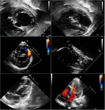
We operated three patients for muscular apical ventricular septal defects (Table 1) and associated cardiac malformation. The median age was 6.3 months (with a range from 2 to 13 months). Their body surface area and weight ranged from 0.24 to 0.45 m2 (median, 0.32 m2) and 4.6 to 9 kg (median, 6.4 kg), respectively (Fig 1).
Preoperative and postoperative echocardiogram.

Patients characteristics.

BSA=body surface area; PM=perimembranous
Surgical technique
The surgery was performed through a median sternotomy with mild hypothermic cardiopulmonary bypass. After aortic cross-clamping, a right atriotomy was performed.
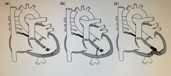
To close the large apical or multiple ventricular septal defects in two patients, we used one or two pericardial patches. The objective was to create a new floor in the right ventricle, leaving the multiple ventricular septal defects on the left side. The first bovine pericardial patch was sutured at the upper edge of the ventricular septal defect and the septum marginalis moderator band using a 6–0 polypropylene running suture. A second pericardial patch was used to close the large apical ventricular septal defect. It was sutured between the moderator band and the anterior wall of the right ventricle with a 6–0 polypropylene running suture (Fig 1).
In the third patient, a 5–0 polypropylene purse-string suture was used to exclude the apical portion of the right ventricle, thereby occluding multiple defects between the muscular bands in the right ventricle (Table 1). A bovine pericardial patch was used to close the perimembranous ventricular septal defect with a 7–0 polypropylene running suture in this patient.
The moderator band was kept intact in all cases.
With both techniques, the apical portion of the right ventricular apex was excluded from the rest of the right ventricular circulation.
Results
The mean cross-clamp time and cardiopulmonary bypass time were 55.3±16.2 and 91.0±13.1 minutes, respectively. There was no hospital mortality. At the postoperative echocardiography evaluation, the left ventricular ejection fraction was within the normal range in all patients, without any residual shunt.
Discussion
The management of patients with large apical ventricular septal defect remains controversial. Interventional catheter techniques, palliative surgery, or primary closure have been used with variable success, but specific management guidelines remain undefined.
The results are less than optimum, although interventional catheter technology is being used for treating these defects.Reference Holzer, Balzer, Cao, Lock and Hijazi 1
Some authorsReference Cetin, Ozkara and Akcevin 2 consider the right atrial approach as the most difficult way to achieve the closure of multiple apical ventricular septal defects; therefore, they recommend a palliative surgery with a pulmonary artery banding to prevent congestive heart failure in early infancy and allow the heart cavities to grow. Pulmonary artery banding operation, however, has high mortality in the early postoperative period and in addition causes progressive right ventricular hypertrophy and diastolic dysfunction.
The muscular septal defects located in the apical portion of the septum are difficult to approach even through the right or left ventricle.Reference Gu, Zhou, Gu and Zhang 3 The presence of the moderator band and the multiple trabeculations in this area hide these defects despite extensive resection of muscular trabeculations.Reference Shin, Jhang and Park 4
In 2003, Ootaki et al.Reference Ootaki, Yamaguchi, Yoshimura, Oka, Yoshida and Hasegawa 5 developed a technique for the closure of trabecular muscular defects by sandwiching the septum between two polyester felt patches placed on the left and right ventricles. The outcome with this technique was satisfactory. Although the surgical access through a left ventriculotomy helps in visualising the defects more easily, this approach has its own complications in the form of ventricular dysfunction, dyskinesis, apical aneurysm, and ventricular arrhythmias.
We used a transatrial approach in order to avoid the disadvantages of ventriculotomy, ventricular dysfunction, arrhythmias, and possible aneurysm formation.Reference Mishra, Shah and Desai 6 When a very large apical ventricular septal defect is present, two patches are needed. The first one is sutured between the upper edge of the apical ventricular septal defect and the septomarginal moderator band, and the second one is sutured between the moderator band and the free anterior right ventricular wall. This approach for complete correction of an apical complex ventricular septal defect offers very good initial results, avoiding complications associated with left ventriculotomy as well as with palliative pulmonary artery banding.
More patients with a longer duration of follow-up are needed to reach a meaningful conclusion. Further experience and precise evaluation of the right ventricular function are also necessary to assess the safety and efficacy of this approach in small infants (Fig 2).
Surgical technique. (a) Anatomical view of an apical muscular VSD. (b) Closure of an apical VSD with a pericardial path. (c) Closure of an apical VSD with a suture.

Acknowledgements
The authors thank the perfusionist, cardiologist, anestesist, and ICU doctors of their unit.
Financial Support
This research received no specific grant from any funding agency or from commercial or not-for-profit sectors.
Conflicts of Interest
None.