Introduction
The West Nile virus (WNV) belongs to the genus Flavivirus Flaviviridae serocomlex that was first separated from the blood of a feverish woman in Uganda in 1937. Until 1999, the geographical distribution of WNV was restricted to Africa, the Middle East, western and central Asia, India and Europe. WNV has spread in the western hemisphere since 1999, and also spread in the United States. In recent years, it has also been found in migratory birds in some regions of the Xinjiang Uygur Autonomous Region of China (Petersen and Roehrig, Reference Petersen and Roehrig2001; Cui et al., Reference Cui, Raymond, Berthomieu and Alout2006). WNV is arthropod borne and is transmitted to vertebrates by infected mosquito or tick vectors. Wild birds are the reservoir host of WNV. Since WNV can be transmitted through blood transfusion, dissemination of the infection brought a huge threat to the safety of transfused blood (Nicolle et al., Reference Nicolle, Gutkin and Smart2004; Zhang, Reference Zhang2004).
Neither a vaccine nor an effective therapy has been developed for WNV infection in humans. Development of an effective assay method is essential for prevention of WNV. For forecasting the infectivity and epidemiology of WNV in humans, horses and other animals, we need to establish a monitoring system for blood products, mosquitoes and birds, etc. (Bakonyi et al., Reference Bakonyi, Hubalek and Rudolf2005; Bakonyi and Ivanics, Reference Bakonyi and Ivanics2006).
This study developed a real-time polymerase chain reaction (RT-PCR) using the intercalating dye SYBR Green I. The assay should be economical for large-scale routine testing of clinical samples and blood products in China.
Materials and methods
Virus gene and sample
The WNV cDNA sample was kindly provided by Dr Li of the Transfusion Medicine Department of Haematology, Cambridge University. The live vaccine of JEV (Japanese encephalitis virus) is kept in our laboratory.
Reagents
The Real Master Mix (SYBR Green I) was bought from Tiangen Biotech (Beijing) Co. Ltd (Beijing, China); the AMV system, plasmid kit, gel extraction kit and pGEMT-Easy Vectors were all bought from the Promega Corporation (Madison, Wisconsin, USA); the DNA/RNA nucleic acid extraction kit was bought from AXYGEN Scientific, Inc. (San Francisco, California, USA).
Primer design
Specific RT-PCR primers were designed according to the conserved region of the capsid gene of WNV. The length of amplified segment was 158 bp. The primer designing software was Primer Premier 5.0 (Premier Biosoft International Company, Canada). The primers were synthesized by Beijing Sunbio Biotech Co. Ltd. The sequences were:
The upstream sequence (WP1): 5′-TCTAAGAAACCAGGAGGGCC-3′;
The downstream sequence (WP2): 5′-AAGAACGCCAAGAGAGCCAAC-3′.
Preparation of standard plasmid of WNV
The RNA extraction of WNV was performed according to the manual of the AXYGEN RNA extraction kit. The amplified segments were subcloned into pGEM-T Easy Vectors. Sequencing of the amplified segment was carried out by Beijing Sunbio Biotech Co. Ltd. Tenfold successive dilutions of positive WNV DNA (107–103 copies/μl) were used as a standard plasmid of WNV.
RT-PCR amplification
The standard templates (107–103 copies/μl) were amplified by real-time PCR. The total reaction mixture of 20 μl contained 9 μl of Real Master Mix (SYBR Green I), 1.5 μl WP1 and WP2 respectively, 1 μl template, 7 μl H2O. The RT-PCR profile was: 2 min at 94°C; followed by 40 cycles of 94°C for 30 s, 60°C for 30 s and 68°C for 30 s. The RT-PCR products were analysed by melting curves and 2% agarose gel electrophoresis.
Specificity, sensitivity and reproducibility of the RT-PCR assay
The specificity of the assay system was determined by amplified WNV gene segments and JEV gene segments, with the same primers and reaction system of WNV. To measure the sensitivity of RT-PCR assay for WNV, the assay was performed using serially diluted standard templates. The intra- and inter-assay reproducibility were evaluated by coefficient of variation (CV).
Results
The result of amplified standard templates
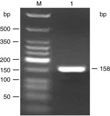
Electrophoresis of RT-PCR products showed a 158 bp segment as expected, indicating that the amplification was successful (Fig. 1). The positive segments were subcloned into pGEMT-Easy Vectors and quantitative assays of duplicates gave 2.607×10¹⁰ copies/μl.
The amplification results of the cDNA segment. Lanes: M, low molecular weight DNA ladder; 1, amplified fragment of standard sample.

Detection limit of the real-time PCR assay
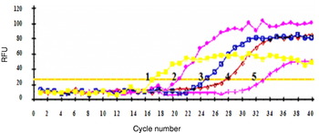
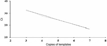
Tenfold serial dilutions of the standard templates were used in the real-time PCR assay. The cycle threshold (Ct) values for each dilution were measured in duplicate (Fig. 2). The log-linear regression plot generated from the collected data showed a strong linear relationship (Fig. 3). The regression equation is y=−3.982x+47.977; r=0.996. The detection limit of the assay was determined to be 102 copies/μl.
The amplification results of standard samples of WNV. 1, 1×10⁷ copies/μl; 2, 1×10⁶ copies/μl; 3, 1×10⁵ copies/μl; 4, 1×10⁴ copies/μl; 5, 1×10³ copies/μl. RFU, relative fluorescence unit. (See online for a colour version of this figure.)

Regression curves of standard samples of WNV. Ct, cycle threshold.

Specificity of the real-time PCR assay
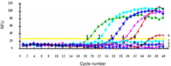
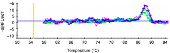
To determine assay specificity, JEV live vaccine strains and WNV genes were tested in the real-time PCR assay. The result indicated that the assay system cannot amplify JEV gene segments (Fig. 4). At the same time, primer-dimers were produced during the amplification reaction (Fig. 5). Thus, the results indicated a high specificity for assay of WNV.
The sensitivity and specificity of assay systems. Lines 1–7, WNV: 1, 1×10⁶ copies/μl; 2, 1×10⁵ copies/μl; 3, 1×10⁴ copies/μl; 4, 1×10³ copies/μl; 5, 1×10² copies/μl; 6, 1×101 copies/μl; 7, 1×100 copies/μl; Line 8, JEV; Line 9, negative sample. RFU, relative fluorescence unit. (See online for a colour version of this figure.)

The melting curves of standard samples of WNV. (See online for a colour version of this figure.)

Reproducibility of the real-time PCR assay
The inter-assay reproducibility was assessed by testing the standard templates on different days. The intra-assay reproducibility was assessed by testing the standard templates in four repeats. The assay system showed high reproducibility with CV <2% (Table 1).
Analysing the reproducibility of RT-PCR assay for WNV

Discussion
Real-time fluorescence quantitative PCR was developed based on traditional PCR, and has several advantages over conventional PCR. RT-PCR is more sensitive and yields more information. Detection, classification and quantification take place within one tube. RT-PCR has already been used extensively for detection of many kinds of pathogens (Hadfield et al., Reference Hadfield, Turell and Dempsey2001; Lo et al., Reference Lo, Tilgner and Shi2003; Parida et al., Reference Parida, Posadas, Inoue, Hasebe and Morita2004). In real-time PCR, DNA-binding dyes are used as fluorescent reporters to monitor the real-time PCR reaction. The fluorescence of the reporter dye increases as the product accumulates with each successive cycle of amplification. By recording the amount of fluorescence emission at each cycle, it is possible to monitor the PCR reaction during the exponential phase.
SYBR Green is the most widely used double-strand DNA-specific dye reported for real-time PCR. It binds to the minor groove of the DNA double helix. In solution, the unbound dye exhibits very little fluorescence. This fluorescence is substantially enhanced when the dye is bound to double-stranded DNA. SYBR Green remains stable under PCR conditions and the optical filter of the thermocycler can be arranged to harmonize the excitation and emission wavelengths (Hadfield et al., Reference Hadfield, Turell and Dempsey2001).
With the increase of human travel, the migration of birds and the use of foreign blood products, the risk of human WNV infection in China has become more and more serious. So it was necessary to establish an assay method to detect WNV in China. The segment amplified in this study was the most conserved capsid area of WNV genes. Through analysing the primers with the software Primer Premier 5.0, the possibility of producing primer-dimers is notably decreased.
In conclusion, the RT-PCR described here for detection and quantification of WNV has been shown to be sensitive and specific, making it an excellent large-scale screening tool for many kinds of samples.
Acknowledgement
This work was supported by the scientific funds of the National High Technology Research and Development Program (‘863’ Program) of China.