Introduction
As one of the most economically significant vegetable crops, tomato (Solanum lycopersicum L.) is cultivated globally. Its production is severely threatened by the highly destructive phloem-feeding pest, whitefly Bemisia tabaci (Gennadius) (Hemiptera: Aleyrodidae), which causes disastrous consequences by feeding and virus transmission (Gilbertson et al., Reference Gilbertson, Batuman, Webster and Adkins2015). For a long period, chemical control methods based on pesticides have been employed to manage B. tabaci. Unfortunately, the indiscriminate use of these chemicals has led to the development of resistance within B. tabaci populations (Horowitz et al., Reference Horowitz, Ghanim, Roditakis, Nauen and Ishaaya2020; Zheng et al., Reference Zheng, Xie, Fu, Xiao, Tan, Ji, Cheng, Wang, Liu, Yang, Guo, Wang, Wu, Xu, Zhou and Zhang2021). Additionally, growing concerns about environmental pollution from these chemicals have increased the emphasis on utilising whitefly-resistant varieties in integrated pest management strategies (Suhag et al., Reference Suhag, Yadav, Chaudhary, Subramanian, Jaiwal and Jaiwal2021; van den Oever‐van den Elsen et al., Reference van den Oever‐van den Elsen, Lucatti, van Heusden, Broekgaarden, Mumm, Dicke and Vosman2016).
Conventional resistance breeding, which involves introducing traits from highly resistant plant accessions into target crop cultivars, has been reported primarily in tomato. The first tomato cultivar with resistance, Motelle, carrying the Mi-1 gene, was developed by introgressing this gene from the wild species Lycopersicon peruvianum into the cultivated variety S. lycopersicum Moneymaker (Ho et al., Reference Ho, Weide, Ma, Vanwordragen, Lambert and Koornneef1992). Initially, the Mi gene was identified for its resistance to nematodes; subsequent studies confirmed that Motelle exhibited lower susceptibility to whitefly compared to Moneymaker (Jiang et al., Reference Jiang, Nombela and Muniz2001; Nombela et al., Reference Nombela, Beitia and Muniz2000). Following this, several studies have reported that wild relatives of cultivated tomato serve as sources of whitefly resistance. For example, introgressing traits associated with whitefly resistance from the wild tomato species Solanum pimpinellifolium TO-937 into Moneymaker resulted in enhanced whitefly resistance (Escobar-Bravo et al., Reference Escobar-Bravo, Alba, Pons, Granell, Kant and Moriones2016; Rodríguez-López et al., Reference Rodríguez-López, Garzo, Bonani, Fereres, Fernández-Muñoz and Moriones2011). Additionally, mini tomato cultivars of S. lycopersicum were intercrossed with S. pennellii LA-716, yielding several lines of mini tomatoes with high levels of whitefly resistance (Maciel et al., Reference Maciel, Almeida, da Rocha, Andaló, Marquez and Santos2017). These resistant lines, developed through conventional breeding, have since been used to generate new resistant varieties. For instance, two tomato lines, both carrying the Mi gene but differing in acyl-sugar content, were crossed to produce several lines with significantly enhanced whitefly resistance (Gouveia et al., Reference Gouveia, de Oliveira, Ribeiro and Maluf2018). Although the utilisation of resistant varieties is a promising method for whitefly management, the evolution of specific whitefly sibling species that adapt to overcome these resistance traits significantly reduces the long-term effectiveness of crop resistance (Elbaz et al., Reference Elbaz, Halon, Malka, Malitsky, Blum and Aharoni2012). Therefore, investigating the molecular mechanisms by which whiteflies initiate resistance responses in plants could help extend the duration of crop resistance, thereby improving the sustainability of agricultural production.
Plants can activate inducible defence mechanisms to resist herbivore infestations. Resistant plants typically exhibit augmented induced defence responses, such as increased production of reactive oxygen species (ROS) (Kerchev et al., Reference Kerchev, Fenton, Foyer and Hancock2012; Shoala et al., Reference Shoala, Edwards, Knight and Gatehouse2018), phloem protein plugging (Medina-Ortega and Walker, Reference Medina-Ortega and Walker2015; Tjallingii, Reference Tjallingii2006), and callose deposition (Liu et al., Reference Liu, Du, Ding, Zhou, Xie and Wu2017; Villada et al., Reference Villada, González, López-Sesé, Castiel and Gómez-Guillamón2009). One of the key resistance mechanisms against herbivorous insects, including whiteflies, is the accumulation of ROS (Kempema et al., Reference Kempema, Cui, Holzer and Walling2007; Liu et al., Reference Liu, Williams, Nemacheck, Wang, Subramanyam, Zheng and Chen2010). The generation of ROS is catalysed by various enzymes, with NADPH oxidases being among the most studied (Mittler et al., Reference Mittler, Vanderauwera, Gollery and Van Breusegem2004). Among the genes encoded respiratory burst oxidase homolog (RBOH) in tomato, the expression level of RBOH1 was most abundantly expressed in leaves (Nie et al., Reference Nie, Wang, Xia, Zhou, Shi, Chen and Yu2013). In contrast, plants employ a range of ROS-scavenging enzymes, such as catalase (CAT), superoxide dismutase (SOD), and ascorbate peroxidase (APX), to maintain redox homeostasis by removing excess ROS (Mittler et al., Reference Mittler, Vanderauwera, Gollery and Van Breusegem2004). Phloem protein plugging occurs rapidly in response to herbivore attack, although knowledge of the specific mechanisms behind sieve element protein plugging remains limited (Garzo et al., Reference Garzo, Fernández-Pascual, Morcillo, Fereres, Gómez-Guillamón and Tjallingii2018; Knoblauch et al., Reference Knoblauch, Froelich, Pickard and Peters2014). Persistent callose deposition, a β-1,3-glucan, is induced not only by wounding and pathogen infection but also by insect infestation (Luna et al., Reference Luna, Pastor, Robert, Flors, Mauch-Mani and Ton2011; Voigt, Reference Voigt2014). Whitefly infestation has been shown to induce the expression of callose synthase (CalS) genes, promoting callose deposition in Arabidopsis thaliana (Kempema et al., Reference Kempema, Cui, Holzer and Walling2007). Twelve CalS genes in A. thaliana are responsible for callose synthesis (Verma and Hong, Reference Verma and Hong2001). Among these, CalS1 and CalS7 are both upregulated following aphid infestation, with CalS1 also induced by whitefly infestation and CalS7 by wounding (Kempema et al., Reference Kempema, Cui, Holzer and Walling2007; Kuśnierczyk et al., Reference Kuśnierczyk, Winge, Jørstad, Troczyńska, Rossiter and Bones2008; Sun et al., Reference Sun, Voorrips, Steenhuis-Broers, Van’t Westende and Vosman2018; Xie et al., Reference Xie, Wang, Zhu, Zhang and Hong2011). Additionally, Cals12, which is responsible for wound and papillary callose formation, is upregulated by both pathogen infection and insect feeding (Jacobs et al., Reference Jacobs, Lipka, Burton, Panstruga, Strizhov, Schulze-Lefert and Fincher2003; Lü et al., Reference Lü, Sun, Zhang, Zhang, Qian, Wang, Gao and Dong2011). Alongside callose formation and deposition, its degradation also acts as an influential role in plant resistance. β-1,3-Glucanases (GLU), which degrade callose, act as susceptibility factors in interactions between the brown planthopper and rice (Hao et al., Reference Hao, Liu, Wang, Chen, Tang, Du, Zhu and He2008), the predatory mirid bug Nesidiocoris tenuis (Reuter) (Hemiptera: Miridae) and tomato (Depalo et al., Reference Depalo, Gallego, Ortells-Fabra, Salas, Montalt, Urbaneja and Pérez-Hedo2024; Urbaneja et al., Reference Urbaneja, Ortells-Fabra and Pérez-Hedo2024), as well as between the bird cherry-oat aphid and barley (Mehrabi et al., Reference Mehrabi, Åhman and Jonsson2016). Plant resistance to whiteflies manifests at multiple tissue levels, from the epidermal surface to the phloem sieve elements. The electrical penetration graph (EPG) technique is a valuable tool for studying whitefly probing and feeding behaviour, providing insights into plant resistance mechanisms. EPG detects whitefly activity on plants through distinct waveforms (Walker and Janssen, Reference Walker, Janssen, Walker and Backus2000), which can be used to identify specific resistance factors encountered by whiteflies (Jiang et al., Reference Jiang, Nombela and Muniz2001; Jiang and Walker, Reference Jiang and Walker2007).
In previous study concerning the effects of tomato anthocyanins (a kind of flavonoid) on resistance to abiotic and biotic stresses, Qiu et al. (Reference Qiu, Wang, Gao, Guo, Huang and Du2016) developed and compared near-isogenic lines (NILs) of cultivated tomato in which anthocyanins are over-produced (line NIL-purple hypocotyl [PH]) or under-produced (line NIL-green hypocotyl [GH]). Subsequent research showed that, in addition to reduced anthocyanin levels, NIL-GH also had lower levels of other flavonoids compared to NIL-PH (Su et al., Reference Su, Chen, Mescher, Peng, Xie, Wang, Wu, Liu, Li, Wang and Zhang2018; Yang et al., Reference Yang, Zhang, Shen, Xue, Tian, Zhang, Hu, Tong, Zhang and Su2023a). Our previous work demonstrated that high flavonoid production in tomato contributes to both direct and indirect defences against whiteflies, with NIL-PH lines exhibiting greater resistance than NIL-GH lines (Su et al., Reference Su, Chen, Mescher, Peng, Xie, Wang, Wu, Liu, Li, Wang and Zhang2018; Yang et al., Reference Yang, Shen, Huang, Yao, Hu, Tang, Zhang, Tong, Wu, Zhang and Su2023b). To further investigate the mechanism underlying the increased resistance of NIL-PH tomato to whiteflies, we examined the biochemical interactions between the whiteflies and both tomato NILs, which involves not only whitefly feeding activity but also constitutive and induced plant resistance mechanisms. Our results indicated that whitefly feeding triggered stronger ROS accumulation and callose deposition in resistant NIL-PH plants, leading to significantly decreased phloem feeding and reduced whitefly performance, compared to the susceptible NIL-GH plants.
Materials and methods
Plant growth and whitefly rearing
The two tomato NILs, NIL-PH and NIL-GH, used in this study were obtained from the initial cross of cultivar FMTT271 (lacking anthocyanin pigments due to loss of function of the anthocyanin synthesis gene AH (Hoffman’s anthocyaninless; Solyc09g065100)) with wild-type S. pennellii LA716 (purple pigmentation in the hypocotyl) followed by four cycles of combined recurrent crosses with cv. FMTT271 and subsequent selfing steps with selection for high or low anthocyanin production by anthocyanin quantification (Qiu et al., Reference Qiu, Wang, Gao, Guo, Huang and Du2016). Compared to NIL-PH plants, NIL-GH plants are deficient in anthocyanins and other flavonoids, including rutin, quercetin, kaempferol, myricetin, chlorogenic acid, and quinic acid (Su et al., Reference Su, Yang, Hu, Peng, Huang, Tong, Zhang, Yang, Zhou, Liang, Zhu, Wu, Zhang and Guo2025). Plants were grown in a whitefly-proof greenhouse at 25 ± 2°C, 50–70% relative humidity (RH), and a 14/10 h light/dark photoperiod as described previously (Su et al., Reference Su, Mescher, Wang, Chen, Xie, Wu, Wang and Zhang2016), and used for experiments when they were 4–5 weeks old and had four fully expanded true leaves.
The colony of whitefly B. tabaci Mediterranean (MED; formerly known as the Q biotype) used in this study was maintained on tomato plants (cv. L402) in individual whitefly-proof screen cages under natural lighting at 26 ± 2°C in a glasshouse, as previously described (Su et al., Reference Su, Peng, Tong, Xie, Wang, Wu and Zhang2019), and newly emerged (0–8 h post-emergence) adults were used in the experiments.
Whitefly performance assays
Groups of 20 adult female whiteflies were restricted to the first leaf (counted from the bottom) of individual NIL-PH and NIL-GH plants within a clip cage (3 cm diameter, one cage/plant) as previously described (Su et al., Reference Su, Peng, Tong, Xie, Wang, Wu and Zhang2019). The number of surviving adults in each clip cage was recorded daily. Whiteflies were removed after 7 days, and the number of eggs laid by females in the clip cages was counted with a stereomicroscope and was used to calculate the average individual fecundity. For whitefly development, 50 adults (a mixture of females and males to 1:1) were released and allowed to feed on NIL-PH and NIL-GH plants in a ventilated cage (13.5 cm diameter, 21.0 cm high; one plant per cage). After 24 h of infestation and oviposition, the adults were removed from the plants. At 21 days after introduction of adults, the number of nymphs and their developmental stages (first through fourth instars) were recorded. Developmental rate was estimated by calculating the proportion of nymphs represented by fourth instars (red-eye stage) on each plant. These experiments were repeated 15 times per plant genotype.
Analysis of whitefly probing and feeding behaviour
We used EPG to analyse whitefly probing and feeding behaviour on NIL-PH and NIL-GH plants according to the method as described in Liu et al. (Reference Liu, Preisser, Chu, Pan, Xie, Wang, Wu and Zhang2013). In brief, individual 2-day-old adult females were placed onto the lower surface of the top leaf and their feeding on plants was monitored continuously for 8 h using a Giga-8 direct-current EPG (DC-EPG) system (Wageningen University, The Netherlands). Relationships between EPG waveforms and feeding behaviour were described by Liu et al. (Reference Liu, Preisser, Chu, Pan, Xie, Wang, Wu and Zhang2013). Fresh plants and whiteflies were used for each replicate, and 16–18 replicates were carried out per plant genotype. All experiments were carried out at 26°C ± 2°C, at 70% RH, and under artificial light (1,500 lx) with a 14/10 h light/dark photoperiod.
ROS accumulation
Groups of 50 adult female whiteflies were placed into a clip cage on one leaflet of individual NIL-PH and NIL-GH plants. Each plant received three clip cages and uninfested control plants received three empty clip cages at the same time. Leaflets from each tomato genotype were harvested at 0, 24, and 48 h after whitefly infestation to measure ROS accumulation using DAB (3,3ʹ-diaminobenzidine) staining as described in Su et al. (Reference Su, Peng, Tong, Xie, Wang, Wu and Zhang2019). Leaflets were examined under an inverted fluorescence microscope and images were then captured with a digital camera. The percentage of the area of brown polymerised deposits, which reflect ROS accumulation on each leaflet, was quantified using ImageJ. The average percentage from three leaflets that were collected from the same plant was used as the data for one biological replicate, and five plants (replicates) per genotype were collected for each treatment.
Assays of antioxidant enzyme activity
Whitefly-infested and uninfested NIL-PH and NIL-GH tomato leaflets were obtained as described in the previous section. Leaf samples were homogenised in liquid nitrogen and were individually completely mixed with 1 mL of phosphate-buffered saline, and the supernatants were collected following microcentrifugation of the extract. The enzyme activities of NADPH oxidase, CAT, SOD, and APX in the supernatant from tomato leaves were analysed using the NADPH oxidase, CAT, SOD, and APX assay kits (Jiancheng, Nanjing, China) by following the manufacturer’s instructions. Soluble protein was quantified by the dye-binding method (Bradford, Reference Bradford1976) with bovine serum albumin as a standard. Results for NADPH oxidase activity are expressed in μM O2.– per mg protein per min, while the activity of other three enzymes are expressed in Units per gram of leaf tissue; treatment values are the mean of six replicates. Analyses were conducted using a fluorescence microplate reader (SpectraMax M2e, Molecular Devices) at room temperature (25 ± 2°C).
Callose deposition
Aniline blue staining was performed to detect callose deposition in whitefly-infested and uninfested NIL-PH and NIL-GH tomato leaflets following the method as described in Su et al. (Reference Su, Preisser, Zhou, Xie, Liu, Wang, Wu and Zhang2015). Callose was extracted as previously described (Yang et al., Reference Yang, Zhang, Xue, Tian, Tong, Hu, Zhang, Tang and Su2022) and quantified by fluorescence spectrophotometry at an excitation wavelength of 400 nm and an emission wavelength of 500 nm, using pachyman (Sigma-Aldrich) as a standard callose source. Each treatment was represented by 10 replicate samples of tomato leaflets.
Gene expression analysis
Total RNA from whitefly-infested and uninfested NIL-PH and NIL-GH tomato leaflets was extracted using the RNeasy plus Mini kit (Qiagen, Hilden, Germany) following the manufacturer’s protocol. First-strand cDNA synthesis and quantitative reverse transcription PCR (qRT-PCR) were performed using the SYBR Green PCR Master Mix (TaKaRa, Dalian, China) on the 7500 Sequence Detection System (Applied Biosystems, Foster City, USA) as previously described (Su et al., Reference Su, Peng, Tong, Xie, Wang, Wu and Zhang2019, Reference Su, Yang, Zhang, Tong, Hu, Zhang, Xie, Wang, Wu and Zhang2020). Expression levels of target genes were normalised to the housekeeping genes Actin and Ubiquitin from tomato. Primers used for the target genes are listed in Table S2. Each gene was analysed in triplicate for each of four biologically independent treatments.
Statistical analysis
Before analysis, data were checked for normality and homogeneity of variance using the Kolmogorov–Smirnov test and Levene’s test, respectively. Differences in whitefly survival, fecundity, development as well as probing and feeding behaviour parameters between the two tomato genotypes were conducted with Student’s t-test. For data on ROS accumulation, antioxidant enzyme activity, callose deposition, and gene expression, analyses of variance (ANOVAs) were used to assess differences between treatments. If the ANOVA was significant, means were compared with a Tukey’s honestly significant difference test. All statistical analyses were performed using SPSS, version 17.0 (SPSS Inc., Chicago, USA).
Results
High flavonoid-producing tomato exhibits strong defence against whiteflies
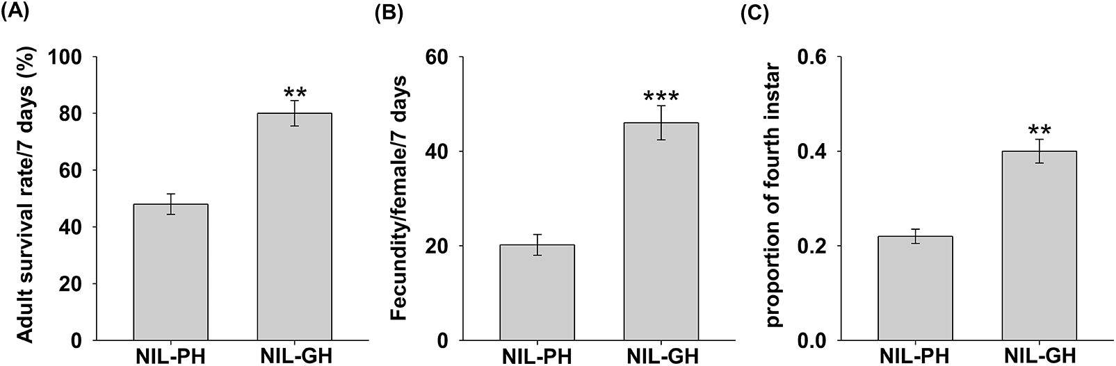
To confirm the resistance of NIL-PH tomato against B. tabaci, we compared B. tabaci performance on NIL-PH and NIL-GH tomato plants. Seven days after releasing adult female whiteflies, adult survival on NIL-GH plants was significantly higher than that on NIL-PH plants (fig. 1A). Likewise, the mean number of eggs laid per female on NIL-GH plants was 2.3-fold higher than that on NIL-PH plants (fig. 1B). Moreover, the proportion of fourth instars was significantly higher on NIL-GH plants than on NIL-PH plants at 21 days after whitefly infestation (fig. 1C).
High flavonoid-producing tomato exhibits a strong defence against whiteflies. Adult whiteflies were allowed to feed on high flavonoid-producing NIL-PH and low flavonoid-producing NIL-GH tomato plants for seven days before their survival (A) and fecundity (B) were determined. (C) The proportion of fourth nymphs at 21 days after adult whiteflies were placed on NIL-PH and NIL-GH tomato plants. Values are the means ± SEs (n = 15). Asterisks in (A)–(C) indicates significant differences between the two plant genotypes (**P < 0.01 and ***P < 0.001 respectively, Student’s t-test).

High flavonoid-producing tomato deters whitefly phloem-feeding efficiency
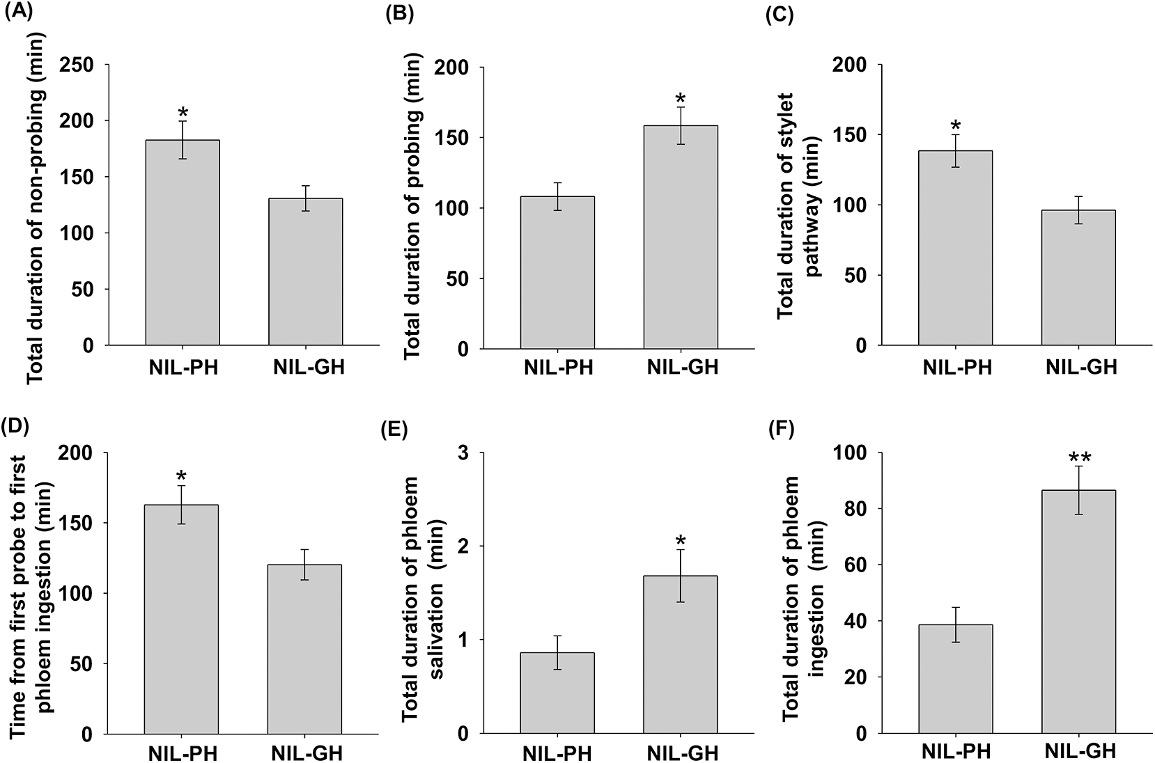
The EPG analysis showed that whiteflies exhibited significantly different probing and feeding behaviours on NIL-PH and NIL-GH plants (Table S1). Based on the EPG parameters, whiteflies did not prefer to feed on NIL-PH plants as shown by a significantly longer duration of non-probing and shorter duration of probing events (fig. 2A, B and Table S1). The longer duration of non-probing events on NIL-PH plants was mainly caused by a longer time from start to first probe (Table S1). Moreover, the duration of stylet pathway and the time from first probe to first phloem ingestion were significantly longer in NIL-PH plants than in NIL-GH plants (fig. 2C, D and Table S1). In addition, the duration of phloem salivation and ingestion by whiteflies on NIL-PH plants was significantly shorter than that of whiteflies on NIL-GH plants (fig. 2E, F and Table S1).
Feeding behaviour parameters of whiteflies fed on high flavonoid-producing NIL-PH and low flavonoid-producing NIL-GH tomato plants. Total duration of electrical penetration graphing waveform types (A–F) for whiteflies fed on NIL-PH and NIL-GH tomato plants over a 8-h recording period. Values are means ± SEs (n = 16–18). Asterisks in (A)–(F) indicates significant differences between the two plant genotypes (*P < 0.05 and **P < 0.01 respectively, Student’s t-test).

Whitefly infestation induces ROS accumulation in high flavonoid-producing tomato
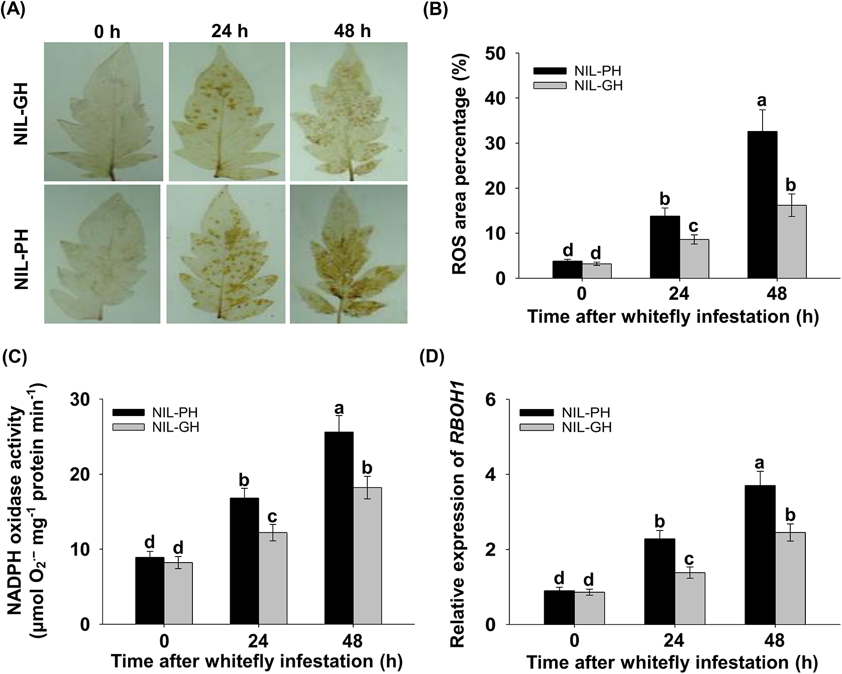
To investigate possible differences in the accumulation of ROS in NIL-PH and NIL-GH plants when whiteflies were present, leaflets where whiteflies had been feeding for 0, 24, and 48 h were collected and stained for ROS accumulation. No notable ROS accumulation was observed in leaves from NIL-PH and NIL-GH plants without whitefly infestation (fig. 3A). However, NIL-GH leaves accumulated fewer ROS compared with that of NIL-PH leaves at 24 and 48 h after whitefly infestation (fig. 3A), which was reflected by a significantly lower percentage of DAB staining area on the NIL-GH leaf (fig. 3B). Next, the enzyme activities of NADPH oxidase, CAT, SOD, and APX in NIL-PH and NIL-GH plants at 0, 24, and 48 h after whitefly infestation were measured. As shown in fig. 3C, the activity of ROS-generating NADPH oxidase did not differ significantly between NIL-PH and NIL-GH plants without whitefly infestation, but significantly increased in NIL-PH plants compared with that in NIL-GH plants at 24 and 48 h after whitefly infestation. The similar trend was also been found in the expression levels of RBOH1. The expression levels of RBOH1 did not differ significantly between the two genotypes, while significantly increased in NIL-PH plants than in NIL-GH plants at 24 and 48 h after whitefly infestation (fig. 4D). The activities of ROS-scavenging enzymes like CAT, SOD, and APX significantly increased in whitefly-infested plants compared to uninfested plants, while these enzyme activities did not differ significantly between NIL-PH and NIL-GH plants (fig. S1).
ROS accumulation in high flavonoid-producing NIL-PH and low flavonoid-producing NIL-GH tomato leaves in response to whitefly infestation. (A) Representative images of DAB (3,3′-diaminobenzidine) staining to show ROS accumulation in NIL-PH and NIL-GH tomato leaves at 0, 24, and 48 h after whitefly infestation. (B) The percentage of stained DAB area from NIL-PH and NIL-GH tomato leaflet at 0, 24, and 48 h after whitefly infestation was calculated. Values are means ± SEs (n = 5). (C) The NADPH oxidases activity was determined in NIL-PH and NIL-GH tomato leaves at 0, 24, and 48 h after whitefly infestation. Values are means ± SEs (n = 6). (D) Transcript levels of RBOH1 in NIL-PH and NIL-GH tomato leaves at 0, 24, and 48 h after whitefly infestation were analysed using quantitative real time (qRT)-PCR. Values are the means ± SEs (n = 4). Means with different letters in (B)–(D) indicate significant differences between each treatment (P < 0.05, two-way ANOVA followed by Tukey’s post hoc test).

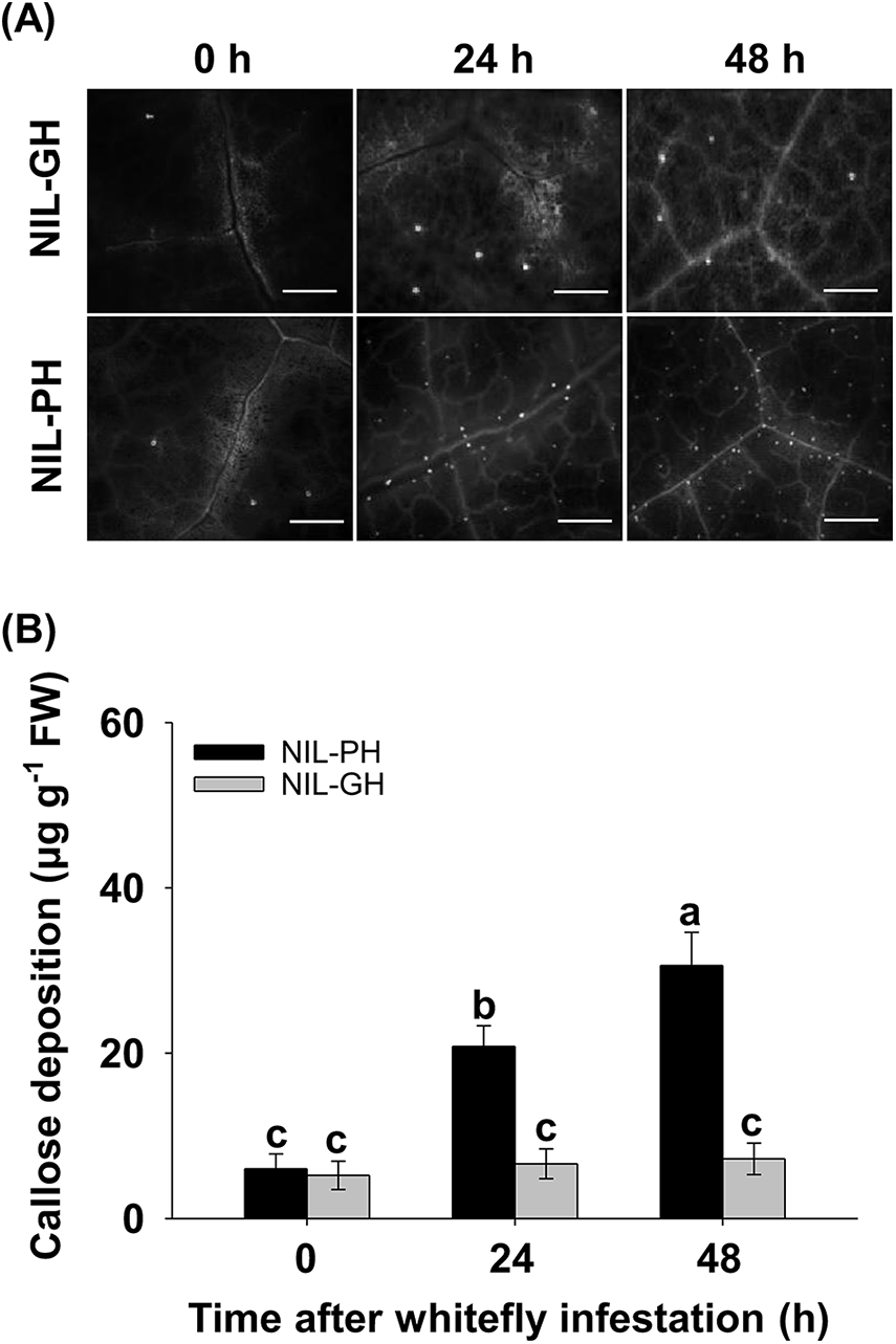
Callose deposition in high flavonoid-producing NIL-PH and low flavonoid-producing NIL-GH tomato leaves in response to whitefly infestation. (A) Histochemical assay for callose deposition in NIL-PH and NIL-GH tomato leaves at 0, 24, and 48 h after whitefly infestation was measured by aniline blue staining. Representative microscope images are shown and callose deposits appear as bright spots on a dark background. Scale bars = 100 μm. (B) Callose deposition in NIL-PH and NIL-GH tomato leaves at 0, 24, and 48 h after whitefly infestation. Values are means ± SEs (n = 10). Means with different letters in (B) indicate significant differences between each treatment (P < 0.05, two-way ANOVA followed by Tukey’s post hoc test).

Whitefly infestation enhances callose deposition in high flavonoid-producing tomato
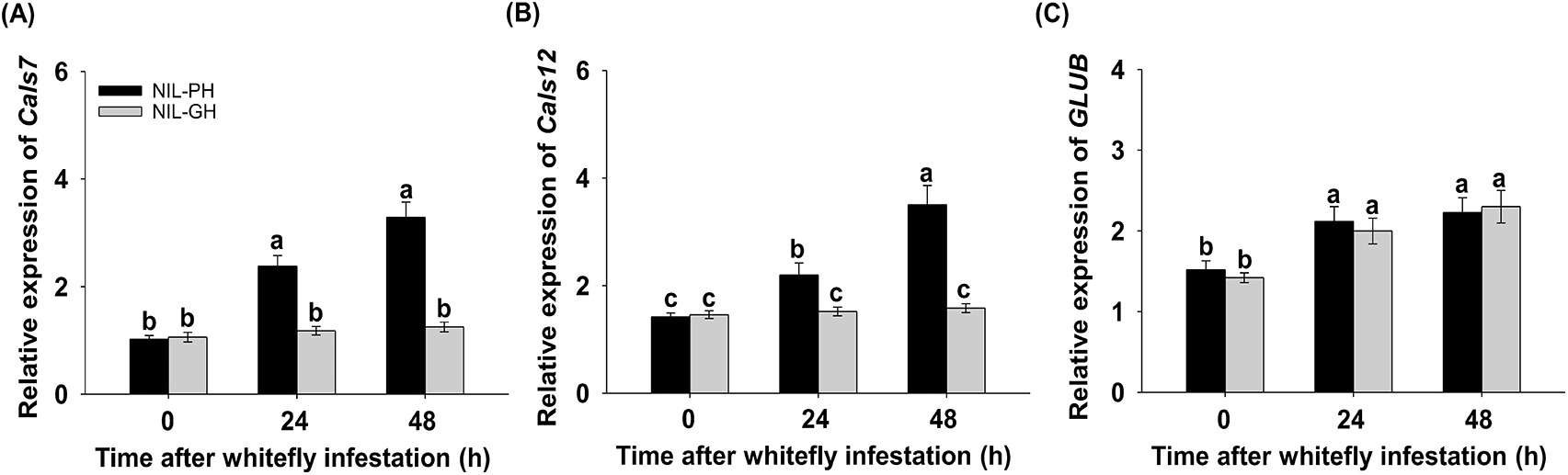
The formation of callose was examined to explore differences in defence after infestation with whiteflies on NIL-PH and NIL-GH plants. No obvious callose signal was detected in uninfested NIL-PH and NIL-GH leaves or in whitefly-infested NIL-GH leaves, while whitefly infestation significantly enhanced callose signal in the vascular tissue of NIL-PH leaves (fig. 4A). Consistently, callose deposition was increased in NIL-PH leaves but not in NIL-GH leaves after whitefly infestation (fig. 4B). To shed light on the regulation of the callose deposition, we compared the expression of callose synthase (CalS) genes and callose degradation GLU genes in whitefly-infested leaves with those of uninfested leaves. Among the eight CalS genes, only CalS7 and CalS12 showed a clear change in transcript accumulation upon whitefly infestation (figs 5A, B and S2). In the NIL-GH leaves, the expression of the CalS7 and CalS12 genes remained stable during 48 h of whitefly infestation compared to uninfested leaves (fig. 5A, B). No difference in expression was detected for both CalS7 and CalS12 genes between NIL-PH and NIL-GH leaves without whitefly infestation, but expression of these two genes were significantly up-regulated in NIL-PH leaves at 24 and 48 h after whitefly infestation (fig. 5A and b). Among the seven GLU genes examined, six of them (GLU1, 3, 5, 6, 7, and 13) were either stable or little induced by whitefly infestation in NIL-PH and NIL-GH leaves (fig. S3). On the other hand, GLUB was significantly induced in NIL-PH and NIL-GH leaves at 24 and 48 h after whitefly infestation (fig. 5C). However, there was no significant difference in expression of all these genes between NIL-PH and NIL-GH leaves (fig. S3).
Expression analysis of callose synthase and callose degradation genes in response to whitefly infestation. Transcript levels of Cals7 (A), Cals12 (B), and GLUB (C) in NIL-PH and NIL-GH tomato leaves at 0, 24, and 48 h after whitefly infestation were analysed using quantitative real-time (qRT)-PCR. Values are the means ± SEs (n = 4). Means with different letters in (A)–(C) indicate significant differences between each treatment (P < 0.05, two-way ANOVA followed by Tukey’s post hoc test).

Discussion
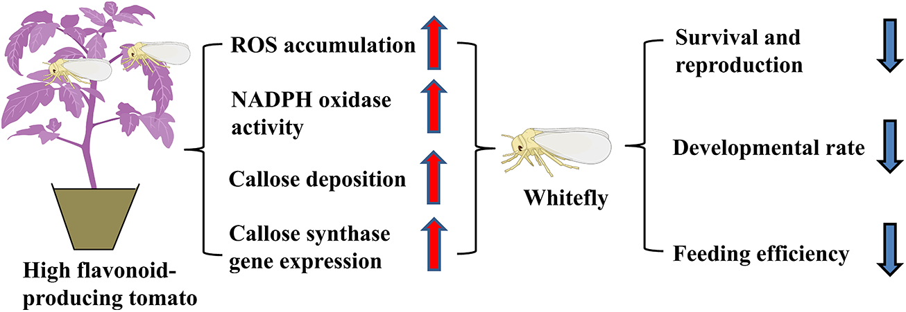
Our previous studies demonstrated that high flavonoid-producing NIL-PH tomato exhibited stronger resistance to whiteflies compared to the low flavonoid-producing NIL-GH tomato (Su et al., Reference Su, Chen, Mescher, Peng, Xie, Wang, Wu, Liu, Li, Wang and Zhang2018; Yang et al., Reference Yang, Shen, Huang, Yao, Hu, Tang, Zhang, Tong, Wu, Zhang and Su2023b), while the mechanism underlying this resistance in NIL-PH tomato remains unclear. In the present study, we observed that whiteflies encountered difficulties in the ingestion of phloem, which triggered a robust defence response in the resistant NIL-PH tomato. This response included the accumulation of ROS and callose deposition, ultimately leading to a reduction in whitefly performance on this tomato line (fig. 6). Similar defence mechanism, such as the production of acyl-sugars, has been reported in ABL 14-8 and BC5S2 tomato lines, which also confer resistance to whiteflies (Escobar-Bravo et al., Reference Escobar-Bravo, Alba, Pons, Granell, Kant and Moriones2016; Rodríguez-López et al., Reference Rodríguez-López, Garzo, Bonani, Fereres, Fernández-Muñoz and Moriones2011). Furthermore, BC5S2 tomato plants fed upon by the predatory mirid bug N. tenuis, which is used for B. tabaci management in southern Spain, exhibited an increased density of type IV trichomes and enhanced acyl-sugar production, traits that have been shown to contribute to effective whitefly suppression (Riahi et al., Reference Riahi, Urbaneja, Fernández-Muñoz, Fortes, Moriones and Pérez-Hedo2023).
A proposed model of high flavonoid-producing tomato increases whitefly resistance. When feeding on high flavonoid-producing NIL-PH tomato, B. tabaci triggers a robust defence response in the resistant NIL-PH tomato, including the accumulation of ROS and callose deposition, ultimately leading to a reduction in whitefly performance on this tomato genotype.

The application of EPG technique enables the in-depth investigation of the feeding behaviour of phloem-feeding insects and could specify the possible resistance factor encountered by these insects during the feeding process on plants (Jiang and Walker, Reference Jiang and Walker2007; Tjallingii, Reference Tjallingii2006). In this study, we detected many differences of whitefly feeding between NIL-PH and NIL-GH plants through the EPG recordings, and these differences were apparent at almost all phases, although not for all parameters (Table S1). The non-probing period of whiteflies was longer on NIL-PH plants compared to NIL-GH plants, while the duration of probing was shorter in NIL-PH plants (fig. 2A, B). Moreover, whiteflies spent longer time for stylet pathway and from the first probe to the first phloem ingestion in NIL-PH plants than in NIL-GH plants (fig. 2C, D). Once the whitefly stylet reached the phloem, the duration of phloem salivation and ingestion was shorter in NIL-PH plants than in NIL-GH plants (fig. 2E, F). As a result, whiteflies feeding on resistant NIL-PH plants face difficulties in initiating and sustaining phloem sap ingestion. These feeding challenges of whiteflies caused by the phloem-based resistance are likely to grow more slowly, have lower fecundity and higher mortality due to the problems they experience with taking up insufficient nutrition, which is in line with our observations of whitefly performance (fig. 1). In addition to its potential to control whitefly populations, phloem-based resistance may reduce the transmission of persistent viruses, because whiteflies typically cannot acquire these viruses during short feeding periods (Fereres and Moreno, Reference Fereres and Moreno2009; Jiang et al., Reference Jiang, De Blas, Barrios and Fereres2000). Consequently, the decreased salivation and phloem ingestion with NIL-PH plants reduced the efficiency of virus transmission by whiteflies, as observed in our previous study that NIL-PH plants significantly reduced the spread of Tomato yellow leaf curl virus (Yao et al., Reference Yao, Peng, Tong, Yang, Xing, Wang, Zheng, Zhang and Su2019).
The mechanism underlying phloem-based resistance may involve the occlusion of phloem vessels in response to whitefly feeding, potentially caused by the accumulation of ROS and callose deposition. ROS accumulation occurs more rapidly and earlier than callose deposition following pathogen or insect attack (Miller et al., Reference Miller, Schlauch, Tam, Cortes, Torres and Shulaev2009). Compared to NIL-GH plants, whitefly infestation induced a stronger defence response in NIL-PH plants, accompanied by more ROS accumulation (fig. 3A, B). ROS accumulation in plants upon insect interaction has been well-documented, and variations in ROS levels are associated with differences in plant resistance (Liu et al., Reference Liu, Williams, Nemacheck, Wang, Subramanyam, Zheng and Chen2010; Moloi and van der Westhuizen, Reference Moloi and van der Westhuizen2006). ROS is believed to function as a local toxin and deters attackers (Liu et al., Reference Liu, Williams, Nemacheck, Wang, Subramanyam, Zheng and Chen2010). It is plausible that the strong ROS accumulation in resistant NIL-PH tomato leaves is localised in the phloem vessels, as indicated by the distribution of stained spots along leaf veins (fig. 3A). NADPH oxidases have been implicated in ROS production during plant–insect interactions (Foyer et al., Reference Foyer, Rasool, Davey and Hancock2016; Torres, Reference Torres2010), and higher NADPH oxidase activity has been observed in aphid-infested resistant plants, such as wheat and maize, compared to susceptible varieties (Sytykiewicz, Reference Sytykiewicz2016). Our analysis showed increased NADPH oxidase activity and SlRBOH1 expression in NIL-PH plants upon whitefly infestation (fig. 3C, D), suggesting that NADPH oxidase-mediated ROS production may be crucial to the defence response of resistant NIL-PH plants against whiteflies.
Whitefly infestation induced a stronger callose signal in the leaf veins of resistant NIL-PH plants compared to susceptible NIL-GH plants (fig. 4), suggesting that one or more genes from the tomato CalS genes or GLU genes may contribute to the observed differences between resistant NIL-PH and susceptible NIL-GH plants in response to whitefly infestation. We conducted quantitative real-time PCR analysis to examine whether increased callose deposition is linked to the enhanced expression of CalS genes. Among the eight CalS family genes, only CalS12 and CalS7 were significantly induced in NIL-PH leaves at 24 and 48 h after whitefly infestation, while expression levels of these genes remained constant in NIL-GH leaves during the 48 h of whitefly infestation (figs 5A, B and S2). Our previous study also confirmed that whitefly infestation induced the expression of tomato CalS12 gene (Yang et al., Reference Yang, Zhang, Xue, Tian, Tong, Hu, Zhang, Tang and Su2022), which is essential for callose formation in tomato (Adkar-Purushothama et al., Reference Adkar-Purushothama, Brosseau, Giguere, Sano, Moffett and Perreault2015). The CalS7 gene, the only phloem-specific callose synthase, mediates the callose biosynthesis in developing sieve elements and in mature phloem by mechanical wounding (Xie et al., Reference Xie, Wang, Zhu, Zhang and Hong2011). Similar to its role in pepper plants, CalS7 expression can also be induced by other phloem-feeding insects like the green peach aphid Myzus persicae (Sulzer) (Hemiptera: Aphididae) (Sun et al., Reference Sun, Voorrips, Steenhuis-Broers, Van’t Westende and Vosman2018), suggesting the common role in callose deposition triggered by phloem-feeding pests. The upregulation of both CalS genes upon whitefly attack in resistant NIL-PH leaves but not in susceptible NIL-GH leaves, which leads us to speculate that CalS7 and/or CalS12 genes are responsible for the increased callose deposition observed in the resistant NIL-PH tomato after whitefly feeding. Interestingly, previous studies confirmed the strong connection between ROS (e.g. H2O2) and callose deposition, and the heightened accumulation of callose is tightly associated with the transcriptional regulation of Cals12 (Vellosillo et al., Reference Vellosillo, Jorge, Satish, Mats and Carmen2010; López-Cruz et al. Reference López‐Cruz, Crespo‐Salvador, Fernández‐Crespo, García‐Agustín and González‐Bosch2017). Furthermore, application of chemical inhibitor ascorbic acid which could remove H2O2 significantly inhibited the accumulation of callose, reinforcing the notion that ROS could act as stress signal to promote plant defence (Hosseini-Zahani and Parissa, Reference Hosseini-Zahani and Parissa2023).
The GLU protein, commonly known as pathogenesis-related protein 2, is responsible for hydrolysing callose to destabilise the cell wall of pathogens and releases immune elicitors that trigger defence responses (Funnell et al., Reference Funnell, Lawrence, Pedersen and Schardl2004; Gupta et al., Reference Gupta, Ravi and Sharma2013). Previous studies have shown that aphid infestation induced the accumulation of GLU protein or its gene transcript in the leaves of barley and Arabidopsis (De Vos et al., Reference De Vos, Van Oosten, Van Poecke, Van Pelt, Pozo, Mueller, Buchala, Métraux, Van Loon and Dicke2005; Forslund et al., Reference Forslund, Pettersson, Bryngelsson and Jonsson2000). Moreover, the predatory mirid bugs N. tenuis could induce callose deposition in tomato plants, while GLU protein likely facilitated the degradation of callose (Depalo et al., Reference Depalo, Gallego, Ortells-Fabra, Salas, Montalt, Urbaneja and Pérez-Hedo2024; Urbaneja et al., Reference Urbaneja, Ortells-Fabra and Pérez-Hedo2024). It is thought that the accumulation of GLU protein weakens the feeding barrier imposed by callose deposition in susceptible plants, while low GLU expression allows callose deposition to be maintained in resistant plants. However, contrary to this hypothesis, the expression of GLU genes remained stable or increased in both NIL-PH and NIL-GH tomato leaves during the 48 h of whitefly feeding, with no significant difference observed between the two genotypes (figs 5C and S3). This suggests that the relationship between the expression of callose synthesis and degradation genes is delicately balanced. The observed accumulation of GLU could indicate the plant’s need to degrade callose in the phloem, as excessive callose deposition could interfere with the transport of assimilates.
In conclusion, high flavonoid-producing NIL-PH tomato exhibits phloem-based whitefly resistance according to the EPG recordings. This resistance may be associated with ROS accumulation and callose deposition in the sieve elements. The increased activity of NADPH oxidase and the up-regulated expression of CalS7 and CalS12 genes in NIL-PH tomato also support this finding. Further detailed studies are necessary to elucidate the mechanism underlying the interaction between the whitefly and two tomato NILs.
Supplementary material
The supplementary material for this article can be found at https://doi.org/10.1017/S0007485325100205.
Acknowledgements
This work was supported by the National Key R & D Program of China (2022YFD1401200), the National Natural Science Foundation of China (32172388), China Agriculture Research System (CARS-24-C-02), the Outstanding Youth Science and Technology Innovation Team Project of Colleges and Universities in Hubei Province (T2022009), the Science and Technology Innovation Program of the Chinese Academy of Agricultural Sciences (CAAS-ASTIP-IVFCAAS), and the Beijing Key Laboratory for Pest Control and Sustainable Cultivation of Vegetables.
Author contributions
Yujie Li: Writing – review & editing, Writing – original draft, Visualization, Methodology, Investigation, Formal analysis, Data curation, Conceptualization. Tianyu Huang, Fengbo Yang, Rong Zhang, Weina Gu, Peng Lu, Hong Tong: Methodology, Investigation, Formal analysis, Data curation. Yuting Yang, Zhixiong Zhou: Methodology, Data curation, Conceptualization. Qingjun Wu, Youjun Zhang: Project administration, Funding acquisition. Qi Su: Writing – review & editing, Writing – original draft, Visualization, Supervision, Resources, Project administration, Funding acquisition, Conceptualization. All authors have read and agreed to the published version of the manuscript.
Competing interests
The authors declare that they have no competing interests.