Introduction
Panspermia requires that life survive travel through space. Man has explored Earth's moon, and most of the unmanned craft sent to planets and beyond have been contaminated by microorganisms. Exospermia, the dissemination of Earth's life through space (Tepfer & Leach Reference Tepfer and Leach2006), is thus clear, but the capacity of life to thrive in a remote environment has not been demonstrated. No other Earth-like environment is known in the Solar System, and exoplanets are only just being discovered. Conditions on these exoplanets are likely to be inhospitable or extremely deleterious to life as we know it.
The origin of life on Earth is unknown, but fossilized bacteria are found in rocks from 3.43 Gy ago, suggesting that complex life either evolved in the first gigayear of Earth's existence or was imported. The first case, spontaneous generation, could have occurred through incremental steps, starting with simple chemistry. The second case, the introduction of life from outside the Earth or introspermia (Tepfer & Leach Reference Tepfer and Leach2006), requires that life resist conditions in space. Destructive UV light is a potential liability in both cases.
Theories of the origin of life are difficult to test, but we can use terrestrial models to study how organisms currently resist UV radiation, and we can hopefully improve this resistance. The environment on Earth and the chemistry of prebiotic life 4 Gy ago are not known with certitude (Leach et al. Reference Leach, Smith and Cockell2006). Thus, attempts to spontaneously generate life must be made under assumed conditions of simulation. On the other hand, the plausibility of panspermia can be tested by examining the ability of contemporary terrestrial life to withstand space travel. However, proof that life came to Earth via panspermia (introspermia) will require finding life elsewhere (on Mars, for instance) and showing that it uses DNA and the same genetic code as terrestrial life forms. Unfortunately, contamination (as a result of Man's space exploration) potentially complicates the search for extra-terrestrial life.
On Earth, life survives in extreme conditions, e.g. close to undersea fumaroles and in the desiccated Atacama desert. The mechanisms whereby extremophiles adapt to these environments are as diverse as the occupied niches, but they all serve, either directly or indirectly, to protect the biological information encoded in DNA. Some of the mechanisms for conserving the genome are starting to be understood. For instance, Deinococcus radiodurans survives desiccation and radiation by rebuilding a functional genome from the fragments produced during stress, using a novel genome reconstruction strategy (Zahradka et al. Reference Zahradka, Slade, Bailone, Sommer, Averbeck, Petranovic, Lindner and Radman2006). In photosynthetic organisms, damage from UV radiation can be attenuated by UV shields that filter out deleterious UV radiation, but still transmit the wavelengths required to fix CO2.
In exoenvironments, UV radiation would be a major danger for organisms dependent on light to fix carbon. Solar irradiance diminishes between 300 and 130 nm, but since photon energy increases and DNA absorbs in this range, short-wave Solar UV radiation remains a liability for unprotected organisms. On Earth, present-day organisms are shielded from these wavelengths by stratospheric ozone, but before the accumulation of atmospheric oxygen, as a result of photosynthesis, life probably had to cope with the full spectrum of Solar UV radiation. We reasoned that by examining UV shields in present-day organisms, it might be possible to understand how life could have adapted to conditions on primitive Earth, and eventually how to better prepare it for survival in exohabitats.
A UV shield should absorb incident photons and dissipate their energy, if possible without molecular damage. Physical distance between the shield and sensitive targets, such as DNA, would thus help cope with the energy absorbed. Extracellular coats or sheaths are well known in the microbial world, and plant seeds are surrounded by a seed coat. Chromosomal DNA is localized in the interior, as far as possible from incident light and other deleterious components of the environment, and is surrounded by cellular substances that help protect against UV. We assume here that an ideal UV shield for DNA would absorb UV radiation in a similar manner as DNA. We thus evaluate the potential suitability of UV shields according to the degree of similarity between their VUV–UV absorption spectrum and that of DNA. This approach allows us to propose concepts that can be experimentally verified.
In this and the following paper (Zalar et al. Reference Zalar, Tepfer, Hoffmann, Kollmann and Leach2007), we measure VUV–UV spectra of thin films of potential and demonstrated UV shields, using synchrotron light between 125 and 340 nm, extending the energy range of existing spectra that were generally not known below 200 nm. We begin by examining absorption by DNA and its components, and then we establish absorption curves for potential universal UV screens, i.e. those found in all forms of life. (The accompanying paper (Zalar et al. Reference Zalar, Tepfer, Hoffmann, Kollmann and Leach2007) is devoted to demonstrating UV screens in specific prokaryotic and eukaryotic organisms.) The structures of the substances examined here are given in Fig. 1.
Chemical structures of molecules used in VUV–UV absorption spectroscopy, including DNA, proteins (and their subunits) and potential UV screens: (a) A/G/T/C nucleobases (adenine/guanine/thymine/cytosine) and nucleotides (deoxyribonucleoside triphosphates, dNTPs); (b) amino acids (valine, Val; arginine, Arg; aspartate, Asp; tryptophan, Trp); (c) naturally occurring polyamines (putrescine, Put; spermidine, Spd; spermine, Spm) and the aromatic amine, tyramine.

Results
VUV–UV absorption by DNA and its components
DNA
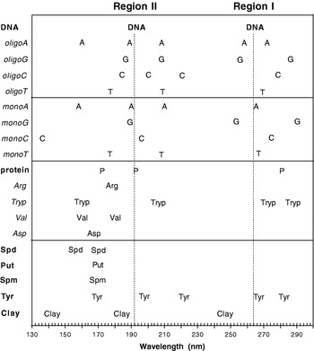
In the dry salmon sperm DNA sample, absorption maxima (Fig. 2; see also Fig. 9) were observed at 264 nm (region I) and 192 nm (region II), with absorbance climbing to 125 nm at the lower end of the spectrum (region III). In the hydrated sample, the peak at 264 nm was shifted to 260 nm, and the peak at 192 nm was obscured by absorbance by the solvent (Fig. 2). These peak behaviours are similar to those observed in comparing the heavy-water solution spectra of native DNA and DNA denatured by heat (Falk Reference Falk1964).
VUV–UV absorption by hydrated and dry salmon sperm DNA.

Oligonucleotides (polymerized)
All oligonucleotides (homopolymers of 20 nucleotides) showed major absorption peaks grouped in three regions: 255–286 nm (region I), 159–220 nm (region II) and in a third region (III) of absorbance, increasing to the highest observed value at 125 nm, the shortest wavelength measured (Fig. 3; see also Fig. 9). The oligonucleotides showed more spectral features than DNA, with three peaks or shoulders detected for oligoT, five for oligoA and four for oligoG. The purines (A, G) thus showed more features than the pyrimidines (C, T), as expected from their more complex structures. A peak or shoulder was observed at 208 nm in all but oligoC, where a peak was seen instead at 220 nm and an inflection around 200 nm. The two purines had peaks at 255–257 nm, while the pyrimidines had peaks at 268 (oligoT) and 278 nm (oligoC).
VUV–UV absorption by polymerized oligonucleotides (A, T, G and C) with dry DNA as a reference.

The broad salmon sperm DNA peaks at 192 and 264 nm are thus resolvable into oligonucleotide spectral families, with the peak interval greater in the pyrimidines than in the purines. As noted above, in the purines, the region I peak occurred at 255 nm for oligoG and at 257 nm for oligoA, with shoulders at 286 and 271 nm, respectively. In oligoC, the region I peak occurred at 278 nm, but in oligoT it was found at 268 nm. In addition, the oligoG spectrum showed a strong shoulder at 286 nm. Thus, in DNA sequences rich in GC base pairs, absorption by region I should shift to longer wavelengths. A similar analysis for the region II family (150–230 nm) showed that absorption by the AT pair should occur at shorter wavelengths, compared with the GC pair, but this difference should be less dramatic than for region I.
These observations suggest that the overall absorption spectrum of DNA depends on its state of hydration and the relative frequencies of the GC versus the AT base pairs. DNA sequences composed uniquely of AT will absorb more at the short wavelengths, and those made up of GC more at the longer wavelengths.
Nucleotide monomers (unpolymerized)
The unpolymerized monomers that make up DNA could also serve as UV filters. These include the four bases alone and their ribose derivatives: nucleosides (without the phosphate) or nucleotides (with the phosphate). A potential advantage of these small molecule UV screens would be their ability to diffuse or be transported through cellular compartments of the organism.
We therefore determined absorption spectra for the four monomer nucleotides (Fig. 4; see also Fig. 9), and compared them with our spectra for polymerized (oligo)A/G/C/T, as well as with the published spectra of thin films of the unpolymerized A/G/C/T nucleobases, measured by Yamada & Fukutome (Reference Yamada and Fukutome1968) and also by Isaacson (Reference Isaacson1972) (Table 1). Our peak wavelength assignments generally agreed with previous reports. In addition, the spectra for polymerized A, T and G were similar, in numbers of bands and peak wavelengths, to those of the monomers (see Fig. 9).
VUV–UV absorption by mononucleotides (A, T, G and C) with dry DNA as a reference. Absorbance values are adjusted to make the curves understandable.

Comparison of peak wavelengths of monomer and polymerized A/G/C/T (our measurements) with those of unpolymerized A/G/C/T nucleobases measured by Yamada & Fukutome (Reference Yamada and Fukutome1968) and by Isaacson (Reference Isaacson1972)


(a) Present paper; (b) Yamada & Fukutome (Reference Yamada and Fukutome1968) ; (c) Isaacson (Reference Isaacson1972).
We can conclude for A, T and G that interaction between bases of the same type in an oligonucleotide is relatively small, beacuse polymerization has little effect on the absorption spectrum in the region we measured. Thus, the accumulation in a cell of these mononucleotides could contribute to protecting DNA against UV radiation. (The remarkable similarity between monoA and DNA absorption curves is discussed later.)
In contrast to A, T and G, the peaks in the cytidine nucleotide sample changed with polymerization, with the region I peak shifting to higher wavelengths and the region II peak shifting to lower wavelengths (Fig. 4; see also Fig. 9). A peak at 182 nm in oligoC was absent in the monomer. In water, the cytosine peak wavelengths in the 190–300 nm region are more dependent on pH than the corresponding peaks for A, G and T (Voet et al. Reference Voet, Gratzer, Cox and Doty1963), and absorption spectra for monoC appear to vary for different samples and as a result of ageing (Yamada & Fukutome Reference Yamada and Fukutome1968).
UV absorption by proteins and their components
The amino acid sequence determines protein structure as well as overall charge. Basic proteins (e.g. histones) bind DNA electrostatically. Depending on their amino acid sequence, proteins have the potential to insert into membranes via hydrophobic residues, adhere to membrane phospholipids through electrostatic interactions or bind specifically to diverse structures or molecules (e.g. consider the interactions between antigens and antibodies, enzymes and substrates and receptors and their ligands).
We examined the UV absorption spectrum of histones, which are positively charged DNA-binding proteins found in eukaryotic cells. Absorption peaks (Fig. 5; see also Fig. 9) were observed at 170 and 192 nm, and a continuous increase in absorbance occurred below 150 nm. Although there was relatively little absorption above about 230 nm, higher protein concentration showed the characteristic protein absorbance peak at 280 nm (Fig. 5, inset). Histones do not appear to be ideal UV protectors for DNA absorption region I. An identical spectrum was obtained for bovine serum albumin (results not shown), a small globular protein of 585 amino acids.
VUV–UV absorption by histone protein with dry DNA as a reference. Inset gives a higher concentration to show the peak at 280 nm.

Amino acids can be classified according to their side groups. For absorption spectroscopy, we chose one from each of two of the four standard classes. In order to better assess the influence of the aromatic ring in tryptophan, an additional two from the ‘neutral and hydrophobic’ class were selected. (Another criterion in the choice of particular samples was the ability of amino acids to make thin films that produced a minimum of scattering.) The amino acids were: arginine (basic), tryptophan (neutral and hydrophobic), valine (neutral and hydrophobic) and aspartic acid (acidic). Their spectra (Fig. 6; see also Fig. 9) showed that potential protection of DNA absorption regions II and III in the DNA curve is a common feature, but that UV screening for region I is only seen with the aromatic amino acid, tryptophan, where a broad, complex peak at 287 nm was recorded, with a shoulder at 272 nm, reminiscent of the vibrational structure in benzenoid spectra in this region.
VUV–UV absorption by amino acids: arginine (Arg), tryptophan (Trp), valine (Val) and aspartic acid (Asp). Dry DNA is included as a reference.

UV absorption by polyamines and tyramine
Polyamines are small polycationic molecules, found in all living organisms. They are essential for optimal growth and development in eukaryotic and prokaryotic cells (Martin-Tanguy et al. Reference Martin-Tanguy, Tepfer, Paynot, Burtin, Heisler and Martin1990). As they are positively charged at cellular pH, they interact with negatively charged molecules, such as DNA (Basu et al. Reference Basu, Schwietert, Feuerstein and Marton1990; Pohjanpelto & Holtta Reference Pohjanpelto and Holtta1996), membrane phospholipids (Schuber et al. Reference Schuber, Hong, Duzgunes and Papahadjopoulos1983; Beigbeder et al. Reference Beigbeder, Vavadakis, Navakoudis and Kotzabasis1995; Tassoni et al. Reference Tassoni, Antognoni and Bagni1996) and some proteins (Apelbaum et al. Reference Apelbaum, Canellakis, Applewhite, Kaur-Sawhney and Galston1988). Putrescine (Put), spermine (Spm) and spermidine (Spd) are the major polyamines found in most cells, where they are present in high concentrations. In addition to the aliphatic polyamines (Put, Spm and Spd), aromatic amines, such as tyramine (Tyr), naturally occur in prokaryotic and eukaryotic organisms.
Polyamines can be found not only as free molecular bases, but also conjugated to small molecules or bound to proteins. In plants, polyamines and aromatic amines are usually conjugated with phenolic acids such as hydroxycinnamic acids, e.g., t-cinnamic, caffeic, ferulic and p-coumaric acids (Martin-Tanguy Reference Martin-Tanguy1985, Reference Martin-Tanguy1997; Martin-Tanguy et al. Reference Martin-Tanguy, Tepfer and Burtin1991). Polyamines are known in the laboratory to interact strongly with DNA, to stabilize condensed DNA conformations (Flink & Pettijohn Reference Flink and Pettijohn1975) and thus to protect DNA from degradation. In nature they are important in the regulation of growth, but they are not considered to be hormones, because of their elevated cellular concentrations. They accumulate in response to stress, possibly as a way of storing nitrogen produced by the breakdown of proteins. In plants, polyamines and their conjugates are implicated in growth, development and the regulation of flowering and responses to biotic and abiotic stress (Martin-Tanguy Reference Martin-Tanguy1985, Reference Martin-Tanguy1997, Reference Martin-Tanguy2001; Martin-Tanguy et al. Reference Martin-Tanguy, Tepfer and Burtin1991; Tepfer & Martin-Tanguy Reference Tepfer and Martin-Tanguy1991). Polyamine accumulation in plants was induced by UV-B (An et al. Reference An, Liu, Zhang, Chen, Liu, Feng, Xu, Qiang and Wang2004) and by UV-C radiation (Campos et al. Reference Campos, Figueras, Boronat, Pinot, Tiburcio, Galston and Tiburcio1991).
The UV absorption spectra (Fig. 7; see also Fig. 9) for spermidine, putrescine, spermine and tyramine showed a peak in the 170 nm region, with continued absorbance to 123 nm. Tyramine gave peaks at 198 nm, at 221 nm and twin peaks in the region of 282 nm with a shoulder at 267 nm. Tyramine would thus protect DNA in both DNA absorption regions I and II.
VUV–UV absorption by amines: spermine (Spm), putrescine (Put), tyramine (Tyr) and spermidine (Spd). Dry DNA is included as a reference.

UV absorption by clay
Inorganic materials, such as clay, which has been proposed as a catalyst in prebiotic chemistry (Bernal Reference Bernal1951; Ferris et al. Reference Ferris, Hill, Liu and Orgel1996; Rode Reference Rode1999), are obvious candidates for protecting DNA and the organisms that contain it. Adsorption of DNA on soil components, particularly on clays, protects DNA against degradation by nucleases (Romanowski et al. Reference Romanowski, Lorenz and Wackernagel1991; Paget et al. Reference Paget, Jocteur-Monrozier and Simonet1992; Gallori et al. Reference Gallori, Bazzicalupo, Dal Canto, Fani, Nannipieri, Vettori and Stotzky1994; Lorenz & Wackernagel Reference Lorenz and Wackernagel1994; Demaneche et al. Reference Demaneche, Bertolla, Buret, Nalin, Sailland, Auriol, Vogel and Simonet2001) and DNA adsorbed on clay minerals, such as montmorillonite and kaolinite, is partially protected against UV light and X-rays (Ciaravella et al. Reference Ciaravella, Scappini, Franchi, Cecchi-Pestellini, Barbera, Candia, Gallori and Micela2004; Scappini et al. Reference Scappini, Casadei, Zamboni, Franchi, Gallori and Monti2004). We therefore examined montmorillonite clay as a possible UV screen. The absorption spectrum of clay showed peaks at 141, 184 and 247 nm, with strongly increasing absorbance below 135 nm (Fig. 8; see also Fig. 9). Although the features are not as distinct as in other substances analysed, clay absorbed throughout the absorption spectral region of DNA, but the observed peaks were blue-shifted by about 10–20 nm with respect to dry salmon sperm DNA. These montmorillonite clay peaks could be the result of organic contamination.
VUV–UV absorption by montmorillonite clay, with dry DNA as a reference.


Discussion
DNA conserves the information that defines life on Earth. Terrestrial life on primitive Earth or in future space travel must cope with the full spectrum of Solar UV. We therefore examined UV absorption by DNA and by a few of the possible chemical screens that could protect DNA, based on the working assumption that the DNA and UV screen absorption curves should match as closely as possible. We considered molecules common to essentially all forms of life: DNA, DNA subunits, proteins, amino acids, polyamines and tyramine. In addition, we examined a clay as a possible abiotic DNA protector.
Absorption spectra of DNA
Peak wavelengths and intensity ratios vary in published DNA absorption spectra. This could be due to differences in the source of the DNA and how it was prepared. Our salmon sperm DNA sample was extracted with organic solvents to remove proteins and small molecules. Salmon sperm DNA belongs to the AT rich class, having a GC content of 41.2% (Chargaff et al. Reference Chargaff, Lipshitz, Green and Hodes1951). Spectra in the dry and hydrated states were compared (Fig. 2), using a DNA film dried onto a MgF2 window and the same DNA dissolved in water. DNA in films is no longer in the B state, but in a distorted configuration in which the complementary pairing and the parallel stacking of the DNA bases is reduced, but the double helical strands remain entwined (Falk et al. Reference Falk, Hartman and Lord1963; Falk Reference Falk1964).
Table 2, where we compare our results with already published DNA absorption spectra (Inagaki et al. Reference Inagaki, Hamm, Arakawa and Painter1974; Sontag & Weibezahn Reference Sontag and Weibezahn1975; Ito & Ito Reference Ito and Ito1986), gives relative absorption intensities of DNA films at selected wavelengths, with respect to the intensity of the 260 nm band. The DNA samples of Inagaki et al. (Reference Inagaki, Hamm, Arakawa and Painter1974) and those of Sontag & Weibezahn (Reference Sontag and Weibezahn1975) were from calf thymus, but the DNA sample of Ito & Ito (Reference Ito and Ito1986) was specified only as ‘highly polymerized DNA’. Our relative intensities agree with those of Sontag & Weibezahn, who used lamp radiation sources. The relative intensities of Ito & Ito, who used a synchrotron radiation source, have higher values, and those of Inagaki et al. have lower values. The DNA films of Sontag & Weibezahn and Inagaki et al. were on metal mesh screens, whereas those of Ito & Ito were on CaF2 plates.
Comparison of the relative absorption of DNA films at selected wavelengths, relative to the 260 nm absorption peak

(a) Sontag & Weibezahn (Reference Sontag and Weibezahn1975) ; (b) Ito & Ito (Reference Ito and Ito1986) ; (c) Inagaki et al. (Reference Inagaki, Hamm, Arakawa and Painter1974).
The intensity measurements of Inagaki et al. were indirectly determined, partly based on a Kramers–Kronig analysis of the index of refraction of the films. They are thus subject to defects in this treatment. The higher relative intensities recorded by Ito & Ito might be due to greater scattering or to the DNA sample's unreported origin. Although the absorption spectra of the DNA films reported in Table 2 are quite similar, apart from the relative intensity differences mentioned above, they exhibit small differences in peak wavelengths, as might be expected from the different DNA source materials and sample preparations.
Effects of nucleotide composition on UV absorption
Absorption spectra of the polymerized nucleotides (homopolymers) that compose DNA suggest that absorption region I would shift to longer wavelengths in DNA composed uniquely of G and C nucleotides, compared with DNA made up of A and T. Furthermore, DNA in the dry state shows a small red-shift for region I (Fig. 2). Relative GC content varies in different organisms (Marmur & Doty Reference Marmur and Doty1962), e.g. between 31% (Clostridium perfigens) and 72% (Micrococcus lysodeikticus); see Lewis & Johnson (Reference Lewis and Johnson1974). The potential for G use is clear in the genetic code, where in many cases an alternative codon can contribute a supplementary G to the sequence, without changing the protein sequence. We calculate that a hypothetical gene, encoding a protein incorporating one of each of the 20 amino acids, can vary in GC content by more than twofold, without changing the amino acid sequence. (The minimum GC content was 30%; the maximum was 63%.) In theory, nucleotide content (including non-coding sequences) could thus evolve or be engineered to alter the overall absorption characteristics of the genome, but this hypothesis needs to be verified experimentally.
Three hydrogen bonds in the GC pair, as opposed to two in the AT pair, make the double helix of nucleic acids more stable at high temperatures (Galtier & Lobry Reference Galtier and Lobry1997). According to our observations, the red-shift potentially conferred by a sequence rich in G and C should also confer better UV protection by proteins (see below section on proteins and aminoacids as UV screens). In addition, lowering the frequency of the AT pair would reduce the chances of dimerization of adjacent thymines, which are involved in the majority of the mutagenic photo lesions caused by UV radiation.
The existence and degree of a hypothetical red-shift associated with high GC can be determined by studying UV absorption by synthetic DNA sequences that vary in GC content, and the sensitivity of these hybrids to specific UV wavelengths can be measured in vitro. The ecological pertinence of altered GC content could be assessed using a gene encoding a genetic marker, such as an antibiotic resistance, that has been altered to contain more GC base pairs, while encoding the same protein. The biological robustness of this altered sequence could be tested by exposing the gene to UV radiation in vitro (with and without UV screens), then testing its integrity in vivo, by introducing it into bacteria challenged by the antibiotic. It should thus be possible to experimentally evaluate the adaptive significance of changes in the absorption spectrum of a target nucleic acid.
In addition to the above spectroscopic properties, chemical, thermodynamic and conformational attributes could be influenced by overall base composition or interactions between neighbouring bases (DNA sequence). Hydrodynamic (e.g. viscosity) and visible light-scattering properties of DNA probably depend more on gross polynucleotide structure and molecular weight, than on base composition or sequence. These characteristics could nevertheless influence genome survival, and they should be taken into account in studies of DNA stability using synthetic DNA.
ATP as a UV screen
A simple life form could consist of DNA and molecules it encodes, which would replicate the DNA and protect it from UV radiation. The simplest UV screen for DNA would thus be nucleotide monomers, an RNA or a protein that has the same absorption spectrum as DNA. An overview of the absorption curves (Fig. 9) suggests that dry salmon sperm DNA is best protected by nucleic acids and their components, while free amines, histone protein and selected amino acids are less attractive UV screens.
Among the DNA components, the absorption spectrum of adenine mononucleotide best matches that of DNA, with absorption peaks corresponding closely to those of DNA (Fig. 4; see also Fig. 9). Adenine mononucleotide is the DNA form of ATP, which is the primary energy currency in contemporary cells. The two forms have identical spectra (unpublished results). The intracellular ATP steady state pool is about 1 mM in mammalian cells (Wang et al. Reference Wang, Tao, Trumbore and Berger1997), which would provide a 30% attenuation at 260 nm for a tissue thickness of 100 μm. UV protection would be further increased in thicker tissues or if cells are adapted to accumulating higher ATP concentrations. Organisms living before the formation of our oxygen-rich atmosphere were likely exposed to high UVC and VUV, and they could have used adenine mononucleotide or ATP as an element of their UV resistance phenotype. In primitive cells, this attribute might have preceded the use of ATP in energy transfer. Thus, an answer to the question of why ATP is the energy currency of the cell might be that it was essential to accumulate high concentrations of ATP to protect the genome against UV light.
Proteins and amino acids as UV screens
DNA protection by proteins is less obvious, because DNA has an absorption peak at around 260 nm and protein has a peak around 280 nm (Fig. 5; see also Fig. 9). They both absorb around 200 nm (region II), and continue absorbing through the VUV, but there is no obvious way for a protein to efficiently protect DNA at 260 nm (region I), unless the concentration of the screen is so high that the target is protected even at absorption minima. We assume that such a non-specific, generalized opacity to UV radiation would be less efficient than that afforded by a well-matched UV screen, such as ATP.
An examination of four amino acids, representative of the 20 that make up proteins, showed that protection of region I in DNA could be produced by the aromatic amino acid, tryptophan (region I at 272–287 nm), which carries a ring system that is rich in π electrons and reminiscent of the double ring structure found in the purines. We conclude that a DNA sequence with high GC content (which would shift region I to higher wavelengths), encoding a protein carrying aromatic amino acids, such as tryptophan, would be protected for both regions I and II. This hypothesis can be tested experimentally, by designing and expressing such a protein. Similar protein engineering might be used to improve UV protection in vivo in exoenvironments. For instance, storage proteins in seeds might be altered to better serve as UV screens. Proteins rich in aromatic amino acids are found in the Bacillus subtilis spore coat, where they are thought to play a part in UV resistance (Driks Reference Driks1999; Riesenman & Nicholson Reference Riesenman and Nicholson2000).
Polyamines and tyramine as UV screens
Aside from nucleic acid and protein precursors, organisms produce a variety of small metabolites through enzymatic pathways encoded in multiple genes. As the genes and regulatory systems responsible for these pathways are becoming known, they are recognized as candidates for metabolic engineering. Small metabolites constitute possible UV screening material that could diffuse within the cell and be transported across membranes.
The polyamines are an example of small diffusable molecules known to stabilize DNA and for which the molecular genetics are known, but according to their absorption spectra, they would protect regions II and III, but not region I (Figs 6 and 8). However, the aromatic amine, tyramine, carries a benzene ring, and given its UV absorption spectrum, should protect both DNA regions I and II. Tyramine is positively charged at physiological pH, so it will bind electrostatically to DNA. In plants, tyramine conjugation to hydroxycinnamic acids, which also carry a benzene ring, is induced by UV light, and hydroxycinnamic acids conjugate with polyamines, possibly improving their DNA protective capacities. Caffeic, ferulic and t-cinnamic acid have a peak around 290 nm (our unpublished results), but this could change after conjugation. Engineering of these biosynthesis pathways is possible, and as regulatory circuits are discovered that control batteries of genes, these pathways should become easier to manipulate.
Small molecules, including secondary metabolites, such as polyphenols (see the accompanying paper (Zalar et al. Reference Zalar, Tepfer, Hoffmann, Kollmann and Leach2007)), could thus act as UV screens. Some of them, such as the polyamines, have DNA binding and DNA protecting properties. In contemporary organisms, the biosynthesis of such metabolites requires complex enzymatic reactions, but similar compounds might have been present in the prebiotic environment (Bernstein Reference Bernstein2006). Any UV screen that also binds DNA carries the risk that the absorbed energy is transferred to the DNA, causing photosensitization rather than protection. The need to have spatial distance between the screen and the target DNA could have contributed to the evolution of cells surrounded by phospholipid bilayers, and eventually to the formation of cell walls and spore or seed coats.
The binding and UV protective characteristics of small molecules can be tested experimentally in vitro (e.g. using synthetic DNA sequences) and in vivo (e.g. by manipulating enzymatic pathways through metabolic engineering). It should thus be possible to test the predictive validity of our assumption that an ideal UV screen would have an absorption pattern similar to that of the target. Genetic engineering can also direct enzymes into selected cellular compartments or into specific tissues. For instance, a seed storage protein could be made richer in tryptophan, and the resistance of the resulting seeds assessed under space conditions.
Astrobiological considerations
The theory that life is universal (panspermia) was restated by Arrhenius at the beginning of the last century. It has since been championed by (among others) Wickramasinghe and Hoyle (Hoyle & Wickramasinghe Reference Hoyle and Wickramasinghe1986; Wickramasinghe & Wickramasinghe Reference Wickramasinghe and Wickramasinghe2003; Wickramasinghe Reference Wickramasinghe2004) and by Crick and Orgel (Crick & Orgel Reference Crick and Orgel1973; Crick Reference Crick1981). The former thought of life as intrinsic to matter and carried by comets; the latter proposed that extra-Solar System intelligences intentionally disseminated life through the universe (directed panspermia).
In exploring the Solar System and beyond, man is clearly engaged in Crick and Orgel's directed panspermia. To consciously direct it, we will need to adapt life to survival in space. The UV absorption spectra presented here suggest that DNA sequences could be altered to adapt the genome's absorption spectrum to a given UV environment or to given UV screens. UV screen synthesis could be encoded in the DNA as proteins or as enzymes that produce small molecules derived from metabolic processes. Our survey of some obvious candidates singled out adenine mononucleotide as a promising small molecule. It is theoretically possible to design a simple organism starting with the DNA sequence. Features, such as UV screens, could be built in that would make for enhanced survival in space. Similar genetic methods could be used to improve existing organisms for space travel and for possible loss of atmospheric UV protection on Earth.
Methods
Absorption spectra
Absorption spectra were recorded at the UV1 beamline at the synchrotron radiation source ASTRID (Institute for Storage Ring Facilities, University of Aarhus in Denmark). Monochromatized light was passed from the beamline through a LiF window into a vacuum chamber capable of containing up to four MgF2 discs. The light exited the chamber via a MgF2 window and was detected with a UV–VUV sensitive photomultiplier tube (PMT). The discs could be placed in the beam one at a time, and the transmitted light was measured from 125 to 340 nm in 1 nm steps. Typically, three discs with dried samples were placed in the chamber and a fourth disc was kept blank. More details of how to avoid absorption of VUV light in the gap between the exit window and the PMT, and on how to avoid higher-order radiation contaminating the spectrum are described elsewhere (Eden et al. Reference Eden, Limão-Vieira, Hoffmann and Mason2006).
Spectra are based on transmission measurements. They have scattering components in addition to absorption. Scattering due to imperfections in the MgF2 windows was removed by computing the ratio between the transmission of the window plus the sample and that of the blank window. Absorption was thus determined as A=log(I 0/I t) where I 0 and I t are the measured intensity of transmitted light through the blank disc and the sample disc, respectively. Scattering was assumed to be similar for each sample, based on the reproducibility of selected spectra. In published spectra (125–400 nm) of thin films of the five nucleobases (Yamada & Fukutome Reference Yamada and Fukutome1968), scattering was estimated to be 10–30% of the optical density at the shortest wavelengths.
Preparation of samples
Samples were air-dried onto MgF2 windows (diameter, 23 mm). Spectroscopy using dry samples under vacuum avoided solvent absorbance, which is usually observed in absorption measurements below 200 nm (Foldvari et al. Reference Foldvari, Fekete and Corradi1981), and approximated conditions expected in dormant spores or seeds. Sterile, ultrapure water (R>10 MΩ Cm) was made with the Milli-Q apparatus (Millipore).
Salmon sperm DNA (Sigma-Aldrich, France) was extracted twice with a mixture of phenol (saturated with H2O) and chloroform in a 1:1 ratio, in order to remove proteins. After centrifugation, the aqueous phase was extracted three times with chloroform to remove phenol, then precipitated with two volumes of ethanol in the presence of ammonium acetate. The DNA was spooled and washed with ethanol (70%) to remove salts and the remaining traces of organic solvents. The DNA was dried and resuspended in H2O. To facilitate pipetting, viscosity was reduced by passage through a 22-gauge needle.
Oligonucleotides (Genosys-Sigma, France) of adenine (oligoA), thymine (oligoT) and cytosine (oligoC) were purchased in the dry state as 20-mers. Oligonucleotide solutions were prepared in H2O (pH 7.5). 200 μl of oligoA/T/G/C (100 μM each) were air-dried on MgF2 windows. About 0.1 mg (dry weight) of each sample was deposited on each MgF2 window.
Monomer nucleotides (monoA/T/G/C) in the form of deoxyribonucleoside triphosphates (Promega, France) were prepared as 1 mM solutions in water (pH 7.5). 750 μl of dATP and dGTP and 500 μl of dTTP and dCTP were dried onto MgF2 windows, giving films consisting of 0.45 mg of dATP and dGTP and 0.3 mg of dTTP and dCTP.
Samples of unfractioned whole histone from calf thymus, type II-A (Sigma-Aldrich, France), were prepared in water (pH 7.5). 500 μl samples at 0.3 and 1 mg ml−1 were air-dried on MgF2 windows. The dry histone samples consisted of 0.15 and 0.5 mg (high histone concentration) per MgF2 window.
L-Arginine (Arg), L-Aspartic acid (Asp), L-Valine (Val) and L-Tryptophan (Trp) (Sigma-Aldrich, France) were dissolved in water (pH 7.5). Samples, consisting of 400 μl of Arg (2 mg ml−1), 500 μl of Asp (2 mg ml−1), 1 ml of Val (2.6 mg ml−1) and 400 μl of Trp (0.5 mg ml−1) were air-dried on MgF2 windows. The final mass per MgF2 window was 0.8 mg of Arg, 1 mg of Asp, 2.6 mg of Val and 0.2 mg of Trp.
Spermine (Sigma-Aldrich, France), spermidine (Sigma-Aldrich, France) and putrescine (Fluka, France), were freshly prepared in water (pH 7.5). The solutions of 1 ml of spermine (1.1 mg ml−1), 500 μl of spermidine (3 mg ml−1) and 500 μl of putrescine (15 mg ml−1) were air-dried, so that final quantities were 1.1, 1.5 and 7.5 mg per MgF2 window, respectively. Tyramine (Sigma-Aldrich, France) was prepared as 0.3 mg ml−1 solution in 100% methanol, and 500 μl of this solution was dried on the MgF2 window. The solid film contained 0.15 mg of tyramine per MgF2 window.
The clay mineral (Na-montmorillonite/SWy-1, from Crook Country, Wyoming, USA, from the clay minerals repository, University of Missouri, Columbia, USA) was kindly provided by R. Prost. Clay particles were suspended in water, followed by centrifugation at 3000G for 5 min, which caused visible layering. The upper band of particles in the pellet was removed with a spatula and the process was repeated five times. A 500 μl sample (0.5 mg ml−1) of Na-montmorillonite/SWy-1 was air-dried on a MgF2 window.
Acknowledgements
Financial support was provided by the Centre National d'Etudes Spatiales and the University of Aarhus (EC Integrated Activity on Synchrotron and Free Electron Laser Science, contract number R113-CT-2004-50608). R. Prost generously provided the clay sample.