Introduction
Duck-billed dinosaurs, or hadrosaurs, rank among the most diverse and successful dinosaur groups. They have been found across Europe, Asia, North and South America, and Antarctica (Case et al., Reference Case, Martin, Chaney, Reguero, Marenssi, Santillana and Woodburne2000; Lund and Gates, Reference Lund and Gates2006; Prieto-Márquez, Reference Prieto-Márquez2010b) concentrated in a 15 million year period toward the end of the Late Cretaceous (Campanian–Maastrichtian; Horner et al., Reference Horner, Weishampel and Forster2004). These herbivorous browsers are split into a diphyletic origin of hollow-crested and solid-crested species (Horner, Reference Horner1990), and their remains are often prolific, ranging from isolated elements to complete articulated skeletons. With the addition of comprehensive ontogenetic stages including eggs and embryos along with a vast array of trace fossils, hadrosaurs provide some of the most extensive data sets of any dinosaur group (Horner et al., Reference Horner, Weishampel and Forster2004).
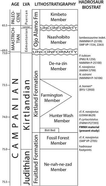
For over a century, vertebrate fossils have been collected from the San Juan Basin (SJB) of New Mexico (upper Campanian, Late Cretaceous; Fig. 1; Williamson, Reference Williamson2000). Although the hadrosaurid record of the SJB pales in comparison to the well-preserved specimens and growth series from the northern United States and Canada, many enigmatic taxa have been discovered. In 1904, Barnum Brown briefly collected and later described the solid-crested Kritosaurus navajovius (Brown, Reference Brown1910; AMNH 5799), but the SJB remained primarily unexplored until Charles Hazelius Sternberg arrived in 1921. Sternberg, aged 71, collected for three consecutive field seasons discovering the hollow-crested hadrosaurids Parasaurolophus tubicen (Wiman, Reference Wiman1931; PMU R.1250) and Parasaurolophus cyrtocristatus (Ostrom, Reference Ostrom1961, Reference Ostrom1963; FMNH P27393), the ceratopsian Pentaceratops sternbergii (Osborn, Reference Osborn1923; AMNH 6325), and vast amounts of isolated material. More recent excavations have continued to expand the diversity of the region with the additions of the solid-crested Anasazisaurus horneri (Hunt and Lucas, Reference Hunt and Lucas1993; BYU 12950) and Naashoibitosaurus ostromi (Hunt and Lucas, Reference Hunt and Lucas1993; NMMNH P-16106), but simultaneously complicated the taxonomy (Prieto-Márquez, Reference Prieto-Márquez2013; Sullivan and Lucas, Reference Sullivan and Lucas2014).
Stratigraphic distribution of hadrosaurid taxa and key specimens in the San Juan Basin, New Mexico (modified from Sullivan and Lucas, Reference Sullivan and Lucas2014). Prieto-Márquez (Reference Prieto-Márquez2013) considered A. horneri as a junior synonym of K. navajovius. LVA=North American Land Vertebrate Age; Hadrosaur Biostrat=Hadrosaur Biostratigraphy.

Here we present the first descriptive account of isolated hadrosaurid material at the Field Museum of Natural History (FMNH) from C. H. Sternberg’s 1922 expedition of the SJB. Although detailed locality information was inconsistently documented, Sternberg’s correspondences with FMNH likely place this collection within the Hunter Wash Member of the lower Kirtland Formation (Sullivan and Lucas, Reference Sullivan and Lucas2011; personal communication, R. Sullivan, 2014).
Materials and methods
Hadrosaurid material including both cranial and appendicular elements in the collection of the Field Museum of Natural History in Chicago were examined and compared. The data matrix from Prieto-Márquez et al. (Reference Prieto-Márquez, Wagner, Bell and Chiappe2014) was used for comparative analysis. Characters (Prieto-Márquez, Reference Prieto-Márquez2010a) used for materials in the present study are given in Supplemental Data. Character state scores of the Sternberg SJB specimens used herein are also listed in Supplemental Data. Selected measurements are listed in Table 1.
Selected measurements (in millimeters) of FMNH hadrosaurid Sternberg material. DCC=depth of caudal constriction; DCVF=depth of caudoventral flange; DPCL=length of deltopectoral crest; DPCW=width of deltopectoral crest; DRC=depth of rostral constriction; DW=distal width; MW=midshaft width; PCW=width of proximal constriction; PPBW=width of prepubic blade; PPW=width of pubic process; PW=proximal width; TL=total length; NW=width of minimum constriction of neck.

* Incomplete measurement.
Repositories and institutional abbreviations
AMNH, American Museum of Natural History, New York, New York; BYU, Brigham Young University, Provo, Utah; FMNH, The Field Museum of Natural History, Chicago, Illinois; NMMNH, New Mexico Museum of Natural History and Science, Albuquerque, New Mexico; PMU, Museum of Evolution, University of Uppsala, Uppsala, Sweden; SMP, State Museum of Pennsylvania, Harrisburg, Pennsylvania; USNM, United States National Museum of Natural History, Washington, D. C.
Systematic paleontology
Superorder Dinosauria Owen, Reference Owen1842
Order Ornithischia Seeley, Reference Seeley1887
Suborder Cerapoda Sereno, Reference Sereno1986
Infraorder Ornithopoda Marsh, Reference Marsh1881
Suborder Iguanodontia Dollo, Reference Dollo1888
Family Hadrosauridae Cope, Reference Cope1870
Subfamily Saurolophinae Brown, Reference Brown1914 (sensu Prieto-Márquez, Reference Prieto-Márquez2010a)
Tribe Kritosaurini Lapparent and Lavocat, Reference Lapparent and Lavocat1955
Materials
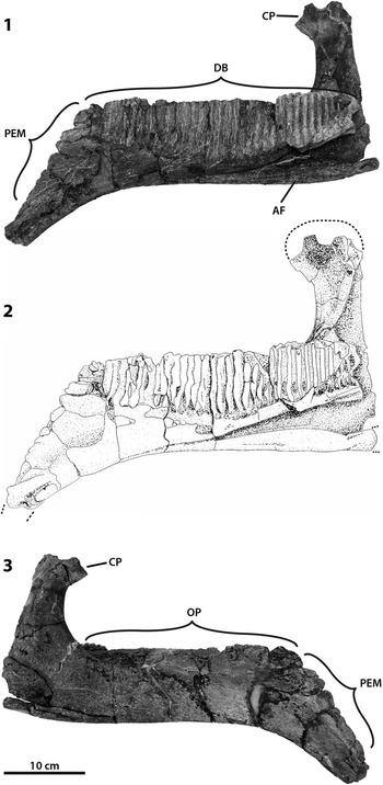
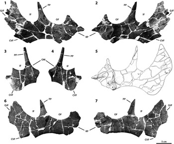
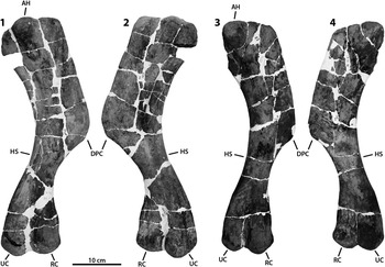
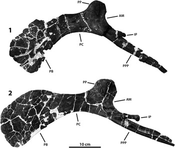
PR 2248, right dentary (Fig. 2); PR 1203a, right jugal (Fig. 3.1, 3.2); PR 1203b, right jugal (Fig. 3.5, 3.6); PR 1252, right jugal (Fig. 3.3, 3.4); PR 1300, right humerus, Sternberg 1-22, No. 8 (Fig. 4.1, 4.2); PR 1301, right humerus, Sternberg 1-22, No. 12 (Fig. 4.3, 4.4); PR 1202, left pubis, Sternberg 1-22, No. 33; PR 1290 (Fig. 5.1); left pubis, Sternberg 1-22, No. 75 (Fig. 5.2).
Right dentary (PR 2248) in (1) medial; (2) medial illustration; and (3) lateral views. AF=angular facet; CP=coronoid process; DB=dental battery; OP=occlusal plane; PEM=proximal edentulous margin.

Right jugals. PR 1203a in (1) lateral; (2) medial; and (5) medial illustration views. PR 1203b in (3) lateral and (4) medial views. PR 1252 in (6) lateral and (7) medial views. CVF=caudoventral flange; IF=infratemporal fenestra; OF=orbital fenestra; OM=orbital margin; PP=postorbital process; QJF=quadratojugal flange; RP=rostral process.

Right humeri. PR 1300 in (1) lateral and (2) medial views. PR 1301 in (3) lateral and (4) medial views. AH=articular head; DPC=deltopectoral crest; HS=humeral shaft; RC=radial condyle; UC=ulnar condyle.

Left pubes. (1) PR 1202 in lateral view; (2) PR 1290 in lateral view. AM=acetabular margin; IP=ischial peduncle; PB=prepubic blade; PC=proximal constriction; PP=pubic process (iliac peduncle); PPP=postpubic process.

Occurrence
The referred specimens were collected by Charles Hazelius Sternberg during his 1922 expedition of the San Juan Basin in New Mexico, southwest United States, and come from the lower Hunter Wash Member of the Kirtland Formation (late Campanian; Fig. 1). FMNH acquired the collection in 1924 from C. H. Sternberg.
Cranial element descriptions
Dentary.—
PR 2248 (Fig. 2) is a nearly complete right dentary. The dental battery extends the entire length of the occlusal surface preserving 32 tooth rows, but no teeth. The tooth rows are slightly inclined caudally with respect to their long axis, but this may be a result of mediolateral compression. The caudal tooth rows are narrower than the rostral tooth rows. The rostral tooth rows appear to have undergone more mediolateral compression than the caudal rows. The occlusal plane of the dental battery is straight and runs parallel to the lateral surface in dorsal view. The rostroventral margin has a 38° angle of deflection similar to Kritosaurini. The origination of the ventral deflection of the dentary occurs near the rostral end at approximately the same point at which the edentulous portion begins. The medial and lateral profiles of the dorsal margin of the rostral edentulous region are straight, lacking any pronounced concavity, a state shared with Kritosaurus navajovius, Saurolophini, and Edmontosaurus. The symphyseal end of the dentary projects lingually at a 33° angle to the cranio-caudal axis of the dentary. The coronoid process is well preserved except for the dorsal margin, and the long axis is subvertical in relation to the alveolar margin. The apex of the coronoid process has well-developed expansions of both the caudal and rostral margins, with the rostral margin being more prominently expanded. A dorsoventrally elongated ridge on the medial side of the coronoid process is absent. The occlusal plane is rostrocaudally straight and lacks any lingual arching. The caudal end of the dental battery ends slightly rostral of the caudal margin of the coronoid process. The coronoid process is laterally offset relative to the tooth row, with the presence of a laterodorsal concave platform separating the base of the process from the dental battery. The dentary ramus rostral to the coronoid process displays a slight ventral convexity in lateral view, a feature shared with Kritosaurini and Brachylophosaurini as well as Lambeosaurinae.
Jugal.—
The collection consists of three gracile right jugals: two nearly complete (PR 1252 and PR 1203a) and one partial (PR 1203b). PR 1203 has been split here into PR 1203a and PR 1203b to better discern between the two jugals.
PR 1203a (Fig. 3.1, 3.2, 3.5) is a nearly complete right jugal. The rostral apex of the rostral process is well preserved and is positioned within the dorsal half of the rostral process. The rostrodorsal margin of the rostral process forms a steep angle with the horizontal axis. The dorsoventral expansion of the caudodorsal margin of the rostral process is dorsoventrally shallow and rostrodorsally directed. The triangular caudoventral margin of the rostral process is ventrally pointed and as deep as its proximal end is wide, which it has in common with Brachylophosaurini and Kritosaurini. The caudoventral apex of the rostral process relative to the caudodorsal articulation with the lacrimal is located caudoventral to the caudal margin of the lacrimal process. The medial articular surface of the rostral process faces medially and is bound only caudally by a rim of bone. The orbital and infratemporal margins are approximately equal in width. The caudal constriction is deeper than the rostral constriction. The lateral profile of the quadratojugal flange is mostly preserved, missing only small pieces along the caudal margin. PR 1203a is auricular in shape with transitioning concave to nearly straight dorsal and convex ventral margins that converge dorsally into a dorsally directed postorbital process, but the dorsal region of the flange is rostrocaudally narrow. This character is shared with Edmontosaurus, Prosaurolophus, Saurolophus, and the Kritosaurini clade. The caudoventral margin is not well preserved, but the general outline resembles a similar shape to that of PR 1252.
PR 1203b (Fig. 3.3, 3.4) is a partial right jugal preserving only the portion between the orbital fenestra–ventral margin and infratemporal fenestra–caudoventral flange constrictions. The ventral expansion of the caudoventral flange displays a moderately expanded flange and a shallow curvature of the ventral margin of the jugal between the caudoventral flange and area approaching the rostral process. The caudal constriction is deeper than the rostral constriction. The postorbital process is slightly inclined caudally.
PR 1252 (Fig. 3.6, 3.7) is a nearly complete right jugal with a poorly preserved rostral process missing the dorsal, rostral, and caudoventral apices. The dorsoventral expansion of the caudodorsal margin of the rostral process is dorsoventrally shallow, but more steeply recurved in lateral view (Fig. 3.6) than in PR 1203a. The caudoventral apex of the rostral process relative to the caudodorsal articulation with the lacrimal is located caudoventral to the caudal margin of the lacrimal process. The medial articular surface of the rostral process faces medially and is bound caudally by a worn rim of bone. The orbital margin is wider than the infratemporal margin, but this may be due to an unclear termination of the dorsal apex of the rostral process. The caudal constriction is deeper than the rostral constriction. The lateral profile of the quadratojugal flange is mostly preserved, missing only small pieces along the caudal margin. PR 1252 is auricular in shape with transitioning concave to nearly straight dorsal and convex ventral margins that converge dorsally into a dorsally directed postorbital process, but the dorsal region of the flange is rostrocaudally narrow. The caudoventral margin located between the caudoventral and quadratojugal flanges is wide with a well-pronounced concavity, a characteristic shared with Kritosaurini and Brachylophosaurini.
Appendicular element descriptions
Humerus.—
The collection consists of two nearly complete right humeri (PR 1300, PR 1301) consistent in size with subadults. Both are exceptionally preserved displaying prominent muscle scar sites and have relatively low and smooth shapes.
PR 1300 (Fig. 4.1, 4.2) is a nearly complete right humerus missing only a small piece along the dorsal margin of the proximal end. The deltopectoral crest is deflected anterolaterally and extends about half the total length (0.55), in common with Kritosaurini and Brachylophosaurini, before tapering to meet the anteroposteriorly compressed and transversely expanded distal shaft. It terminates with well-developed ulnar and radial condyles. The minimal width of the shaft is approximately half the width (0.55) of the deltopectoral crest (measured at the maximum curvature of the dorsal margin). The ventral margin of the deltopectoral crest extends abruptly from the humeral shaft to display a distinct angular profile.
PR 1301 (Fig. 4.3, 4.4) is a nearly complete right humerus with an eroded dorsal expansion of the proximal end. PR 1301 is slightly smaller in overall size than the more robust PR 1300. Although the terminating distal region of the deltopectoral crest is slightly eroded, it would likely not have been as prominent as in PR 1300. The ulnar condyle is not completely preserved in lateral view, but it too is less robust relative to PR 1300. PR 1301 shares most other characteristics with PR 1300 with minor exceptions. The deltopectoral crest is deflected anterolaterally extending along half the total length (0.52), and the minimal width of the shaft is about half the width (0.57) of the deltopectoral crest.
Pubis.—
The collection consists of two nearly complete left pubes (PR 1202, PR 1290) consistent in size with subadults.
PR 1202 (Fig. 5.1) has heavily weathered margins and is missing the distal portions of most processes. Only the central body of the pubis surrounding the pubic process preserves true margins. The ventral region of the dorsoventral expansion of the prepubic blade is more expanded than the dorsal region directing the distal expansion ventrally and medially. The region of the prepubic blade just distal of the proximal constriction is curved downward at a greater degree than PR 1290, but this is likely a result of preservation. The dorsoventral expansion in lateral view has a subrectangular shape, is craniocaudally longer than dorsoventrally tall, and has generally straight profiles of the dorsal and ventral proximal margins in common with Kritosaurini and Saurolophini. The dorsoventral expansion of the distal region of the prepubic blade is deeper than the width of the acetabular margin of the pubis. The maximum ventral concavities of the dorsal and ventral margins of the prepubic blade are located ventral to the maximum dorsal concavity. Although the lateral margin of the pubic process shows mediolateral compression, the pubic process progressively disappears ventrally into the lateral surface of the region adjacent to the acetabular margin. The obturator foramen is absent, and the proximal postpubic ramus lacks a dorsocaudally oriented process. The ischial peduncle is strongly weathered distally and does not preserve a total length. The postpubic process is not complete distally but is primarily straight, showing minimal concavity near the proximal origin in lateral view.
PR 1290 (Fig. 5.2) is well preserved, missing only the flush anterior margin of the prepubic blade and a distal portion of the postpubic process. The central body of the pubis preserves muscle scars surrounding the pubic process and acetabular margin. The pubic process is intact and preserves true margins unlike the mediolaterally compressed PR 1202. Except for a slightly eroded distal margin of the dorsoventral expansion of the prepubic blade, the prepubic blade has a subrectangular shape, is craniocaudally longer than dorsoventrally tall, and maintains a smoothly curved dorsal margin leading proximally into the proximal constriction. The ischial peduncle is complete, has a lateroventral protuberance on the proximal region, and is craniocaudally broader than dorsoventrally tall. PR 1290 shares remaining characteristics with PR 1202.
Discussion
In contrast to specimens from northwestern North America, many of the hadrosaurid specimens collected in the San Juan Basin are referable only to the subfamily level (Williamson, Reference Williamson2000). Although the specimens of this collection are isolated, they are complete and of better quality than many excavated in the San Juan Basin. Many of the elements described here retain taxonomically informative characters.
The majority of the Sternberg SJB specimens appear to be saurolophine, with some of the specimens, including the humeri, only referable to the subfamily level. The humeri possess a moderately expanded deltopectoral crest, with a ratio between the width of the humerus across the distal fourth of the deltopectoral crest and the width of the distal shaft at the point of maximum curvature between 1.65 and 1.90, an unambiguous synapomorphy of all saurolophines (with the exception of Brachylophosaurini; Prieto-Márquez, Reference Prieto-Márquez2010a). The prepubic blade of the pubis has a subrectangular and ventrally deflected distal blade, characteristic of Gryposaurus and Prosaurolophus (Prieto-Márquez, Reference Prieto-Márquez2010a, Reference Prieto-Márquez2013). The jugals are the most taxonomically definitive. Both jugals in this collection have a wide and strongly concave margin between the caudoventral and quadratojugal flanges and a ventral spur of the rostral process that is as deep as, or slightly deeper than, it is wide proximally; these are both unambiguous synapomorphies for the Kritosaurini (Prieto-Márquez, Reference Prieto-Márquez2013). These specimens cannot be conclusively identified as Kritosaurus or more specifically Kritosaurus navajovius. Kritosaurus is diagnosed as having a unique combination of cranial characters; these jugals possess one of these characters by having an orbital constriction that is deeper than the infratemporal constriction. Likewise, K. navajovius is characterized by a unique group of cranial characters, of which this dentary possesses one by having at least 35° of ventral deflection originating near the rostral end of the dentary (Prieto-Márquez, Reference Prieto-Márquez2013).
Efforts to understand variation in the hadrosaurid faunas of North America require a comprehensive picture of taxonomic diversity, something that remains difficult in the San Juan Basin. The recent systematic reevaluation of Kritosaurus navajovius, in which Anasazisaurus horneri is regarded as a junior synonym, potentially extends the range of K. navajovius into the Hunter Wash Member where these specimens are presumed to have been found (Prieto-Márquez, Reference Prieto-Márquez2013). Although many of these specimens show affinities to Kritosaurini, they do not conclusively belong to Kritosaurus navajovius, suggesting that the saurolophine diversity in the SJB could be greater than currently recognized and further suggesting that the proposed cladogenesis within hadrosaurids occurred as a result of Laramide tectonic events (Gates et al., Reference Gates, Prieto-Márquez and Zanno2012). The dominance of saurolophines in this collection of specimens reflects the general picture of diversity previously recognized in this part of the section (Williamson, Reference Williamson2000; Sullivan and Lucas, Reference Sullivan and Lucas2014). However, comparisons of overall hadrosaurid diversity in the SJB and northern North America remain difficult due to the effect of preservation.
This collection illustrates the value of reexamining historical specimens such as those collected by C.H. Sternberg in the early 1920s. These newly described specimens are the most taxonomically informative of the collection housed at FMNH. Future examination of currently unprepared material will add to our developing understanding of the ambiguous hadrosaurid diversity of the San Juan Basin.
Acknowledgments
We thank P. Makovicky and W. Simpson for access to specimens. R. Sullivan provided further detail on locality information. We also thank V. Lotesto for specimen illustrations. P. Bell and an anonymous reviewer offered additional comments that improved the quality of this paper.
Accessibility of supplemental data
Data available from the Dryad Digital Repository: http://doi.org/10.5061/dryad.503d9