Introduction
Abdominal angiostrongyliasis (AA), which is caused by Angiostrongylus costaricensis, was first described in Costa Rica and has long been endemic in southern Brazil. Adult nematodes reside in the mesenteric arterial system of wild rodents, which are the definite hosts of the pathogen and are infected after ingesting contaminated terrestrial molluscs, which are the intermediary hosts. Infected rodents shed first-stage larvae (L1) in their faeces, and those L1 remain in the environment, eventually infecting other molluscs. In the molluscs, L1 undergo two changes, thus reaching the infective stage (L3), and are shed together with mucous (Morera & Cespedes, Reference Morera1971).
Humans are infected with A. costaricensis L3 through the consumption of food and accidentally participate in the life cycle because they do not eliminate the larvae (Graeff-Teixeira et al., Reference Graeff-Teixeira, Camillo-Coura and Lenzi1987). The main histopathological manifestations of AA are vascular lesions, with eosinophilic infiltrate and perivascular granulomas that can be associated with thrombi, as well as parasites in the vascular lumen (Agostini et al., Reference Agostini, Peixoto, Caleffi, Dexhaimer and Camargo1983; Iabuki and Montenegro, Reference Iabuki and Montenegro1979; Sierra and Morera, Reference Sierra and Morera1972).
Methods for the diagnosis of AA include enzyme-linked immunosorbent assay, for the detection of IgG antibodies through recognition of a surface antigen produced by the female worms (Graeff-Teixeira et al., Reference Graeff-Teixeira, Camillo-Coura and Lenzi1997), and polymerase chain reaction, which can detect parasite DNA in paraffin-embedded tissue samples (Rodriguez et al., Reference Rodriguez, Silva, Müller, Alves, Graeff-Teixeira and Fornari2014). A test for the detection of nucleic acids in serum, amplified by polymerase chain reaction, has shown satisfactory results and could be of great utility for the diagnosis of AA (da Silva et al., Reference Da Silva, Graeff-Teixeira and Zaha2003). The histopathological examination provides a definitive diagnosis through the identification of adult worms or eggs (Graeff-Teixeira et al., Reference Graeff-Teixeira, Camillo-Coura and Lenzi1991). The manifestations of AA have been studied in experimental models using Swiss mice, which develop the disease and show clinical symptoms similar to those seen in humans, although mortality is higher in such models than in the human form of the disease (Morera, Reference Mentz and Graeff-Teixeira1985; Ishih & Nishimura, Reference Iabuki and Montenegro1997).
Anthelmintics have not proved efficient in treating AA, because of the clinical manifestations that occur after parasite maturation and because the worms can cause lesions after their death inside the blood vessels (Mentz & Graeff-Teixeira, Reference Liu, Bair, Chang, Lin and Chan2003). One of the main presentations of AA is the formation of thrombi, and the anticoagulant enoxaparin, used in order to prevent ischemic intestinal lesions in mice, has not been found to have a protective effect against AA, even at high doses, and its use is not recommended as a treatment for the disease (Rodriguez et al., Reference Rodriguez, Porto, Ferrari, Marcolan, da Silva, Graeff-Teixeira and Fornari2011; Sandri et al., Reference Rodriguez, Silva, Müller, Alves, Graeff-Teixeira and Fornari2017).
It is not yet clear whether the size of the infective dose is related to the severity of lesions in experimental animal models. We hypothesized that higher doses would result in greater lesion severity in rodents. Our objective was to analyse the histopathological changes and mortality in Swiss mice infected with different doses of L3 of A. costaricensis.
Materials and methods
Animals
We obtained 32 male Swiss mice, at 8–10 weeks of age, from the animal facilities at the Biological Sciences Institute of the University of Passo Fundo, located in the city of Passo Fundo, in the state of Rio Grande do Sul, as described by Hermes et al. (Reference Gundamaraju and Vemuri2018). The animals were housed in polypropylene cages and maintained on a 12/12-hour light/dark cycle with ad libitum access to water and standard chow. The study was approved by the University of Passo Fundo Committee on Animal Research and Ethics (reference number 034/2016).
Isolation of A. costaricensis L3
The L3 were obtained from Biomphalaria glabrata molluscs infected with A. costaricensis L1 at the Laboratory of Parasitology of the Pontifical Catholic University of Rio Grande do Sul. The molluscs were euthanized and macerated, after which they were digested overnight in 0.3% pepsin solution and 0.7% hydrochloric acid, at 37°C. The resulting solution was analysed under a magnifier, and the mice were inoculated with L3.
Infection of mice
Mice were inoculated with A. costaricensis L3 by gavage (Bender et al., Reference Bender, Maurer, da Silva, Bem, Terraciano, da Silva and Graeff Teixeira2003). They were randomly divided into four groups (n = 8/group): control, comprising uninfected mice; LD-Inf, comprising mice infected at a low dose (five L3); ID-Inf, comprising mice infected at an intermediate dose (15 L3); and HD-Inf, comprising mice infected at a high dose (30 L3).
Histopathological analysis
A necropsy was performed. All organs (lungs, heart, kidneys, liver, spleen and digestive tract) were fixed in 10% buffered formalin for 24 h, to prevent decomposition, after which they were embedded in paraffin and sectioned in a microtome (at a thickness of 5 μm) for microscopic analysis. The slides were stained with haematoxylin and eosin (H&E) and analysed under light microscopy (Junqueira & Carneiro, Reference Ishih and Nishimura1995). The following aspects were investigated: infarction; the presence of parasites (larvae, adults and eggs); mild (focal), moderate (multifocal) or severe (diffuse) eosinophilic infiltrate; mild (focal), moderate (multifocal) or severe (diffuse) granuloma formation; mild (focal), moderate (multifocal) or severe (diffuse) vasculitis; thrombosis; pancreatitis; and bronchopneumonia. The lesions were subjectively evaluated by an observer who was blinded to their intensity. We also investigated the following: infarction; the presence of parasites (larvae, adults and eggs); thrombosis; pancreatitis; and bronchopneumonia. The eosinophilic infiltrate, the granulomatous reaction and vasculitis were graded, by intensity, as follows (Fan et al., Reference Fan, Lin, Du and Su2003): absent (0); mild/focal (1+); moderate/multifocal (2+); or severe/diffuse (3+).
Statistical analysis
Qualitative data were organized in contingency tables. Descriptive statistics provided the relative and absolute frequencies. The likelihood ratio test was employed in 3 × 2 and 3 × 4 tables to determine the associations between categorical variables when the expected frequencies were lower than 5. The data were considered significantly different at a probability lower than 5% (P < 0.05). The statistical analysis was performed with the SPSS Statistics software package, version 20.0 (IBM Corporation, Armonk, NY, USA).
Results
Macroscopic and microscopic findings
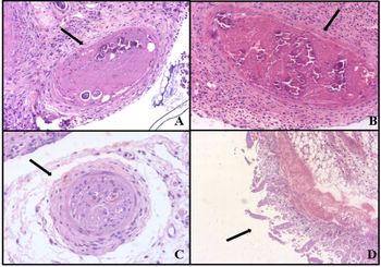
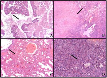
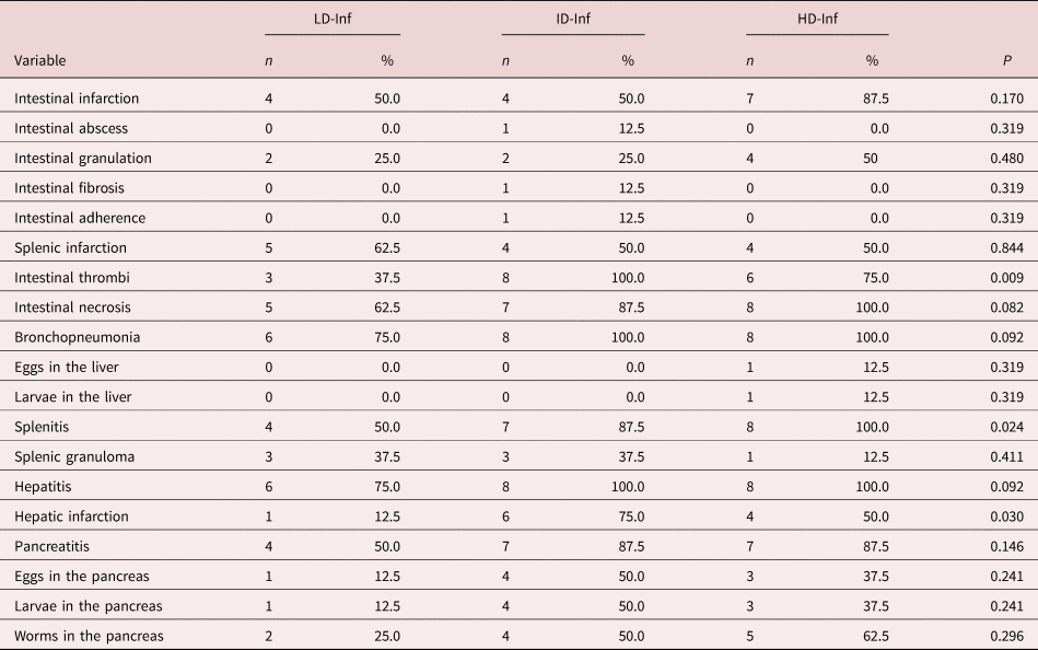
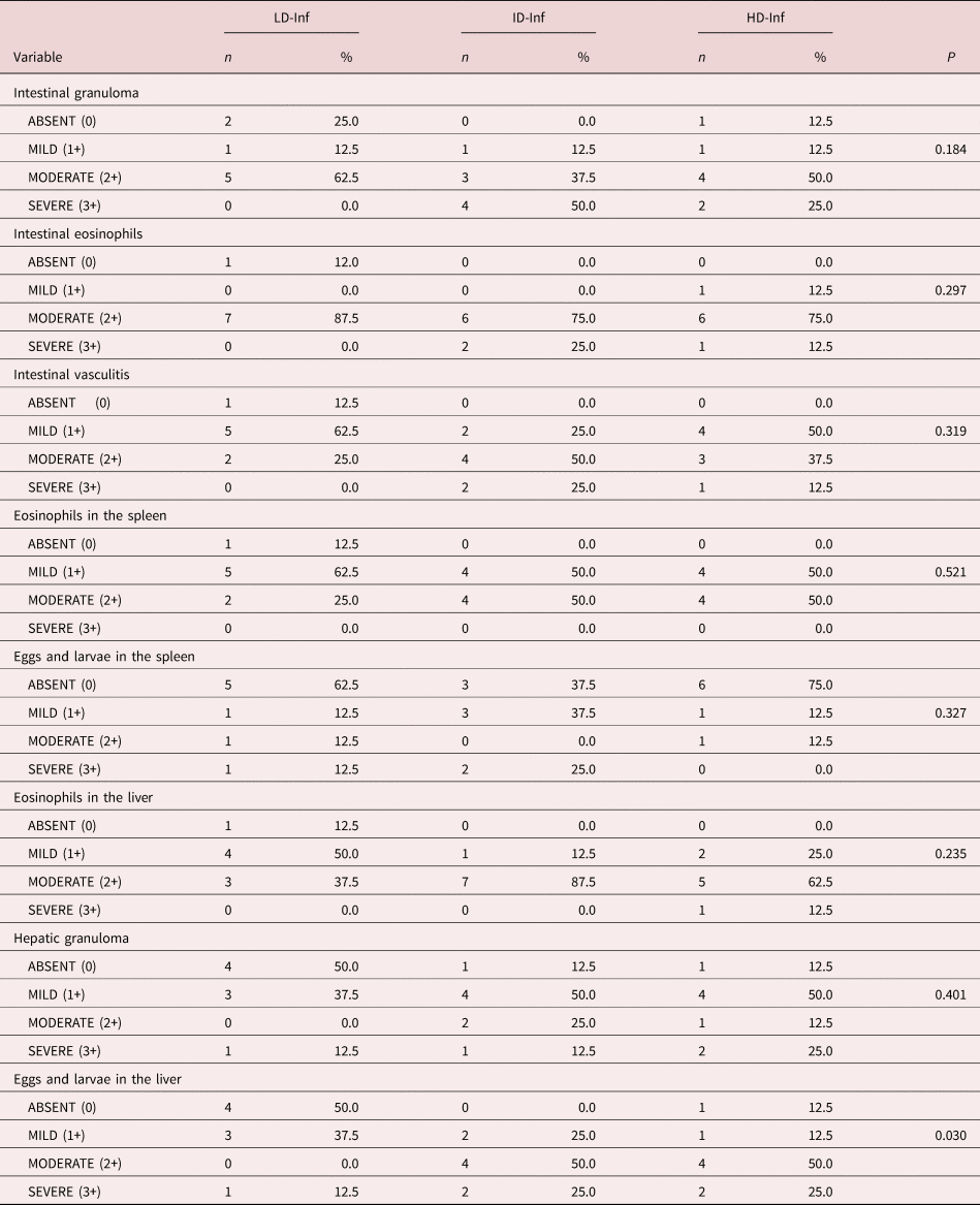
In the intestine, thrombi were present in all of the ID-Inf group mice (fig. 1a, b), compared with 37.5% of the LD-Inf group mice and 75.0% of the HD-Inf group mice (fig. 1c), with significant differences among the groups. Intestinal infarction, as depicted in fig. 1d, was identified in 87.5% of the HD-Inf group mice. Pancreatitis, as depicted in fig. 2a, was observed in 50% of the LD-Inf group mice and in 87.5% of the ID-Inf and HD-Inf group mice (table 1). However, in the ID-Inf group, 62.5% of the mice presented intestinal granuloma and 87.5% presented intestinal eosinophils, both of moderate intensity and both being proportionally more common than in the two other experimental groups (table 2).
(A) ID-Inf group: arterial thromboses with eggs and calcification (H&E; magnification, ×100); (B) ID-Inf group: thrombosis in the mesenteric artery (H&E; magnification, ×100); (C) HD-Inf group: recanalized artery with thrombosis (H&E; magnification, ×200); (D) HD-Inf group: transmural intestinal infarction with coagulative necrosis (H&E; magnification, ×50).

(A) HD-Inf group: acute pancreatitis with parenchymal necrosis (H&E; magnification, ×50); (B) HD-Inf group: spleen infarction with thrombi (H&E; magnification, ×50), (C) LD-Inf group: bronchopneumonia (H&E; magnification, ×50); (D) ID-Inf group: bronchopneumonia (H&E; magnification, ×100).

Macroscopic and microscopic findings associated with infection with different doses of Angiostrongylus costaricensis in Swiss mice.

Microscopic findings associated with infection with different doses of Angiostrongylus costaricensis in Swiss mice.

Splenitis was present in all experimental groups. However, the proportion of mice with splenitis was significantly higher in the ID-Inf and HD-Inf groups than in the LD-Inf group (87.5% and 100.0%, respectively, vs. 50.0%; P = 0.024 for both). Splenic infarction, as depicted in fig. 2b, was seen in 50.0% of the LD-Inf and ID-Inf group mice and in 62.5% of the HD-Inf group mice (table 1).
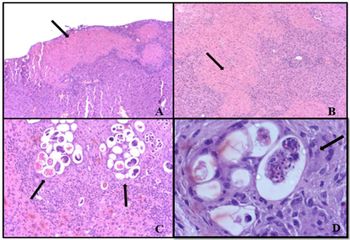
Bronchopneumonia was present in 75.0% of the LD-Inf group mice (fig. 2c), 100.0% of the ID-Inf group mice (fig. 2d) and 100.0% of the HD-Inf group mice. Hepatitis was present in 75.0% of the LD-Inf group mice, 100.0% of the ID-Inf group mice and 100.0% of the HD-Inf group mice. The proportion of mice with hepatic infarction in the ID-Inf group (fig. 3a), HD-Inf group (fig. 3b) and LD-Inf group was 75.0%, 50.0% and 12.5%, respectively, being significantly higher in the first two groups than in the last (P = 0.030; table 1). The majority of the mice in the ID-Inf and HD-Inf groups showed moderate deposition of eggs and larvae in the liver (fig. 3c and d, respectively), whereas the majority of those in the LD-Inf group showed mild deposition, and the proportion of mice showing such deposition differed significantly among the groups (table 2).
(A) ID-Inf group: hepatic infarction (H&E; magnification, ×50); (B) HD-Inf group: hepatic infarction with coagulative necrosis (H&E; magnification, ×50); (C) ID-Inf group: several eggs surrounded by granulomas in the liver (H&E; magnification, ×100); (D) HD-Inf group: granulomas and giant cell (arrow) surrounding eggs in the hepatic parenchyma (H&E; magnification, ×40).

Discussion
Here we show that it is possible to maintain the A. costaricensis life cycle with the inoculation of only five L3 in Swiss mice, reproducing with the same efficacy as the ten L3 inoculation currently used as standard. Similar findings were previously reported by Hermes et al. (Reference Gundamaraju and Vemuri2018), who showed that survival was longer among mice infected with five L3 of A. costaricensis than among those infected with higher doses, as was the time to eliminate L1 in faeces. These findings indicate that it is possible to inoculate mice with lower doses in order to maintain the life cycle of the parasite in animal facilities, despite the high mortality rates reported (Garrido et al., Reference Garrido, Morassutti, Barradas and Graeff-Teixeira2017).
Because of its clinical and histopathological manifestations, as well as its difficult-to-treat character, AA is an important disease, which makes the maintenance of the parasite life cycle in laboratory animals indispensable. Therefore, there is a need for further studies to elucidate its pathogenesis. To that end, we analysed histopathological alterations in Swiss mice infected with different doses of A. costaricensis.
In the present study, all of the mice in the ID-Inf group (infected with 15 L3) showed formation of intestinal thrombi. In a previous study, Graeff-Teixeira et al. (Reference Graeff-Teixeira, Camillo-Coura and Lenzi1991) reported the main histopathological manifestations of AA, including intestinal thrombi, as well as mesenteric lesions with eosinophilic infiltrate and perivascular granulomas.
Intestinal infarction, which was observed mainly in our HD-Inf group mice (infected with 30 L3), is considered another major complication of AA and could lead to death (Graeff-Teixeira et al., Reference Graeff-Teixeira, Camillo-Coura and Lenzi1991). In cases of intestinal obstruction or infarction in patients with AA, surgical treatment is necessary (Rodriguez et al., Reference Rodriguez, Dequi, Peruzzo, Mesquita, Garcia and Fornari2008), because the use of an anti-inflammatory does not provide satisfactory results (Mentz & Graeff-Teixeira, Reference Liu, Bair, Chang, Lin and Chan2003). High doses of enoxaparin also do not prevent the formation of intestinal lesions, nor do they promote an increase in the survival of mice experimentally infected with the parasite (Sandri et al., Reference Rodriguez, Silva, Müller, Alves, Graeff-Teixeira and Fornari2017).
Given that all of the mice in our ID-Inf group presented intestinal thrombi, we can suppose that those mice would develop infarction, leading to peritonitis and finally to sepsis, resulting in death. Rodriguez et al. (Reference Rodriguez, Porto, Ferrari, Marcolan, da Silva, Graeff-Teixeira and Fornari2011) found that sepsis was a major cause of death in Swiss mice infected with ten L3 of A. costaricensis. However, in our HD-Inf group, 50% of the mice developed intestinal granuloma of moderate intensity and 100% developed intestinal vasculitis. Vascular granuloma can lead to obliteration and ischemia of the affected organ. Rodriguez et al. (Reference Rodriguez, Porto, Ferrari, Marcolan, da Silva, Graeff-Teixeira and Fornari2011) also demonstrated that vasculitis and granulomas can result in vascular occlusion and tissue ischemia, emphasizing the importance of those inflammatory reactions in the pathogenesis of AA. The intestinal infarction after mesenteric thrombosis seen in the present study would hasten death among the mice, whereas the vascular obliteration caused by intestinal granuloma accompanied by eosinophilic vasculitis would have slowed the progression to death.
Previous studies have shown that mice infected with five L3, 15 L3 and 30 L3 die at different time points, mortality peaking between 20 and 40 days after infection. Mice infected with 15 L3 have been shown to die sooner than do those infected with 30 L3 (Hermes et al., Reference Gundamaraju and Vemuri2018). The genetic variability of each mouse and its resistance to infection are also factors that affect survival (Ishih & Sano, Reference Ishih and Sano1989; Garrido et al., Reference Garrido, Morassutti, Barradas and Graeff-Teixeira2017). Our histopathological findings contribute to elucidating these temporal differences. Acute pancreatitis, which was seen in 50.0% of our LD-Inf group mice and in 87.5% of our ID-Inf and HD-Inf group mice, is another important histopathological finding in AA. Acute pancreatitis is an inflammatory disease that affects the exocrine pancreatic parenchyma and can vary in a self-limited way from a mild to a more severe form (Singh et al., Reference Sierra and Morera2016). Although biliary calculi and alcohol use are the most common causes of the disease, it is estimated that approximately 10% of cases are caused by viruses, bacteria or parasites (Economou & Zissis, Reference Economou and Zissis2000). Studies have shown that Ascaris lumbricoides is the most important nematode related to pancreatitis (Khuroo et al., Reference Junqueira and Carneiro1992). In rare cases, pancreatitis has been associated with infection with helminths of the genus Taenia (Liu et al., Reference Khuroo, Zargar, Yattoo, Koul, Khan, Dar and Alai2005). There have also been reports of pancreatitis caused by other parasites, such as Opisthorchis spp. (Gundamaraju & Vemuri, Reference Graeff-Teixeira, Agostini, Camillo-Coura and Ferreira-da-Cruz2014), Fasciola hepatica (Veerappan et al., Reference Singh, Jain and Kiran1991) and Echinococcus granulosus (Ozmen et al., Reference Morera and Cespedes2005).
We found that, regardless of the amount of A. costaricensis L3 inoculated, all of the infected mice developed AA. However, a dose of five L3 has been shown to be sufficient to maintain the life cycle of the parasite in Swiss mice, those infected with that dose developing signs of AA, eliminating L1 in their faeces and remaining alive for longer than those infected with higher doses (Hermes et al., Reference Gundamaraju and Vemuri2018).
To our knowledge, this is the first study to correlate different doses of A. costaricensis L3 with the severity of AA in an experimental animal model. It is also the first to highlight the importance of pancreatitis as a histopathological finding. The histopathological aspects of AA in Swiss mice vary in relation to the infective dose of A. costaricensis, the severity of the pathology correlating positively with the parasite burden.
Financial support
This study was funded by the University of Passo Fundo, in the city of Passo Fundo, Brazil.
Conflicts of interest
None.
Ethical standards
The authors assert that all procedures contributing to this work comply with the ethical standards of the relevant national and institutional guides on the care and use of laboratory animals.