Introduction
Antibacterial substances have been found in nearly all larger groups of arthropods and other invertebrates. Nowadays, several classes of antibacterial compounds were characterized among insects, including lysozyme, cercopin, attacin, diptericin, abaecin, apidaecin, and coleoptericin (Xylander, Reference Xylander2009).
A huge number of beetle species produce defensive secretions against potential predators (Blum, Reference Blum1981), with special emphasis on the family Silphidae (Nicrophorus spp.), whose oral and anal secretions have established antimicrobial properties in order to protect their own organism from micropopulations within their ecosystems (Hoback et al., Reference Hoback, Bishop, Kroemer, Scalzitti and Shaffer2004; Degenkolb et al., Reference Degenkolb, Düring and Vilcinskas2011). In addition, involvement of three terpene alcohols may be responsible for the antimicrobial properties (Roach et al., Reference Roach, Eisner and Meinwald1990). Percentage of palmitic acid within oral and anal secretions of Nicrophorus vespilloides Herbst, 1783 could be responsible for the antimicrobial potential (Degenkolb et al., Reference Degenkolb, Düring and Vilcinskas2011). The presence of phenylacetic, isobutyric and caprylic acids contributes to the antimicrobial properties of secondary metabolites of Silphidae. Most of the primary and secondary metabolites of adult N. vespilloides must have a role in ecological function (Degenkolb et al., Reference Degenkolb, Düring and Vilcinskas2011). Within other Coleoptera Polyphaga, certain antimicrobial agents were identified among larvae of Aleocharinae (family Staphylinidae) and adults of Tenebrionidae (Ghilarov, Reference Ghilarov1958; Francke & Dettner, Reference Francke, Dettner and Schulz2005).
Antibacterial substances against gram-positive and gram-negative bacteria within other soil-dwelling arthropods have been described from the haemolymph and the defensive secretion of myriapods, likewise (Xylander, Reference Xylander2009; Makarov et al., Reference Makarov, Dimkić, Antić, Vujisić, Stević, Mitić, Tomić, Ilić, Ćurčić and Stanković2014). The application of haemolymph of different centipede and millipede species inhibits the growth of gram-negative bacteria Enterobacter cloacae and Escherichia coli, and gram-positive Micrococcus lysodeikticus, with reference to the fact that in general the centipedes showed higher antibacterial activity than the millipedes (Xylander, Reference Xylander2009). Antifungal activity of defensive secretions of certain millipede species has been investigated some time ago (Roncadori et al., Reference Roncadori, Dufey and Blum1985), while the defensive secretion of Pachyiulus hungaricus (Karsch, 1881) was recently evaluated for exploration of its antimicrobial activity against several certain gram-negative and gram-positive bacteria and fungi (Makarov et al., Reference Makarov, Dimkić, Antić, Vujisić, Stević, Mitić, Tomić, Ilić, Ćurčić and Stanković2014).
Chemical compounds from pygidial gland secretions such as benzoic or phenylacetic acids are well known for their antimicrobial and preservative capabilities within the suborder Adephaga (Dettner, Reference Dettner1985). The family Carabidae represents the largest and the most abundant taxon within the suborder Adephaga (order Coleoptera), which includes 40 000 species (Beutel, Reference Beutel1997).
Laemostenus (Pristonychus) punctatus (Dejean, 1828) is a troglophilic ground beetle species, which produces a secretion by the paired pygidial glands. The qualitative and quantitative content of the pygidial secretion has been determined by us recently (Vesović et al., Reference Vesović, Ćurčić, Vujisić, Nenadić, Krstić, Perić-Mataruga, Milosavljević, Antić, Mandić, Petković, Vučković, Marković, Vrbica, Ćurčić and Makarov2015). A pair of abdominal pygidial glands is a feature characteristic for several beetle families, but its presence within Carabidae is inevitable. The structure of the glands involves two sets of secretory lobe aggregations, collecting canals, reservoirs where the secretion is deposited, and efferent ducts, which serve to discharge the products of these gland outwards (Forsyth, Reference Forsyth1972). Pygidial glands are variable both in structure and the function of produced substances (Thiele, Reference Thiele1977). The main functions of pygidial glands are probably defence against predators, producing alarm messages and antimicrobial/antifungal activity (Blum, Reference Blum1981; Giglio et al., Reference Giglio, Brandmayr, Talarico and Zetto2011). The latter potential function was of particular interest of our current study. The aim of the current paper was to determine the antimicrobial (both antibacterial and antifungal) activity of the pygidial secretion of a ground beetle species, since the data on the antimicrobial properties of the secretion in Carabidae are scarce (Giglio et al., Reference Giglio, Brandmayr, Talarico and Zetto2011). The additional task was to describe the morphology of the pygidial glands.
Materials and methods
Collecting and handling of ground beetle specimens
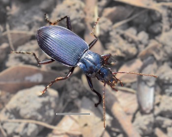
A total of 30 adult specimens of both genders of L. (P.) punctatus were collected by hand on 22 March 2015 in the Ogorelička Pećina Cave, village of Sićevo, near Niš, Svrljiške Planine Mts., Southeastern Serbia (detected environmental conditions: temperature 12–14°C, aerial humidity 60–81%) by S. Ćurčić, N. Vesović, and M. Nenadić (fig. 1). After catching, the pygidial gland secretion of the beetles was extracted via stimulation of the abdomen by milking into vials with solvent. Afterwards the live beetle specimens were stored in laboratory-controlled conditions in portable plastic refrigerators a few days before taking samples of the pygidial glands in the laboratory. These conditions implied constant temperature (10°C), total darkness, and occurrence of moist soil taken from the site of collecting. Water was sprayed over the specimens and soil from time to time to maintain a high level of humidity, similar to that present in the cave from which they originated. The beetles were fed on earthworms. After performed analysis, the dry ground beetle specimens were deposited in the collection of the Institute of Zoology, University of Belgrade – Faculty of Biology, Belgrade, Serbia. The volume value of the discharged pygidial gland secretion of L. (P.) punctatus was measured on the analytical balance Sartorius Model 2405 for weighing up to 30 g maximum, with 0.001 mg resolution (Sartorius Group Göttingen, Germany) at the University of Belgrade – Faculty of Chemistry.
Habitus of L. (P.) punctatus. Scale bar 5 mm.

Morphological analysis
Beetles’ abdomens have been carefully dissected in saline solution. Afterwards the pygidial gland reservoirs and the secretory lobes were observed morphologically. Zeiss Stereo Discovery.V8 with Zeiss Axio Cam ICc 1 attached and AxioVision Microscope Software, as well as Canon G10 digital camera were used for photographing morphological features of the pygidial glands.
Antimicrobial activity
Biological material
The pygidial gland secretions of 30 specimens of L. (P.) punctatus were dissolved in 5% DMSO-water solution [Dimethyl sulfoxide, (CH3)2SO), Sigma-Aldrich, Munich, Germany] and stored in a refrigerator at 4°C in the laboratory.
Antibiotics and antimycotics
Commercial antibiotics
Streptomycin (Sigma-Aldrich, Munich, Germany) and Ampicillin (Panfarma, Belgrade, Serbia) were applied as positive controls (1 mg ml−1).
Commercial antimycotics
Ketoconazole (Zorka Pharma, Šabac, Serbia) and Bifonazole (Srbolek, Belgrade, Serbia) were applied as positive controls (1 mg ml−1).
Tested microorganisms
The laboratory and clinical strains of tested bacteria (four gram-positive and four gram-negative species) and micromycetes (eight species) are tested. These include the following gram-positive bacteria: Bacillus cereus (clinical isolate), Micrococcus flavus (ATCC 10240), Staphylococcus aureus (ATCC 6538) and Listeria monocytogenes (NCTC7973), as well as the following gram-negative bacteria: E. coli (ATCC 35210), Pseudomonas aeruginosa (ATCC 27853), E. cloacae (clinical isolate) and Salmonella typhimurium (ATCC 13311). The tested micromycetes were Aspergillus fumigatus (ATCC 1022), Aspergillus versicolor (ATCC 11730), Aspergillus ochraceus (ATCC 12066), Aspergillus niger (ATCC 6257), Trichoderma viride (IAM 5061), Penicillium funiculosum (ATCC 36839), Penicillium ochrochloron (ATCC 9112) and Penicillium verrucosum var. cyclopium (food isolate). The microorganisms are deposited in the Mycological Laboratory, Department of Plant Physiology, Institute for Biological Research ‘Siniša Stanković’, University of Belgrade, Belgrade, Serbia.
In vitro assays for determination of antimicrobial activity
Preparing of overnight cultures of bacteria
The bacterial strains were stocked in glycerol and seeded on 2 ml of Tryptic Soy Broth (TSB) (Biolife, Italy) medium, then incubated in thermostat for 24 h at 37°C. Overnight cultures have 1.0 × 10⁹ cells ml−1 each. The final solution of culture and medium (1.0 × 10⁸ cells ml−1) was obtained by dissolving 100 ml of a primary culture in 900 ml of TSB.
Preparing of cultures of micromycetes
The micromycetes were cultivated on Malt agar medium for 21 days at 26°C, and stored in thermostat at 4°C. Inoculum was prepared via spores rinsing from the substrate surface using sterile solution 0.85% NaCl and 0.1% Tween 80 (vol/vol). Final concentration of spores was 1.0 × 10⁵ cells ml−1 of medium. Inoculum was cultivated at 4°C.
Microdilution method
The antimicrobial assay was performed using a microdilution (Hanel & Raether, Reference Hanel and Raether1988; Daouk et al., Reference Daouk, Dagher and Sattout1995). We have determined minimal inhibitory (MIC), minimal bactericidal (MBC) and minimal fungicidal concentrations (MFC) of the analyzed pygidial secretion for each species of studied microorganisms by serial dilutions of the tested secretion dissolved in 5% DMSO-water solution.
Microplates with inoculated micromycetes, bacteria and tested solutions of the pygidial secretion were incubated for 72 h at 28°C for fungi and for 24 h at 37°C for bacteria. Minimal concentration, which provoked growth inhibition of bacteria and micromycetes is determined as MIC.
MBC and MFC were determined by reinoculation of 2 µl of inoculum in 100 µl of liquid medium and incubated for 24 h at 37°C (bacteria) and for 72 h at 26°C (micromycetes), after which the growth was not detected. In order to visualize bacterial colonies, we have used INT stain [p-iodonitrotetrazolium violet [2-(4-iodophenyl)-3-(4-nitrophenyl)-5-phenyltetrazolium chloride; (2 mg ml−1 H2O), Sigma-Aldrich, Munich, Germany]. The stain was applied on microplates, while afterwards the stained colonies were incubated for 2 h at 37°C. Unstained chambers of microplates have indicated the presence of bactericidal effect, while pale yellowish color indicated inhibition.
Statistical analysis
The results were expressed as mean values and standard errors, and analyzed using one-way analysis of variance (ANOVA), followed by Tukey's HSD Test with α = 0.05. This analysis was performed via Statgraphics Centurion XVI, v. 16.2 software.
Results
Morphology of the pygidial glands of L. (P.) punctatus
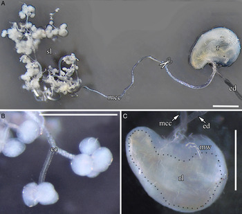
The pygidial gland reservoirs are reniform, but slightly asymmetric, with one broad and one narrow part (not efficiently differentiated to be lobes). Also, a depression on the inner medial zone is present (fig. 2A). L. (P.) punctatus has reservoirs about 2 mm in length and about 1.5 mm in width.
Morphological structure of pygidial glands of L. (P.) punctatus. (A), right pygidial gland apparatus; (B), groups of secretory lobes; (C), appearance of a pygidial reservoir; r, pygidial reservoir; rl, reservoir lumen; mw, muscular wall; ed, efferent duct; cc, collecting canal; mcc, main collecting canal; sl, secretory lobes. Black dots on the pygidial reservoir (r) (fig. 2C) show the edge of the yellowish reservoir lumen (rl). Scissors sign indicates that the main collecting canal (mcc) is longer than showed in fig. 2A (a part of mcc is missing). Scale bars 1 mm.

A muscle-coated but tender efferent duct extends from the reservoir and narrows towards its opening. The length of the efferent duct in L. (P.) punctatus is up to 4 mm.
The secretory lobes are spherical from outside, but slightly reniform in cross section (figs 2A and B). The glands are constituted of 60–70 of these globules (Forsyth, Reference Forsyth1972) (fig. 2A). Secretory lobes are 150–200 µm in diameter.
A long collecting canal links the lobes with the reservoir in its middle, concave zone called hilum (Rossini et al., Reference Rossini, Attygalle, Gonzalez, Smedley, Eisner, Meinwald and Eisner1997) (figs 2A and C). Length of main collecting canal is between 10 and 15 mm.
We did not notice any differences between sexes in terms of glandular structure.
Volume of the discharged pygidial gland secretion of L. (P.) punctatus
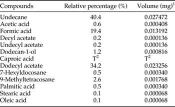
The volume value of the discharged pygidial gland secretion was measured from 30 specimens of L. (P.) punctatus. These were milked into an empty vial in the laboratory. The weight of the vial was measured on an extra-precise analytical balance before (empty vial) and after the secretion discharging (with the secretion inside). The difference of the weight represented the volume of the released secretion of 30 individuals: 0.068 mg ± 0.001 mg (SD). The volume of each isolated compound from the pygidial gland secretion is presented in table 1. The experiment has been repeated three times in a few weeks in order to confirm validity of the results.
Components of the pygidial gland secretion of L. (P.) punctatus and the volume in the secretion (modified after Vesović et al., Reference Vesović, Ćurčić, Vujisić, Nenadić, Krstić, Perić-Mataruga, Milosavljević, Antić, Mandić, Petković, Vučković, Marković, Vrbica, Ćurčić and Makarov2015).

1 Measured from 30 specimens.
2 Trace.
Antibacterial and antifungal activity of the pygidial gland secretion of L. (P.) punctatus
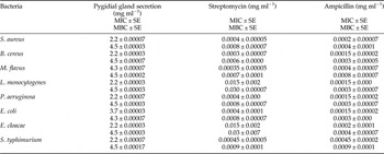
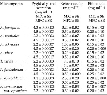
The pygidial gland secretion of L. (P.) punctatus has achieved antibacterial activity against all tested bacteria. M. flavus proved to be more resistant compared with other bacterial strains (MIC 4.3 mg ml−1 and MBC 4.5 mg ml−1), with special emphasis on E. coli (MIC 3.7 mg ml−1 and MBC 4.3 mg ml−1), which proved to be the most sensitive bacteria. Commercial antibiotics Streptomycin and Ampicillin, applied as positive controls, showed much higher antibacterial properties for all bacterial strains (table 2). A. fumigatus proved to be the most resistant (MIC 4.3 mg ml−1 and MFC 4.5 mg ml−1), while P. ochrochloron (MIC 1.1 mg ml−1 and MFC 2.2 mg ml−1) and P. verrucosum var. cyclopium (MIC 1.1 mg ml−1 and MFC 2.2 mg ml−1) – the most sensitive micromycetes. Commercial antifungal drugs Ketoconazole and Bifonazole, applied as positive controls, showed much higher antifungal properties for all fungi species, except for P. ochrochloron, which proved to be more resistant to synthetic antimycotic Ketoconazole compared with pygidial gland secretion of L. (P.) punctatus (MIC 1.1 mg ml−1 and MFC 2.2 mg ml−1) (table 3).
Minimum inhibitory (MIC) and minimum bactericidal concentrations (MBC) of the pygidial gland secretion of L. (P.) punctatus compared with commercial antibiotics Streptomycin and Ampicillin.

SE, standard error. All concentration values show significant differences (P < 0.05).
Minimum inhibitory (MIC) and minimum fungicidal concentrations (MFC) of the pygidial gland secretion of L. (P.) punctatus compared with commercial antifungicides Ketoconazole and Bifonazole.

SE, standard error. All concentration values show significant differences (P < 0.05).
Discussion
Based on presently available data, the morphological features of Laemostenus Bonelli, 1810 pygidial glands are similar to those of the genera Agonum Linnaeus, 1758, Calathus Bonelli, 1810 and Anchomenus Bonelli, 1810 within the same subfamily (Platyninae) (Bonacci et al., Reference Bonacci, Brandmayr, Zetto, Perrotta, Guarino, Peri and Colazza2011). However, among other subfamilies, some similarities could be set aside with certain species from Harpalinae (communication of secretory lobes with the main collecting canal) and Anthiinae (musculature of the eversible valve) (Forsyth, Reference Forsyth1972).
Our results demonstrate that the ground beetle species L. (P.) punctatus produces pygidial gland secretion, which, apart from the assumed defensive role (Vesović et al., Reference Vesović, Ćurčić, Vujisić, Nenadić, Krstić, Perić-Mataruga, Milosavljević, Antić, Mandić, Petković, Vučković, Marković, Vrbica, Ćurčić and Makarov2015), has both antibacterial and antifungal properties as well.
As has already been recently established, the major component of the pygidial gland secretion in L. (P.) punctatus is undecane (40.4%) (Vesović et al., Reference Vesović, Ćurčić, Vujisić, Nenadić, Krstić, Perić-Mataruga, Milosavljević, Antić, Mandić, Petković, Vučković, Marković, Vrbica, Ćurčić and Makarov2015). The remaining main compounds include acetic (0.6%) and formic acids (19.4%), dodecan-1-ol (1.2%), dodecyl acetate (34.2%), and 9-methyltetracosane (2.6%), respectively (Vesović et al., Reference Vesović, Ćurčić, Vujisić, Nenadić, Krstić, Perić-Mataruga, Milosavljević, Antić, Mandić, Petković, Vučković, Marković, Vrbica, Ćurčić and Makarov2015). It has been previously demonstrated that organic components, such as formic and acetic acids, are correlated with antimicrobial activity at the lower concentrations (1–2%) (Raftari et al., Reference Raftari, Jalilian, Abdulamir, Son, Sekawi and Fatimah2009; Raftar et al., Reference Raftar, Jalilian, Abdulamir, Ghafurian, Sekawi, Son and Bakar2012). Likewise, the involvement of palmitic (0.5%), stearic (0.1%) and oleic acids (0.1%) in composition of the pygidial gland secretion in L. (P.) punctatus is significant and they could additionally contribute to the antimicrobial activity. The antifungal and bactericidal properties of fatty acids are previously described and well known (Glassman, Reference Glassman1948; Kabara et al., Reference Kabara, Swieczkowski, Conley and Truant1972; Desbois & Smith, Reference Desbois and Smith2010). Additionally, dodecan-1-ol exhibits fungistatic features, and it can be converted to a fungicidal agent by other compounds (Fujita et al., Reference Fujita, Fujita and Kubo2007). Its antibacterial activity is confirmed as well (Togashi et al., Reference Togashi, Shiraishi, Nishizaka, Matsuoka, Endo, Hamashima and Inoue2007). We are of opinion that a few compounds (acetic and formic acids, fatty acids, and a long-chain fatty alcohol) show synergistic effect on the antimicrobial and antifungal activities of the pygidial gland secretion of L. (P.) punctatus against analyzed microorganisms.
Previous investigations on the antimicrobial (both antibacterial and antifungal) potential of ground beetles are scarce. Pavan (Reference Pavan1949) reported the case of the pygidial secretion of bombardier beetles (Brachinus spp.) (mainly consisting of quinones) (Lečić et al., Reference Lečić, Ćurčić, Vujisić, Ćurčić, Ćurčić, Nikolić, Anđelković, Milosavljević, Tešević and Makarov2014), which exhibits antibacterial features, apart from the defensive role. The pygidial gland secretions of other Adephaga living in freshwater (Hydradephaga) have antimicrobial properties and are mainly applied to hydrofuge body regions important for aquatic respiration. The secretions are supposed to inhibit the growth of microorganisms, thus preventing the contamination and wetting of the cuticular respiratory structures (Kovac & Maschwitz, Reference Kovac and Maschwitz1990). Chemically the secretions are characterized either by phenylacetic acid (some Dytiscidae, Noteridae, Haliplidae), by benzoic acid, hydroxybenzaldehyde and related compounds (some Dytiscidae), or by certain aromatic compounds (Amphizoidae) (Francke & Dettner, Reference Francke, Dettner and Schulz2005). We are of opinion that in the case of L. (P.) punctatus pygidial gland secretion a few additional compounds (acetic and formic acids, fatty acids, a long-chain fatty alcohol) might be responsible for the antimicrobial features, and these serve to protect the beetle from microbial contamination from soil and guano and diseases, something that the future studies in progress will confirm.
Conclusions
Pygidial gland morphological analysis of L. (P.) punctatus presented in the current paper is the first data given for the aforementioned species. Apart from this, taxa within the subfamily Platyninae are poorly known in this sense. Forsyth (Reference Forsyth1972) and Bonacci et al. (Reference Bonacci, Brandmayr, Zetto, Perrotta, Guarino, Peri and Colazza2011) gave some morpho-histological data on the genera Agonum, Calathus and Anchomenus, but sparingly.
In summary, this study has indicated that pygidial gland secretion of ground beetle species L. (P.) punctatus has showed anti-gram-negative and anti-gram-positive bacterial activity, as well as antifungal potential against human pathogens. Compared with commercial antibiotics Streptomycin and Ampicillin and antimycotics Ketoconazole and Bifonazole, the tested secretion had a varied impact, depending on the observed strain of bacteria/fungi. Videlicet, commercial antibiotics and antimycotics showed much higher antimicrobial potential for all bacteria and fungi, except for micromycete P. ochrochloron, which proved to be more resistant to Ketoconazole compared with the pygidial gland secretion. The antimicrobial features of the secretion are probably caused by a few constituents combined that act synergistically.
The observed inhibition and bactericidal/fungicidal effect on the tested bacteria/fungi strains might inspire design of novel agents with potential medicinal applications. Further research will be directed towards a better comprehending of the potential toxicity and usage of pygidial gland secretion compounds of ground beetles with antimicrobial properties in therapeutic purposes.
Acknowledgements
The study is financially supported by the Serbian Ministry of Education, Science, and Technological Development (Grants Nos. 173038, 173032, 172053, and 173027).