Introduction
Haemoglobinopathy can be a source of discrepancy between arterial partial pressure of oxygen (PaO2) and estimated oxygen saturation by pulse oximetry (SpO2). The fluctuation of SpO2 and the presence of CHD complicate its diagnosis in patients with haemoglobinopathy.
Case presentation
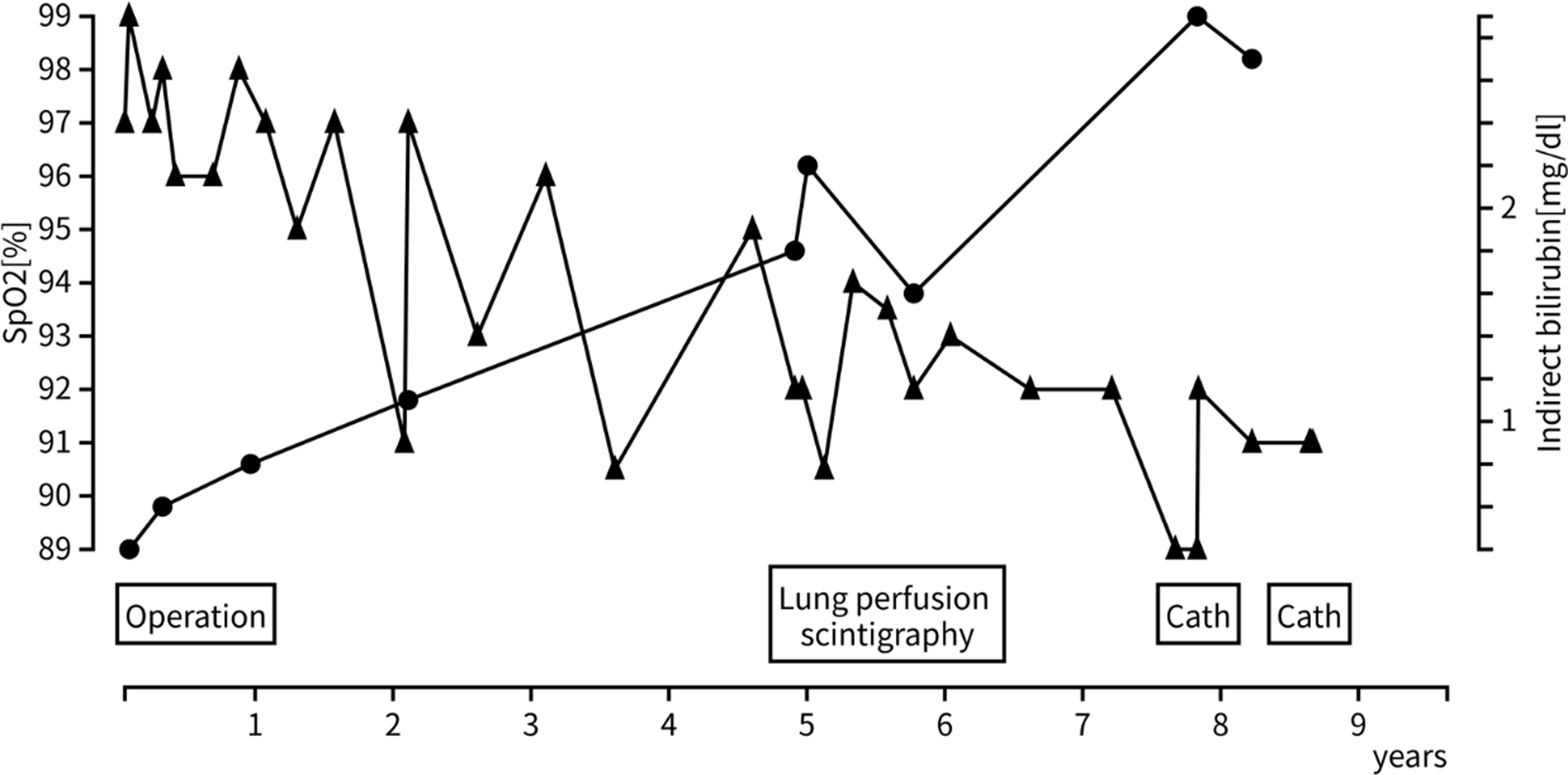
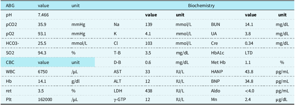
A seven-year-old boy was referred to us because of progressive decline in SpO2. His neonatal history was unremarkable. He was diagnosed with a ventricular septal defect, and his SpO2 was 97–99% when it was surgically closed during infancy. Then, he was followed-up annually. Although he did not report any cardiovascular symptoms or dyspnoea, his SpO2 was 91% when he visited us. His SpO2 was unresponsive to the oxygen challenge test. Echocardiography revealed structurally normal heart, without any signs of pulmonary hypertension. His SpO2, arterial oxygen saturation measured using blood gas analysis (SaO2), and pO2 at ventricular septal defect closure during infancy were 98%, 97.3%, and 104 mmHg, respectively. Thus, we differentiated diseases that may develop progression of right-to-left shunting. Contrast-enhanced CT showed no evidence of pulmonary arteriovenous fistula or patent ductus venosus. Lung perfusion scintigraphy indicated no perfusion defect, with a shunt ratio of 4.5%, which was considered to be within the normal range (Figure 1-a). Reference Ito, Kurihara and Ishibashi1 To further differentiate the source of the desaturation, we performed cardiac catheterisation. At first, the PaO2 under general anaesthesia was 93.1 mmHg, whereas SpO2 was approximately 90–92% and arterial oxygen saturation measured by blood gas analyser (SaO2) was 94.3%, implied the possible discrepancy of SpO2 and PaO2 (Table 1), although we could not exclude measurement errors at this point. Although contrast echocardiography during cardiac catheterisation was negative for pulmonary arteriovenous fistula, we identified significant inferior vena cava stenosis just distal to the hepatic vein, with a pressure gradient of 3–4 mmHg (Figure 1-b). Thus, we performed a second cardiac catheterisation to further investigate the possibility of a portosystemic shunt. While the collateral veins detouring the stenotic inferior vena cava communicated to the azygos vein and portal vein, contrast imaging from the collateral vein was directed to the portal vein; thus, portosystemic shunt and portal hypoplasia were unlikely (Figure1-c). PaO2 in the second cardiac catheterisation was again inconsistent with the value of SpO2. The proportion of methaemoglobin was normal, and an increased fraction of reticulocytes implied rapid red blood cell turnover at the first cardiac catheterisation (Table 1). Taken together, we suspected that the source of low oxygen saturation could be haemoglobinopathy, regardless of his normal SpO2 during infancy. Indeed, blood levels of glycosylated haemoglobin A1c (HbA1c) measured by high-performance liquid chromatography were markedly low, supporting possibility of haemoglobinopathy. Accordingly, we retrospectively summarised changes in SpO2 and found that the serum bilirubin level started to increase when his SpO2 started to decline, implying a possible attribution of intravascular haemolysis (Figure 2). Haemoglobin beta gene sequence identified heterozygous mutation in HBB:c.295G > A, known as haemoglobin Köln disease. At the age of 10 years, he developed his first haemolytic attack. After blood transfusion, his SpO2 rose from 90–93% to 97%, which confirmed the source of desaturation was haemoglobinopathy, rather than right-to-left shunting.
Images to differentiate cyanotic diseases (a): Pulmonary perfusion scintigraphy using 99mTc-macroaggregated albumin indicates no perfusion defects in the lungs. The calculated right to left shunt ratio was 4.5% (cut-off value: <10%).(b): Contrast image from the inferior vena cava (IVC) is shown. IVC obstruction was observed at the junction of the hepatic vein, with a pressure gradient of 3 mmHg (solid arrow). A collateral vein detouring the stenotic IVC is shown.(c): Contrast image from the collateral vein (solid arrow) is shown. The venous catheter was inserted from the right femoral vein and located on the collateral vein between the IVC and the left ascending lumbar vein. The contrast agent was directed downward and then ascended to the portal vein. Note that all the contrast was directed to the portal vein (dotted arrow), and no apparent countercurrent blood flow from the portal vein toward the systemic vein was observed, suggesting no portosystemic shunt.

Trend of arterial oxygen saturation measured by pulse oximetry and serum levels of indirect bilirubin. ●: serum levels of indirect bilirubin.▲: arterial oxygen saturation measured by pulse oximetry. Arterial oxygen saturation measured by pulse oximetry (SpO2) gradually declined with the elevation of serum levels of indirect bilirubin, one of makers of haemoglobinopathy. His SpO2 for a few years after surgery exhibited temporal decline without increase in indirect bilirubin, and it recovered to more than 95% at the subsequent clinical visit. Although this decline was regarded as the result of frequent respiratory infections related to entering nursery school, it might be associated with haemoglobinopathy.

Laboratory data at the first cardiac catheterisation

Abbreviations: ALT = alanine aminotransferase; Aldo = Aldosterone; AST = aspartate aminotransferase; BNP = B type natriuretic peptides; BUN = Blood Urea Nitrogen; Cre = creatinine; D-B = direct bilirubin; γ-GTP = γ-Glutamyl Transpeptidase; Hb = haemoglobin; HANP = human atrial natriuretic peptide; LDH = lactate dehydrogenase; LTD = lower than detectable; Met Hb = methaemoglobin; Mn = Manganese; Plt = platelet count = ret = reticulocytes; T-B = total bilirubin; UA = uric acid; WBC = White blood cell count.
Discussion
We observed progressive decline of SpO2 in the patient with haemoglobin Köln disease. Although there was a discrepancy between PaO2 and SpO2, we could not be sure of haemoglobinopathy as the SpO2 was often as high as >95% during the first 5 years, as shown in Figure 2. The existence of inferior vena cava stenosis also complicates the diagnosis, as it requires us to differentiate from a portosystemic shunt, which might be associated with a pulmonary atrial-venous fistula or pulmonary hypertension, ultimately right-to-left shunting.
Pulse oximetry estimates oxygenated haemoglobin saturation based on the translucency of wavelengths 660 nm and 940 nm. Haemoglobin Köln’s disease is an unstable haemoglobin syndrome, in which the 98th amino acid of the β-chain is replaced from valine to methionine, Reference Katoh, Miyake and Arai2 absorbs more at 660 nm owing to an upward shift in absorbance properties at wavelengths between 600 and 900 nm, resulting in low SpO2. Reference Verhovsek, Henderson, Cox, Luo, Steinberg and Chui3 As haemoglobin F, which is the predominant form of haemoglobin during infancy, does not have a β-chain, oxygen saturation did not decrease during infancy. The progressive decrease in SpO2 was partly related to the disappearance of haemoglobin F, but the reason why SpO2 was often as high as 95% at 5 years of age was unclear. SpO2 of 90–93% at his haemolytic attack was slightly higher than usual, and it rose to 97% after blood transfusion. SpO2 of reported adult haemoglobin Köln cases were approximately 90%. Reference Cheung and Silverman4–Reference Alshurafa, Elsabagh, Obaid, Elhaji and Yassin6 Mild haemolysis and/or early turnover of old haemoglobin in early childhood might have attenuated 660 nm absorbance of oxygenated haemoglobin, resulting in relatively high SpO2 for years. Reference Kuruvilla, Widness and Nalbant7 Indeed, Karabaliev et al demonstrated that changes in red blood cell volume and shape alter absorption spectrum of haemoglobin. Reference Karabaliev, Tacheva, Paarvanova and Georgieva8 The SpO2, SaO2, and pO2 values at cardiac catheterisation were 90–92%, 94.3%, and 93.1 mmHg, respectively. Blood gas analysis directly titrates pO2 with the electrode method, whereas it uses the translucency of at least four wavelengths in the measurement of SaO2, which allows relatively robust estimation of pO2 as compared with SpO2. Reference Brugger, Santafe-Marti and Lakhal9
In addition to progressive oxygen desaturation, stenosis of the inferior vena cava and the presence of a collateral vein communicating with the portal vein further complicate the differential diagnosis. At the time of ventricular septal defect surgery, the central venous line was placed in the superior vena cava, and the location of the venous cannulation of the cardiopulmonary bypass was far proximal to the inferior vena cava stenosis; thus, iatrogenic inferior vena cava stenosis was unlikely. The portal vein was not hypoplastic, and the catheter angiogram indicated collateral vein flows directed towards the portal vein, suggesting no portosystemic shunt (Figure 1). Interestingly, inferior vena cava stenosis and Budd-Chiari syndrome are often associated with haemoglobinopathies. Reference Bahr, Schubert and Bleck10,Reference Sah, Maurya and Weerarathna11 Paroxysmal nocturnal haemoglobinuria is an acquired haematopoietic stem cell disease characterised by intravascular haemolysis and venous thrombosis. In a patient with paroxysmal nocturnal haemoglobinuria, recurrence of inferior vena cava stenosis has been reported after liver transplantation for Budd-Chiari syndrome, Reference Bahr, Schubert and Bleck10 suggesting that haemoglobinopathy and related hypercoagulability might contribute to inferior vena cava constriction. As hypercoagulability is also reported in patients with haemoglobin Köln disease, Reference Okubo, Tamagawa and Yamada5 pathology that related low SpO2 to inferior vena cava stenosis or Budd-Chiari syndrome, such as portosystemic shunt, needed to be differentiated in this case.
Conclusions
We report for the first time the gradual suppression of SpO2 in a patient with Hb Köln disease. Although discrepancy between PaO2 and SpO2, theoretically, can be observed in haemoglobinopathy, a gradual decrease in SpO2 over the years has hindered its diagnosis due to complications of inferior vena cava stenosis. Our case highlights the importance of follow-up SpO2 in cases in which low SpO2 was clinically unexplained, and haemoglobinopathy should be differentiated even though high SpO2 was confirmed at some point in the life.
Financial support
None.
Competing interests
None.
Ethical standard
The patient’s parents provided permission to present the details of the patient.