Isolation of an aortic arch artery is a rare arch anomaly, wherein the aortic connection of the isolated arch artery is absent. Unlike other arch anomalies, these malformations rarely manifest as a vascular ring. Reference Kir, Saylam and Karadas1 In most cases, the isolated artery connects to the pulmonary artery causing left to right shunt outside the heart. In the minority, however, the pulmonary end is atretic, and being asymptomatic may evade detection. The isolated carotid artery is the rarest form of arch vessel isolation, with only 15 cases reported thus far. Reference Fong and Venables2–Reference Juergensen, Quezada and Silverman16
Surgical reimplantation of the isolated carotid artery is the preferred option. Nonetheless, the risk of post-operative cerebral hyperperfusion syndrome mandates careful preoperative evaluation and perioperative management. We present a child with an isolated left common carotid artery highlighting the role of careful preoperative planning and post-operative management.
Case report
A five-year-old boy attended the outpatient department for increased precordial activity and failure to thrive since early childhood. He was the elder of two siblings with an uneventful prenatal and perinatal period. He had attained all developmental milestones at an appropriate age. On examination, the patient had a prominent forehead, depressed nasal bridge, hypertelorism, and a short neck. The arterial pulse was high volume, and all the peripheral pulses were palpable. The blood pressure measured 108/56 mm Hg. There was no hepatomegaly, and the jugular venous pulse was normal. Cardiomegaly with hyperdynamic left ventricular apex palpable in the left anterior axillary line was evident on clinical examination. The first heart sound was normal. The second heart sound (S2) was wide and variably split with a loud pulmonary component. A loud left ventricular third heart sound was audible at the apex. A continuous murmur, peaking near S2, was heard at the left infraclavicular region. A short mid-diastolic rumble was also heard at the apex, albeit without pre-systolic accentuation. Chest radiograph showed cardiomegaly with pulmonary plethora. The electrocardiogram showed features of left ventricular volume overload. Clinical diagnosis of a haemodynamically significant post-tricuspid left to right shunt with pulmonary artery hypertension, most likely patent arterial duct, was considered.
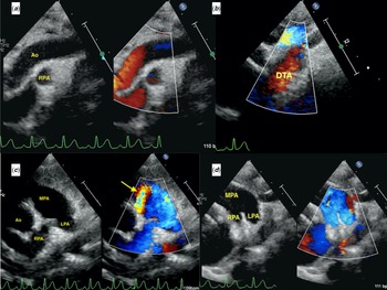
Expectedly, the echocardiogram showed dilated left atrium and left ventricle. The contractile function of the right and the left ventricle was normal. There was no arterial duct. Upon careful evaluation, an abnormal flow was evident in the pulmonary artery. However, the crossed origin of the pulmonary arteries did not permit the exact source of accessory blood flow into the pulmonary artery (Fig 1 and Video 1-2). Despite careful assessment, poorly visualised arch sidedness and branching pattern of arch vessels prompted a diagnosis of arch anomaly. The child also had a non-obstructive sub-aortic membrane with mild aortic regurgitation. Mild mitral regurgitation consequent to mitral valve prolapse was also present. Unusual flow in the pulmonary artery in the clinical setting of left to right shunt prompted a repeat physical examination of the child, which revealed bruit in bilateral carotid arteries, thus making isolation of carotid artery connecting to the pulmonary artery likely.
Echocardiographic images in suprasternal view ( a and b ) showing an apparent lack of continuity between ascending and descending aorta and flow reversal is seen in the descending aorta. An abnormal flow in the pulmonary arteries was evident in high parasternal view (panel c ) but no definite arterial duct could be demonstrated in ductal view (panel d ).

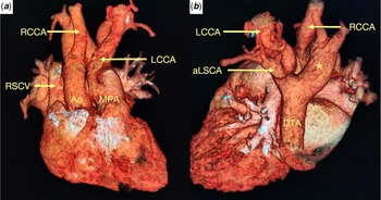
Subsequently, CT angiogram confirmed circumflex right aortic arch (right arch with left descent) with aberrant left subclavian artery and Kommerell’s diverticulum. More importantly, the left common carotid artery was isolated and was connected to the main pulmonary artery instead of the aorta (Fig 2). Pulmonary arteries were confluent with crossed orifices. The arterial duct was not patent. Interestingly, the right coronary artery was arising from the base of the isolated left common carotid artery.
Three-dimensional volume-rendered CT reconstruction images seen from frontal ( a ) and posterior ( b ) aspect showing left common carotid artery (LCCA) arising from the main pulmonary artery (MPA). Circumflex aortic arch is seen (*) with the aberrant left subclavian artery (aLSCA) arising as the last branch. Ao – aorta; DTA – descending thoracic aorta; LCCA – Left common carotid artery; LSCA – left subclavian artery; MPA – main pulmonary artery; RCCA – right carotid artery; RSCA – right subclavian artery; RSCV – right superior caval vein.

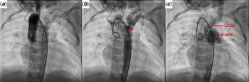
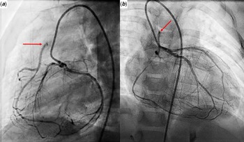
We then performed cardiac catheterisation to measure pulmonary artery pressure, quantify left to right shunt, and determine the adequacy of inter-coronary collaterals given the anomalous origin of the right coronary artery. The pulmonary artery pressure measured 45/30/38 mm Hg and calculated Qp/Qs was 1.7:1. Aortic root angiogram in the late phase of the angiogram showed flow reversal from the isolated left common carotid artery into the pulmonary artery (Fig 3 and Video 3). Left coronary artery angiogram through inter-coronary collaterals filled the right coronary artery, which drained into the left common carotid artery (Fig 4A and B and Video 4). The coronary circulation is equivalent to anomalous right coronary artery from pulmonary artery. A barium oesophagogram revealed mild posterior indentation of the thoracic oesophagus at the level of the aortic arch with the free passage of contrast and no hold-up, indicating a circumflex arch with a loose vascular ring. Fluorescence in situ hybridisation analysis was negative for 22q11 deletion.
Aortic root angiogram in anteroposterior projection in early ( a ), mid ( b ), and late phase ( c ) showing an abnormal orientation of the circumflex aortic arch with the first branch being right common carotid artery (RCCA) and last being aberrant left subclavian artery arising from Kommerell’s diverticulum (*) but no visualisation of the left common carotid artery (LCCA) which is seen draining retrogradely into the main pulmonary artery (MPA) in the late phase.


We planned reimplantation of the left common carotid artery and right coronary artery to the aorta. We assessed the adequacy of intracranial circulation using MRI of the brain. The cerebral parenchyma was normal bilaterally. The circle of Willis was complete, but the intracranial part of the left internal carotid artery was attenuated, and the right posteroinferior cerebellar artery was hypoplastic. Detailed neurological and development assessment revealed no abnormalities.
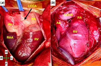
The surgery was performed through median sternotomy without cardio-pulmonary bypass under continuous near-infrared spectroscopy monitoring. The findings were confirmed, including the interarterial course of the right coronary artery before connecting to the base of isolated left common carotid artery (Fig 5A). Dilated left coronary artery with a network of coronary collaterals was evident over the anterior surface of the heart. After careful dissection and systemic heparinisation, we confirmed the physiological adequacy of the intracranial circulation by test clamping of the left common carotid artery for 5 minutes which showed no change in near-infrared spectroscopy. Initially, we harvested the right coronary artery button from the base of left common carotid artery to reimplant to the aortic sinus. Unfortunately, the origin of right coronary artery got injured, and therefore, it was ligated. Subsequently, we divided the isolated left common carotid artery near its origin, and the defect in main pulmonary artery was oversewn. After placing a side-biting clamp over ascending aorta, the ascending aorta was incised at its highest point and anastomosed to the proximal end of left common carotid artery in an end-to-side fashion (Fig 5B). A ductal ligament was also present on the left side that was doubly ligated and divided. The pericardium was left open to avoid compression of reimplanted left common carotid artery. Throughout the procedure, the near-infrared spectroscopy suggested no fall in cerebral oxygenation at any point in time.
Photograph obtained before ( a ) and after ( b ) the surgical reimplantation of the isolated left common carotid artery.

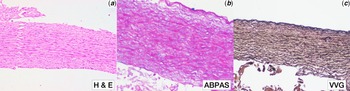
Gross histopathological examination of a resected portion of the isolated left common carotid artery revealed a thin-walled vessel. Microscopic examination after staining with haematoxylin and eosin, Alcian blue periodic acid Schiff stain, and Verhoff-van Gieson stain showed muscular media with well-preserved elastic lamellae characteristic of an elastic artery and not the ductal tissue (Fig 6).
Histopathological sections of the resected proximal portion of the isolated carotid artery with haematoxylin and eosin stain ( a ), Alcian Blue Periodic Acid stain ( b ), and Verhoff-van Gieson ( c ) showing thick smooth muscle media, absence of proteoglycans, and presence of well-preserved medial elastic lamellae (arrow), respectively. These findings are representative of an elastic artery and not arterial ductal tissue.

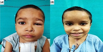
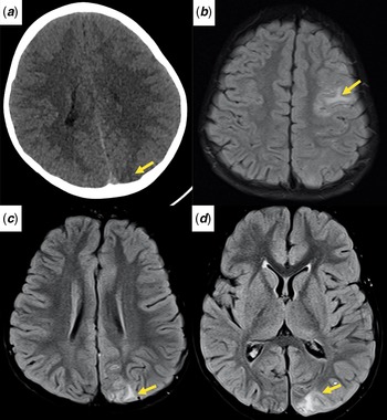
In the post-operative period, anticipating the risk of cerebral hyperperfusion injury, we aggressively controlled the blood pressure to achieve 50-90th percentile using Nitroglycerine (NTG) infusion. The initial post-operative course was smooth with extubation within 4 hours of surgery. On post-operative day 2, we added oral angiotensin-converting enzyme inhibitor (Enalapril), metoprolol, and nitrates, while intravenous NTG infusion was tapered. However, on the fourth post-operative day, the patient developed mild puffiness of the left cheek, which worsened and extended to the periorbital region over the next 24 hours. Besides, the periorbital swelling was painful and warm (Fig 7A). The patient also had drowsiness, left lateral rectus palsy, and projectile vomiting consistent with raised intracranial pressure. He also had a seizure, and blood pressure rose to above 99th centile despite reinstitution of NTG infusion. The echocardiogram showed moderate left ventricular dysfunction (EF 35%) and mild bilateral pleural effusion. Doppler interrogation confirmed normal forward flow in the reimplanted left common carotid artery. CT head demonstrated loss of grey-white matter differentiation suggestive of posterior reversible encephalopathy syndrome (Fig 8A). The left thyroid lobe showed hyperaemic changes, albeit with normal thyroid function test. Fundus examination showed papilledema.
Clinical photograph of the child on post-operative day 5 with accelerated hypertension showing unilateral facial oedema with periorbital puffiness ( a ). The facial swelling completely resolved after few days ( b ).

Non-contrast CT head ( a ) performed on post-operative day 5 shows loss of grey-white matter differentiation (arrow). Subsequently performed MRI of the brain shows loss of grey-white matter differentiation and subcortical hyperintensities in left frontoparietal ( b ) and occipital regions ( c – d ).

Hypertension and raised intracranial pressure were initially managed using 3% saline and sodium nitroprusside infusion. Despite multiple intravenous (sodium nitroprusside and labetalol) and oral (metoprolol and enalapril) antihypertensive agents, however, hypertension was not controlled. Thereafter, based on prior similar reports of cerebral hyperperfusion following carotid endarterectomy, oral clonidine was started which promptly controlled hypertension. Reference Farooq, Goshgarian, Min and Gorelick17 Gradually, the swelling of the face reduced and subsided by the 10th post-operative day (Fig 7B). There were no neurological deficits. After control of systemic hypertension, the ventricular function improved and ejection fraction normalised. Pre-discharge MR angiography with hyperintensities in subcortical white matter in T2/FLAIR sequences along with enhanced adjacent leptomeninges in the left occipital and frontoparietal region confirmed the diagnosis of cerebral hyperperfusion syndrome (Fig 8B-D).
We discharged the patient on furosemide 1 mg/kg/day, metoprolol at 2.5 mg/kg/day, aspirin 75 mg once a day, clonidine 1.5 mcg/kg/dose thrice daily, and oral anti-epileptic levetiracetam. Neurocognitive assessment at discharge was within normal limits. Home blood pressure monitoring, reviewed at 1-month follow-up, showed blood pressure between 50th and 90th centile. At 4 months of follow-up, the patient was doing well, and the echocardiogram showed left ventricular ejection fraction improved to 55%. Ambulatory blood pressure monitoring over 24 hours showed optimal control of blood pressure with medications. After 11 months of post-operative follow-up, the patient is doing well with normal neurocognitive function and well-controlled blood pressure, albeit on antihypertensive medications.
Discussion
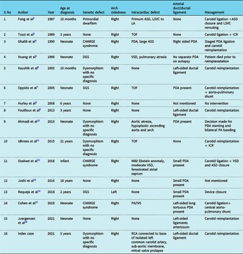
Isolation of the carotid artery is a rare form of aortic arch anomaly, with only 15 cases reported thus far (Table 1). Reference Fong and Venables2–Reference Juergensen, Quezada and Silverman16 All except one case had a right aortic arch with an isolated left carotid artery. Reference Requejo, Strawich, Mouratian and Krings14 In three patients, only the internal carotid artery was isolated, and the external carotid artery was arising from the aorta (8, 11, 14). In all these patients, the ipsilateral subclavian artery was aberrant. Three patients each had DiGeorge syndrome, CHARGE syndrome, and non-specific facial dysmorphism, while one patient had primordial dwarfism. The remaining five patients had no dysmorphism. Reference Huang and Wu5 Nine patients had a coexisting cardiac malformation, while the remaining six had no structural heart disease like our case (Table 1). Interestingly, our case also had an anomalous connection of the right coronary to the base of the isolated left carotid artery, a pattern not reported in the literature.
Summary of all the reported cases of carotid artery arising from pulmonary artery

ASD: Atrial Septal Defect; CHARGE: Coloboma, Heart defect, Atresia of nasal choanae, Retardation of growth, Genital/urinary and Ear abnormalities; DGS: DiGeorge Syndrome; ICR: Intracardiac repair; PDA: Patent Ductus Arteriosus; PA/IVS: Pulmonary Atresia/Intact Ventricular septum; RCA: Right Coronary Artery; TOF: Tetralogy of Fallot; VSD: Ventricular septal defect.
Embryologic considerations
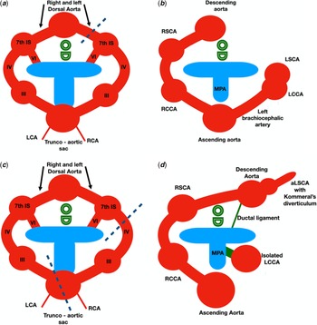
The isolated left carotid artery is usually described based on the Edwards double arch model Reference Edwards18 wherein the third left aortic arch loses its connection to the trunco-aortic sac ventrally and the dorsal aorta posteriorly. The left fourth arch disappears, and the left third arch derivative, the left common carotid artery, connects to the 6th arch through an arterial duct. However, the presence of independent ipsilateral arterial duct or ductal ligament in 9 out of 15 reported cases (60%) as well as in the index case makes the connection between isolated carotid artery with the pulmonary artery unlikely to be via an arterial duct. Reference Tozzi, Hernanz-Schulman and Kiley3,Reference Oppido, Pace Napoleone and Gabbieri7,Reference Fouilloux, Gran and Kreitmann9,Reference Ahmadi, Sabri and Dehghan10,Reference Osakwe, Jones and Hirsch12–Reference Juergensen, Quezada and Silverman16 Some authors have proposed an alternative hypothesis of malseptation of the trunco-aortic sac to explain the isolation of the aortic arch artery, especially the carotid artery. Reference Kaushik, Saba and Rosenfeld6,Reference Fouilloux, Gran and Kreitmann9,Reference Juergensen, Quezada and Silverman16,Reference Manner, Seidl and Steding19 Although not reported previously, the coexistence of anomalous origin of right coronary artery from the isolated left common carotid artery in our case also favours the theory of malseptation of the trunco-aortic sac (Fig 9).
Cartoon showing embryogenesis of normal right aortic arch ( a and b ) and isolated left common carotid artery with right aortic arch ( c and d ) based on Edwards double arch model. The right aortic arch results from regression of the left fourth arch, the disappearance of left dorsal aorta and cranial migration of the left seventh intersegmental artery that forms the left subclavian artery. The isolation of the left carotid artery necessitates regression of the left fourth arch and loss of connection between the ventral aorta and the left third arch explaining aberrant left subclavian artery in all reported cases in the literature ( c ). The persistence of the left dorsal aorta instead of the right explains the right circumflex arch with left descent in our case ( d ). The isolated left carotid artery can connect to the pulmonary artery either directly or by intervening arterial duct. Nonetheless, as shown in panel d , a connection via arterial duct mandates the presence of two arterial ducts on the same side, making it less likely. The histological findings indicating it to be an elastic artery in our case further make the direct connection of the isolated left carotid artery to the pulmonary artery much more likely.

When to suspect isolation of carotid artery?
An abnormal location of flow in the pulmonary artery should prompt suspicion of isolation of an arch artery, particularly in the presence of arch anomaly. The presence of a carotid bruit and increased flow in the contralateral carotid artery are not specific to the isolation of the carotid artery. The lack of discrepancy in left and right upper limb pulses, as seen in our case, on the other hand, practically rules out the isolation of the subclavian artery while indirectly localising the isolation to the carotid artery. On most occasions, there is no intracardiac malformation and lower pulmonary artery pressure permits extracardiac left to right shunt. Coexisting CHD by altering pulmonary artery pressure, however, modifies the flow pattern. For instance, in conditions with reduced pulmonary blood flow, the isolated carotid artery is a source of pulmonary blood flow, thus alleviating arterial desaturation Reference Idhrees, Mathew, Menon, Dharan and Jayakumar11 . In others with pulmonary hypertension, depending upon relative resistances in systemic and pulmonary circulation, the isolated arch vessel may decompress the pulmonary circulation by allowing right to left shunt and manifesting as differential cyanosis. Reference McVadon, Shakti, Knudson, Dodge-Khatami and Baltivala20,Reference Arunamata, Perry, Kipps, Vasanawala and Axelrod21
Management options
Intuitively, surgical reimplantation of the isolated carotid artery is the preferred option and was performed in 6 of the 15 reported cases. Reference Ghalili, Issenberg, Freeman and Brodman4,Reference Kaushik, Saba and Rosenfeld6,Reference Oppido, Pace Napoleone and Gabbieri7,Reference Fouilloux, Gran and Kreitmann9,Reference Idhrees, Mathew, Menon, Dharan and Jayakumar11,Reference Juergensen, Quezada and Silverman16 Nonetheless, in 4 patients, the lower end of the isolated carotid artery was closed (Table 1). Reference Fong and Venables2,Reference Tozzi, Hernanz-Schulman and Kiley3,Reference Joshi, Joshi and Kale13,Reference Cohen, Stanford and Torres15 Although the lower end of the isolated carotid artery can be closed using an occluder device, surgical ligation is preferred as it would also permit ligation and release of arterial duct or ductal ligament. Reference Requejo, Strawich, Mouratian and Krings14 Two infants underwent concomitant aorta-pulmonary shunt for TOF/pulmonary atresia. Reference Oppido, Pace Napoleone and Gabbieri7,Reference Cohen, Stanford and Torres15 Ligation of the isolated carotid artery is an attractive option and would also avoid the risk of cerebral hyperperfusion syndrome but the stump of the carotid artery distal to ligation may have the risk of thrombogenesis. Possible extension of the thrombosis to the ophthalmic artery may lead to blindness. Percutaneous device closure of the isolated vessel may have a similar risk. In our patient, the coexistence of anomalous origin of the right coronary artery from the isolated left carotid artery also favoured surgical reimplantation.
Isolation of common carotid artery is always associated with circumflex aortic arch, producing an incomplete vascular ring. Presence of an ipsilateral ductal ligament results in a complete vascular ring, which can be released by dividing the ductal ligament. If the symptoms due to a vascular ring are present, simultaneous aortic uncrossing procedure is carried out to relieve the airway or vascular compression. It is a complex on-pump surgery and was not contemplated in our patient due to lack of compressive symptoms.
Pre- and post-operative concerns and care
A detailed assessment of the anatomic and physiologic characteristics of the malformation is crucial for meticulous surgical planning. The assessment should also include a neurological evaluation to define the adequacy of the circle of Willis. This is of particular importance since the cerebral circulation on the side of the isolation of the carotid artery is chronically exposed to pressure much lower than the aortic pressure. A possibility of delayed cerebral hyperperfusion mandates careful haemodynamic monitoring. The cerebral hyperperfusion, sometimes also seen following carotid endarterectomy or carotid artery stenting, occurs due to impaired autoregulation or baroreceptor dysfunction. Facial oedema results from venous engorgement due to high flow in the reimplanted left carotid artery or opening up of collateral channels due to high blood pressure, or thyroid gland hyperaemia. Fouilloux and colleagues managed a similar occurrence of facial oedema, headache, and hypertension with propranolol. Reference Fouilloux, Gran and Kreitmann9 Prompt control of blood pressure with centrally acting alpha-agonist clonidine in our case also underscores the importance of seeking prompt guidance from the literature. Reference Farooq, Goshgarian, Min and Gorelick17 High catecholamine levels following carotid endarterectomy explain the benefits of sympatholytic agents such as labetalol and clonidine. In contrast, antihypertensive medications, like nitrates, nitroprusside with a vasodilatory effect, may prove harmful. Reference Farooq, Goshgarian, Min and Gorelick17
Conclusion
Unusual flow in pulmonary arteries should prompt suspicion of isolation of arch vessel, especially in the setting of possible arch anomaly. Cerebral hyperperfusion following surgical reimplantation of the isolated carotid artery is a serious complication. A stepwise multidisciplinary preoperative assessment and careful post-operative care are mandatory for optimal clinical and neurological outcomes.
Supplementary material
To view supplementary material for this article, please visit https://doi.org/10.1017/S1047951121005308
Acknowledgements
Authors acknowledge Dr Sudheer Arava, Additional Professor in Department of Pathology, at All India Institute of Medical Sciences, New Delhi for providing the histopathology images.
Financial support
This research received no specific grant from any funding agency, commercial, or not-for-profit sectors.
Conflicts of interest
None
Ethical standards
The authors assert that all procedures contributing to this work comply with the ethical standards of the ICMR guidelines on human experimentation and with the Helsinki Declaration of 1975, as revised in 2008, and has been approved by the institutional committees (IEC, AIIMS). The consent of the parent was obtained prior to usage of the patient’s data.