Introduction
Typhlocoelum cucumerinum (Rudolphi, 1809) is a trematode of the upper respiratory system (mainly trachea) of waterfowls with worldwide distribution (Asia, Europe, Oceania and Americas) and currently included in the family Cyclocoelidae Stossich, 1902 (Gower, Reference Gower1939; Barry, Reference Barry1959; Schafranski et al., Reference Schafranski, Freitas and Costa1975; Soulsby, Reference Soulsby1982; Tkach et al., Reference Tkach, Kudlai and Kostadinova2016). Infection with these tracheal parasites can cause airway obstruction, resulting in dyspnoea, suffocation and even death (Schafranski et al., Reference Schafranski, Freitas and Costa1975; Soulsby, Reference Soulsby1982; Taylor et al., Reference Taylor, Coop and Wall2017). Despite the potential veterinary importance of typhlocoelosis in domestic anatids, aspects of the biology, epizoology and transmission dynamics of T. cucumerinum are still poorly understood. In fact, the countless reports of infection of birds with this cyclocoelid are based on post-mortem findings (Kinsella and Forrester, Reference Kinsella and Forrester1972; Schafranski et al., Reference Schafranski, Freitas and Costa1975; Hoyos et al., Reference Hoyos, Benavides and Garay2017). To the best of our knowledge, studies involving the in vivo diagnosis of this trematode are lacking, despite the potential importance of this approach to subside the treatment of infection. Moreover, no study reported the occurrence of T. cucumerinum in naturally infected snails so far.
Studies on life cycles of species of the family Cyclocoelidae revealed that these trematodes have a single intermediate host, in which there is the development of rediae, followed by the production of cercariae (Cercariaeum type) and the formation of encysted metacercariae in the same mollusc (Taft, Reference Taft1973, Reference Taft1975, Reference Taft1986; Schafranski et al., Reference Schafranski, Freitas and Costa1975; McLaughlin, Reference McLaughlin1976, Reference McLaughlin1986; Taft and Heard, Reference Taft and Heard1978; Scott et al., Reference Scott, Rau and McLaughlin1982). Infection of birds occurs with the ingestion of infected molluscs (Schafranski et al., Reference Schafranski, Freitas and Costa1975; McLaughlin, Reference McLaughlin1976, Reference McLaughlin1986; Galaktionov and Dobrovolskij, Reference Galaktionov and Dobrovolskij2003), a common food habit known to several species of waterfowls (Lavery, Reference Lavery1970; Middleton and van der Valk, Reference Middleton and van der Valk1987). Information on the molluscs that act as intermediate hosts of T. cucumerinum is scarce and restricted to the experimental life cycle studies carried out several decades ago (Schafranski et al., Reference Schafranski, Freitas and Costa1975; Scott et al., Reference Scott, Rau and McLaughlin1982).
In another aspect, recent molecular studies have contributed to the advancement of knowledge on taxonomy and phylogeny of avian trematodes, including species of the family Cyclocoelidae (Sitko et al., Reference Sitko, Bizos and Heneberg2017; Gomez-Puerta et al., Reference Gomez-Puerta, Salas, Lopez-Urbina and Gonzalez2018; López-Jiménez et al., Reference López-Jiménez, Pérez-Ponce and García-Varela2018; Galosi et al., Reference Galosi, Heneberg, Rossi, Sitko, Magi and Perrucci2019; Li et al., Reference Li, Ma, Lv, Hu, Qiu, Chang and Wang2020; Urabe et al., Reference Urabe, Nor Hashim, Uni, Iwaki, Halim, Marziki, Udin, Zainuri, Omar, Agatsuma, Uga, Takaoka, Azirun and Ramli2020). Despite this, molecular data are scarce for tracheal parasites belonging to the subfamily Typhlocoelinae, which are limited to sequences of isolates identified as Typhlocoelum sp. and Tracheophilus cymbium (Diesing, Reference Diesing1850) from North America and Asia, respectively (Tkach et al., Reference Tkach, Kudlai and Kostadinova2016; Li et al., Reference Li, Ma, Lv, Hu, Qiu, Chang and Wang2020). The acquisition of new molecular information for members of the Typhlocoelinae, especially T. cucumerinum, the type species of the genus, is necessary to access taxonomic issues related to the validity of the genera currently included in this subfamily. Moreover, such molecular data can be useful to confirm the wide geographical distribution previously reported to the species based on morphological data.
In the current study, a host–parasite–environment system involving T. cucumerinum was identified in an urban lake from Brazil. Coproparasitological analysis was used for the first time for the in vivo diagnosis of the infection in free-living domestic waterfowl. Moreover, different parasite developmental stages, including larval forms in molluscs, were subjected to morphological and molecular studies. This integrative taxonomic approach resulted in insights and advances in taxonomy, phylogeny and biology of this avian tracheal trematode.
Materials and methods
Study area, animals and adult parasites
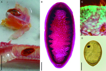
The study area comprises an urban lake located at the Administrative Center of the State of Minas Gerais (‘Cidade Administrativa Presidente Tancredo Neves’), Belo Horizonte city, Minas Gerais, Southeastern Brazil (19°47′06.20″S and 43°57′11.41″W), where a population of about 150 domestic waterfowl was maintained at the study time. During the necropsy of some specimens that died due to an outbreak of botulism between March and April 2016 (Silva et al., Reference Silva, Gómez, Medeiros, Marques, Silva, Mureb, Junior, Favoretto, Lobato and Martins2017), the presence of trematodes in the trachea of Cairina moschata (L.) was verified for the first time (Fig. 1A). Based on this finding, a study was proposed to characterize the host–parasite–environment system present in the locality.
Typhlocoelum cucumerinum found in naturally infected Cairina moschata from Brazil. (A) Alive trematodes found in the trachea during necropsy. (B) Whole view of carmine stained worm. (C) Cirrus sac (arrow). (D) Egg of the parasite containing a developed miracidium. Scale bars: A = 15 mm, B–C = 200 μm, D = 50 μm.

Coproparasitological study
Fecal samples from 61 domestic Anseriformes found in the locality [47 C. moschata (domestic Muscovy ducks), 8 Anas platyrhynchos (L.) (mallards) and 6 Anser cygnoides (L.) (African geese)] were collected between June and August 2018. The nomenclature of the birds followed Clements et al. (Reference Clements, Schulenberg, Iliff, Billerman, Fredericks, Sullivan and Wood2019). For the collection of feces, the birds were attracted by offering food, manually contained and identified with numbered rings on the right leg. After identification, the specimens were individually placed in steel cages (1 m in length and width, 0.6 m in height), with the floor lined with paper for retaining feces. They remained restricted until defecation, usually within no more than 10 min, when samples were collected and placed in plastic containers, fixed in 10% formalin, and sent to the laboratory. The parasitological examination of feces was performed using the spontaneous sedimentation technique (Hoffman et al., Reference Hoffman, Pons and Janer1934). For each animal, five slides containing aliquots of fecal sediment were examined under a light microscope. Samples of eggs found were photographed and subjected to morphometric analysis to confirm the presence of T. cucumerinum. The procedures followed ethical principles approved by the Local Ethics Committee in Animal Experimentation (CEUA-UFMG protocol 68/2017).
Malacological survey
A malacological survey was carried out in the same waterbody frequented by domestic waterfowls. Seven samplings were carried out between June 2018 and May 2019, with a sampling effort of approximately 1 h on each occasion. The molluscs were collected with the aid of a D-shaped hand net (50 × 30 cm, 1 mm mesh, 20 cm depth bag) and sent to the laboratory, where they were washed in running tap water, quantified and placed individually in 24-well polystyrene plates with approximately 2 mL of water. After a routine artificial photostimulation test to detect the emergence of larval trematodes, the gastropods, recognized experimental intermediate hosts of T. cucumerinum (Schafranski et al., Reference Schafranski, Freitas and Costa1975; Scott et al., Reference Scott, Rau and McLaughlin1982), were pressed between glass slides inside a Petri dish and dissected under a stereomicroscope for the search of non-emergent larval stages. The taxonomic identification of the molluscs was based on morphological traits according to different authors (Paraense, Reference Paraense and Cunha1970, Reference Paraense1994; Paraense and Pointier, Reference Paraense and Pointier2003). The sampling of molluscs was made under the permission of the Brazilian Institute of Environment and Renewable Natural Resources (IBAMA, Biodiversity Authorization and Information System – SISBIO permit number 52870-1).
Morphological study of developmental stages and taxonomic identification
A live adult specimen recovered from C. moschata found dead in the locality was processed for morphological study. This trematode was placed in a Petri dish containing 0.9% saline to obtain eggs. Then, the trematode was pressed between glass slides, killed with water at 70°C and fixed in 10% formalin. Subsequently, the parasite was stained with alum acetocarmine, dehydrated in ascending ethanol series, diaphanized with beechwood creosote, and mounted on a permanent slide with Canada balsam. The morphological study of the trematode was performed under a light microscope. Photographs of the specimen in toto were taken using a Samsung ES 70® digital compact camera coupled to a Leica EZ4® stereomicroscope. Details of internal structures were photographed with the aid of a Leica ICC50 HD® digital camera attached to a microscope of the same brand. The captured images were analysed using the software Leica Application Suite® (LAZ EZ) version 2.0. The identification of the parasite was based on morphological criteria according to different authors (Dubois, Reference Dubois1959; Travassos et al., Reference Travassos, Freitas and Kohn1969; Yamaguti, Reference Yamaguti1971; Schafranski et al., Reference Schafranski, Freitas and Costa1975; McDonald, Reference McDonald1981; Kanev et al., Reference Kanev, Radev, Fried, Gibson, Jones and Bray2002). The adult specimen was deposited in the Collection of Trematodes of the Universidade Federal de Minas Gerais (UFMG-TRE 120).
The larval stages of trematodes recovered after mechanical compression of the molluscs were aspirated with the aid of a Pasteur pipette, transferred to another Petri dish containing water. After this, they were wet-mounted between glass slides and coverslip and evaluated with the aid of a light microscope. The photographic record and image analyses were made as described above for the adult. After the morphological study, the larval forms were removed from the slides with the aid of a Pasteur pipette, transferred to 1.5 ml microtubes, and fixed in 95% ethanol for molecular study.
Molecular phylogenetic study
For the molecular study, we used ethanol-fixed eggs shed by the adult parasite obtained from C. moschata and rediae recovered from the molluscs. DNA extraction was performed with the Mini QIAamp® Qiagen® kit (eggs) and Micro QIAamp® Qiagen® (rediae) as recommended by the manufacturer. The concentration of extracted DNA was determined using a spectrophotometer (NanoDrop® ND-1000). For the sample of eggs obtained from an adult, partial regions of the genes 28S (Dig12/1500R primers) (Tkach et al., Reference Tkach, Littlewood, Olson, Kinsella and Swiderski2003), Cox-1 (JB3/COI-R Trema primers) (Miura et al., Reference Miura, Kuris, Torchin, Hechinger, Dunham and Chiba2005) and Nad- 1 (NDJ1/NDJ2A primers) (Morgan and Blair, Reference Morgan and Blair1998) were amplified by polymerase chain reaction (PCR). The amplification reactions were carried out in a final volume of 25 μl. The PCR mixture consisted of 2.5 μl of DNA solution (≈30 ng), 12.5 μl of Go Taq Green Master Mix®, 8.75 μl of ultrapure water and 1.25 μl of a mix of primers (10 μ m). The PCR reactions were performed in a Labcycler® thermocycler (Sensoquest, Germany) using the PCR conditions previously described (Morgan and Blair, Reference Morgan and Blair1998; Tkach et al., Reference Tkach, Littlewood, Olson, Kinsella and Swiderski2003; Miura et al., Reference Miura, Kuris, Torchin, Hechinger, Dunham and Chiba2005). The PCR products were subjected to 1% agarose gel electrophoresis and those samples with the expected band were purified with 20% polyethylene glycol. Sequencing, in both directions, was carried out in ABI 3730 capillary sequencer, using POP7 polymer and Big Dye v3.1 Terminator Cycle Sequencing Ready Reaction Kit (Applied Biosystems®, Foster, USA).
The chromatograms obtained were edited using the ChromasPro program version 2.0.1 (Technelysium Pty Ltd, Australia) and the contigs compared with other sequences available in GenBank, initially by a Blast search (https://blast.ncbi.nlm.nih.gov/Blast.cgi) (Supplementary Table S1). The sequences for each marker were aligned with others available for members of the family Cyclocoelidae through the Clustal W program, implemented in MEGA7.0 (Kumar et al., Reference Kumar, Stecher and Tamura2016). Outgroups were selected based on previously published phylogenies of trematodes (Pérez-Ponce de León and Hernández-Mena, Reference Pérez-Ponce de León and Hernández-Mena2019). Phylogenetic analyses were made by maximum likelihood (ML) and Bayesian inference (BI) using the programs MEGA7.0 and MrBayes 3.1.2 (Ronquist et al., Reference Ronquist, Teslenko, Mark, Ayres, Darling, Hohna, Larget, Liu, Suchard and Huelsenbeck2012), respectively. The best nucleotide substitution models were estimated by the lowest score obtained by the Bayesian information criterion in MEGA 7.0. The selected models were GTR + G, TN93 + G and HKY + G for 28S, Cox-1 and Nad-1 datasets, respectively. For phylogenetic inferences, the CIPRES computational analysis platform (phylo.org) was used (Miller et al., Reference Miller, Pfeiffer and Schwartz2010). The few gaps present in the alignment were eliminated from the analysis. In the ML analysis, the trees were generated by the IQ-TREE platform (phylo.org) and clade supports were estimated by the Bootstrap method (10 000 replications). In the BI analysis, the posterior probability values were determined by the Monte Carlo method via Markov chains, in two runs of four chains, for 106 generations, with sampling of topologies every 100 generations, discarding the first 25% of the trees sampled as ‘burn-in’. The program Fig Tree 1.4.2 (Rambaut, Reference Rambaut2012) was used to visualize the topologies of the trees obtained by BI. The final phylogenetic trees were edited in PowerPoint® (Windows 7®) and Photoshop CS5 Adobe®. Molecular sequences generated in this study were deposited in the GenBank under the accession numbers: MZ262701 (28S); MZ254996–MZ254998 (Cox-1) and MZ268020 (Nad-1).
Results
Identification of the parasite from birds
The morphological analysis of the adult parasite recovered from C. moschata in 2016 enables the identification of this tracheal trematode as T. cucumerinum. The morphology of this parasite is described below.
Adult (N = 1): linguiform, pink-coloured, with very slow vermiform movements when alive (Fig. 1A). Body developed, elliptical, 15 mm long and 7 mm wide, tegument with tiny scales (Fig. 1B). Oral opening subterminal, pharynx rounded, muscular, 461 μm in length by 503 μm wide. Ventral sucker not visualized. Caecum long, with several diverticula internally, united posteriorly. Genital pore in the pharynx region. Cirrus sac median, at the level of the oesophagus, containing a seminal vesicle inside (Fig. 1C). Testes in tandem, extremely lobulated, in the posterior region of the body. Anterior testis on the left side, 2.13 mm long and 1.53 mm wide. Posterior testis 1.27 mm long and 2.60 mm wide. Ovary round, lateral to the anterior testis, 510 μm long and 496 μm wide, in the posterior quarter on the right side. Ootype post-ovarian. Vitelline follicles lateral, from the intestinal bifurcation up to the posterior end, not united posteriorly. Uterus intercecal, sinuous, from the posterior testis to the oesophageal bifurcation. Eggs elliptical to oval-shaped, yellow in coloration, with an operculum (Fig. 1D), 157 ± 8 (131–170) μm long and 87 ± 4 (77–93) μm wide (n = 20) in the terminal uterus of the mounted specimen, presenting a miracidium with eyespot and a formed redia inside.
Remarks
The adult specimen recovered from C. moschata has morphological characteristics compatible with the genus Typhlocoelum, as described by different authors (Dubois, Reference Dubois1959; Schafranski et al., Reference Schafranski, Freitas and Costa1975; Kanev et al., Reference Kanev, Radev, Fried, Gibson, Jones and Bray2002; Hoyos et al., Reference Hoyos, Benavides and Garay2017). Unlike the diagnosis of the genus presented by Kanev et al. (Reference Kanev, Radev, Fried, Gibson, Jones and Bray2002), the presence of a ventral sucker was not observed in the specimen studied here. However, the morphology, including the complete absence of a ventral sucker, as well as the measurements obtained for the evaluated specimen, are in agreement with those reported for T. cucumerinum by different authors (Dubois, Reference Dubois1959; Travassos et al., Reference Travassos, Freitas and Kohn1969; Schafranski et al., Reference Schafranski, Freitas and Costa1975; Hoyos et al., Reference Hoyos, Benavides and Garay2017).
Coproparasitological study
The analysis of fecal samples of the anatids evaluated in the current study revealed the infection of 12/47 (25.53%) C. moschata and 2/8 (25%) A. platyrhynchos. None of the six specimens of A. cygnoides evaluated were found infected. The eggs obtained from formalin-fixed fecal samples measured 163 ± 6 (152–174) μm long by 84 ± 4 (77–90) μm wide (N = 20). They are similar to the eggs present in the uterus of the adult parasite or described by different authors (Schafranski et al., Reference Schafranski, Freitas and Costa1975; Hoyos et al., Reference Hoyos, Benavides and Garay2017).
Identification of the first intermediate host
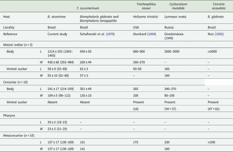
In the malacological analysis, specimens of Biomphalaria straminea (Dunker, 1848) (n = 248), Physa acuta (Draparnaud, 1805) (n = 100) and Pseudosuccinea columella (Say, 1817) (n = 39) were evaluated for the presence of larval trematodes after mechanical compression. Rediae and metacercariae of T. cucumerinum were found in 3 (1.2%) specimens of B. straminea. Of the seven collections made, two were made in 2018 (June and October) and five in 2019 (two collections in March, one in April and 2 in May). Of these, samples of molluscs positive for infection belong to the intervals of March, April and May 2019. Regarding the rediae, few specimens (1, 2 and 3) were found in the infected mollusc. The larval stages obtained are described below. Measures obtained for T. cucumerinum in the present and previous studies and other similar larvae reported in molluscs are shown in Table 1.
Morphometric and morphologic data of rediae, cercariae and metacercariae of Typhlocoelum cucumerinum obtained from Biomphalaria straminea from Brazil

n, number; L, length; W, width.
Data reported for larval cyclocoelids by other authors are shown for comparison.
Values are given in μm by the mean followed by standard deviation and range between parenthesis.
Mature rediae (N = 3) (Fig. 2A, B): elongated, with locomotor appendages in the posterior portion. Mouth in the anterior portion, followed by an oval muscular pharynx. Caecum long, large, with dark-coloured material inside. Presence of cercariae at different stages of development, with those more developed in the anterior half. Germinative masses in the posterior portion.
Larval stages of Typhlocoelum cucumerinum found in Biomphalaria straminea from an urban lake from Brazil. (A) Redia. (B) Details of anterior end redia. (C) Metacercaria. (D) Cercaria. Scale bars: A = 200 μm, B–D = 50 μm.

Cercariae (N = 10) (Fig. 2D): Body oval-shaped, flattened dorsoventrally. Larvae without tail, with a small protuberance at the posterior end. Anterior organ subterminal. Pharynx rounded, immediately below the anterior organ. Ventral sucker absent. Caecum with two branches, united in the posterior part of the body, without reaching the extremity. Excretory vesicle oval, terminal.
Metacercariae (N = 10) (Fig. 2C): encysted form, round to spherical, with a thick cystic wall.
Remarks
In general, the morphology and measurements of the larval forms recovered from B. straminea in the current study agree with those described for T. cucumerinum by Schafranski et al. (Reference Schafranski, Freitas and Costa1975) after experimental infections of molluscs. Considering other species of Cyclocoelidae for which intramolluscan stages are known, the rediae analysed here are significantly smaller than those described for Cyclocoelum mutabile and do not have spines in the anterior third (Ginetsinskaia, Reference Ginetsinskaia1949). The absence of ventral sucker, here confirmed in the cercariae of T. cucumerinum, is also known for larvae of other cyclocoelids, as Ophthalmophagus spp. (Taft and Heard, Reference Taft and Heard1978; Taft, Reference Taft1986) and Morishitium dollfusi (Timon-David, 1950) (Yamaguti, Reference Yamaguti1975). On the other hand, the presence of this structure was well described to the cercariae of Tracheophilus sisowi Skrjabin, 1913 (16 μm) and Cyclocoelum mutabile (Zeder, 1800) (34 × 37 μm) (Szidat, Reference Szidat1932; Stunkard, Reference Stunkard1934; Ginetsinskaia, Reference Ginetsinskaia1949; Yamaguti, Reference Yamaguti1975). In Brazil, a larva found in Biomphalaria glabrata (Say, 1818) in the same locality from the current study (Belo Horizonte) was described as Cercaria acaudata by Ruiz (Reference Ruiz1952). It differs from T. cucumerinum by the presence of a ventral sucker, and possibly a member of the genus Cyclocoelum. Recently, larval stages have been found in B. glabrata from Brazil were tentatively identified as T. cucumerinum (Oliveira et al., Reference Oliveira, Oliveira, Azevedo, Silva, Medeiros, Silva, Cruz and Bogéa2020). Despite the measures of larvae reported by these authors are similar to those here described, experimental or molecular evidence is still necessary for unequivocal identification of the parasite.
Molecular phylogenetic study
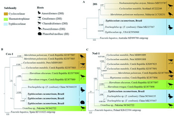
The analysis of sequences 28S (1170 bp) obtained for T. cucumerinum from C. moschata in the current study revealed high similarities with other members of the Typhlocoelinae subfamily, Typhlocoelum sp. (KT956960) (99.3%) and T. cymbium (MK355447) (99.1%), originated from North America and China, respectively. Regarding the recent Chinese isolate of T. cymbium, it is important to note that there is doubt on specific identification of the parasites, given this species is currently considered as belonging to the genus Neivaia Travassos, 1929 (Dubois, Reference Dubois1959; Kanev et al., Reference Kanev, Radev, Fried, Gibson, Jones and Bray2002). Li et al. (Reference Li, Ma, Lv, Hu, Qiu, Chang and Wang2020) did not present morphological data but mention that the parasite was identified according to Tang and Tang (Reference Tang and Tang1978). In this last study, the parasites represented have intestinal diverticula, a morphological trait absent in Neivaia cymbium (Diesing, Reference Diesing1850). We opt by indicating this inconsistency from now on as Tracheophilus sp. (=‘T. cymbium’ of Li et al., Reference Li, Ma, Lv, Hu, Qiu, Chang and Wang2020). Considering other cyclocoelids with sequences available, the percentages of similarity were lower [Morishitium polonicum malayense Urabe, Nor Hashim & Uni (LC520231) (94.7%) and C. mutabile (AY222249) (92.4%)]. In the phylogenetic analysis, the Brazilian isolate of T. cucumerinum grouped in a well-supported clade (1 BI/100 ML) with Typhlocoelum sp. and Tracheophilus sp. (=‘T. cymbium’ of Li et al., Reference Li, Ma, Lv, Hu, Qiu, Chang and Wang2020). Although belonging to different genera, these two last isolates formed a subclade (0.81 BI/94 ML) (Fig. 3A).
Phylogenetic relationships between Typhlocoelum cucumerinum and selected members of the family Cyclocoelidae inferred from partial 28S (1170 bp) (A), Cox-1 (343 bp) (B) and Nad-1 (395 bp) (C) sequences analysed by BI and ML methods. Nodal supports are indicated as ML/BI; values smaller than 0.90 for BI and 70 for ML are indicated by a dash. Asterisks indicate nodes that were not found in ML. New sequences from the current study are shown in bold.

Concerning mitochondrial markers, Cox-1 (802 bp) sequences were obtained for the adult from C. moschata and larval stages from B. straminea, which showed 100% similarity, confirming the conspecificity between these developmental stages. Considering the other two members of the family Cyclocoelidae with data available for comparison, T. cucumerinum showed 82.9 and 92.8% similarity to Uvitellina sp. (NC042722) and Tracheophilus sp. (=‘T. cymbium’ of Li et al., Reference Li, Ma, Lv, Hu, Qiu, Chang and Wang2020) (NC044135), respectively. In the phylogenetic analysis, based on a 343 bp dataset containing eight members of the family Cyclocoelidae, the isolates were partitioned into representative clades containing species from three subfamilies: Cyclocoelinae, Haematotrephinae and Typhlocoelinae. It was verified the grouping of T. cucumerinum with Tracheophilus sp. (=‘T. cymbium’ of Li et al., Reference Li, Ma, Lv, Hu, Qiu, Chang and Wang2020) (NC044135) from China (1 BI/100 ML) (Fig. 3B).
Sequences of the gene Nad-1 (395 bp) were obtained for adult T. cucumerinum and compared with 12 species of cyclocoelids. Again, the Brazilian isolate here characterized showed higher similarity (91.6%) with Tracheophilus sp. (=‘T. cymbium’ of Li et al., Reference Li, Ma, Lv, Hu, Qiu, Chang and Wang2020) from China (MK355447 and NC044135). Considering other genera of the Cyclocoelidae, the percentage of similarity was lower [82.9% to Harrahium tringae (Brandes, 1892) (KU877898); 80.8% to C. mutabile (MH091808-09, KX097823, KU877891); 80.4% to Uvitellina sp. (NC042722) and below of 80% in relation to Harrahium obscurum (Leidy, 1887) (KU877897), Hyptiasmus oculeus Kossack, Reference Kossack1911 (KU877896) and Morishitium polonicum (Machalska, 1980) (KU877893)]. In phylogenetic analysis, T. cucumerinum also clustered in a well-supported clade with Tracheophilus sp. (‘T. cymbium’ of Li et al., Reference Li, Ma, Lv, Hu, Qiu, Chang and Wang2020) (1 BI/98 ML) (Fig. 3C).
Discussion
The occurrence of the tracheal trematode T. cucumerinum in birds and mollusc present in an urban lake from Brazil is described in the current study based on an integrative approach. The data obtained from the analysis of biological samples from anatids and the finding of larval stages in molluscs made it possible to elucidate the natural life cycle of the parasite. The taxonomic identification of T. cucumerinum was based on morphological characteristics of the adult parasite obtained from C. moschata and agrees with data described by different authors (Manter & Williams, Reference Manter and Williams1928; Dubois, Reference Dubois1959; Travassos et al., Reference Travassos, Freitas and Kohn1969; Yamaguti, Reference Yamaguti1971; Schafranski et al., Reference Schafranski, Freitas and Costa1975; Kanev et al., Reference Kanev, Radev, Fried, Gibson, Jones and Bray2002). The only divergence found is about the presence of a poorly developed ventral sucker, a characteristic presented in the diagnosis of the genus Typhlocoelum by Kanev et al. (Reference Kanev, Radev, Fried, Gibson, Jones and Bray2002). We observed the complete absence of this character in T. cucumerinum, as reported by most other authors that previously identified this parasite (Dubois, Reference Dubois1959; Travassos et al., Reference Travassos, Freitas and Kohn1969; Schafranski et al., Reference Schafranski, Freitas and Costa1975; Hoyos et al., Reference Hoyos, Benavides and Garay2017). The absence of a ventral sucker in the adult stage seems to be a characteristic common to the almost absolute majority of the species of Cyclocoelidae. Thus, its presence in Typhlocoelum spp. and Manterocoelum americanum (Manter & Williams, Reference Manter and Williams1928), as described by Kanev et al. (Reference Kanev, Radev, Fried, Gibson, Jones and Bray2002), needs to be reassessed. It is important to comment that M. americanum was originally described as a species of Typhlocoelum and illustrated without a ventral sucker (Manter and Williams, Reference Manter and Williams1928), and previously considered a synonym for T. cucumerinum (Dubois, 1951). Scanning electron microscopy can be an illuminating approach for the unambiguous determination of the presence or absence of ventral sucker in these parasites.
Despite the existence of several reports on the occurrence of T. cucumerinum in birds worldwide (Krull, Reference Krull1940; Branton et al., Reference Branton, Deaton, Gerlach and Ruff1985; Hoyos et al., Reference Hoyos, Benavides and Garay2017; Galosi et al., Reference Galosi, Heneberg, Rossi, Sitko, Magi and Perrucci2019), these studies are restricted to post mortem evaluations, which also occurred at the first moment in this study. This fact reveals gaps in the knowledge on the typhlocoelosis. The impact of parasitism with T. cucumerinum in domestic anatids, either in their natural environment or in creations for consumption, is unknown given the lack of studies involving in vivo detection of the parasite. The sensibility of the spontaneous sedimentation technique for the parasitological diagnosis of T. cucumerinum is demonstrated here for the first time. Among the 61 birds evaluated, eggs of T. cucumerinum were found in the feces of 14 anatids (12 ducks and 2 mallards). We believe that the low cost and speed related to the spontaneous sedimentation method favours its use in future surveys involving not only T. cucumerinum but also other avian parasites worldwide. Additional studies are needed to characterize the performance of coproparasitological methods in the diagnosis of bird parasites and so direct control measures, especially the treatment.
The prevalence of infection with T. cucumerinum (≈25%) here reported in anatids by coproparasitological exams was close to that verified by post mortem studies in the USA (30.5%, 30/78) (Kinsella and Forrester, Reference Kinsella and Forrester1972). On the other hand, a lower prevalence of infection with T. cucumerinum was found, also by necropsy, in Canada [3.8% (53/534) in Aythya marila (Linnaeus, 1761) and 16.7% (90/534) in A. platyrhynchos] (Scott et al., Reference Scott, Rau and McLaughlin1980). More recently, this parasite has been found in 57.1% (24/42) of C. moschata necropsied in Colombia (Hoyos et al., Reference Hoyos, Benavides and Garay2017). The variability verified in the reported prevalence of infection can be related to differences in the transmission dynamics in each location, host susceptibility, besides ecological variables. In the current study, the occurrence of domestic anatids raised in a semi-extensive system in an urban lake may have favoured the occurrence of the parasite. The restriction and long stay of infected hosts in a single aquatic collection containing an abundant population of susceptible intermediate hosts can have potencialized the infection of anatids and snails with T. cucumerinum. However, more in-depth studies are needed for elucidate aspects related to parasite transmission dynamics.
Tracheal trematodes are considered agents that cause respiratory disorders in birds (Barry, Reference Barry1959). Although clinical evaluation was not the focus of the current study, no animal presented respiratory failure, characterizing subclinical infection probably related to low worm burden. However, some anatids were observed performing clearing throat movements during field interventions. Further studies aiming at a quantitative analysis of the eggs released in the feces of birds and its correlation with worm burden can allow us inferences on this aspect. On the other hand, the impact of a subclinical infection cannot be neglected given the existence of reports of growth retardation in chicks infected with N. cymbium in Australia (Barry, Reference Barry1959).
This study represents the first report of a mollusc acting as natural intermediate host to T. cucumerinum. The morphology and measures of the larval stages found in B. straminea in the current study are in accordance with that described for T. cucumerinum after experimental infection of B. glabrata and Biomphalaria tenagophila (d'Orbigny, 1835) (Schafranski et al., Reference Schafranski, Freitas and Costa1975). It is interesting to mention that, although the susceptibility of some species of the families Physidae and Lymnaeidae to T. cucumerinum was previously reported (Scott et al., Reference Scott, Rau and McLaughlin1982), no specimen of P. acuta or P. columella evaluated in our study was found harbouring larval stages of the parasite. However, it is important to note that the experimental susceptibility of molluscs to infection by T. cucumerinum is relatively high [≥90% in Physella gyrina (Say, 1821), ≥70% in Helisoma trivolvis (Say, 1817) and ≥50% in Lymnaea elodes (Say, 1821) (Scott et al., Reference Scott, Rau and McLaughlin1982)]. In a previous experimental study involving other species of Biomphalaria, the rate of infection was not evaluated (Schafranski et al., Reference Schafranski, Freitas and Costa1975). The biology of the asexual phase of members of Cyclocoelidae, in which the formation of the infective stage occurs inside the first intermediate host (without environmental emergence of cercariae), makes it challenging to detect the infection. In fact, most studies involving trematode larvae found in molluscs worldwide are carried out by analysing the active emergence of cercariae, as recommend for most of the species of trematodes with medical and veterinary importance (Dawes, Reference Dawes1968; Frandsen and Christensen, Reference Frandsen and Christensen1984), which can hide the diversity of cyclocoelids. However, this approach does not enable the detection of larval cyclocoelid, suggesting that the diversity of these parasites in molluscs from natural environments is underestimated. In fact, the knowledge on the interaction between cyclocoelids and their first intermediate hosts is practically restricted to experimental data obtained in planorbids [Biomphalaria spp., Planorbis sp. and H. trivolvis], physids [P. gyrina] and lymnaeid (Lymnaea spp.) (Kossak, 1911; Stunkard, Reference Stunkard1934; Ginetsinskaia, Reference Ginetsinskaia1949; Schafranski et al., Reference Schafranski, Freitas and Costa1975; Yamaguti, Reference Yamaguti1975; Scott et al., Reference Scott, Rau and McLaughlin1982). Thus, aiming to highlight this hidden parasite diversity, we suggest that the crushing technique be included in future studies aiming to search for trematodes in molluscs.
Among the few studies reporting close-related larval trematodes in molluscs from Brazil, Ruiz (Reference Ruiz1952) described C. acaudata from B. glabrata also in the Minas Gerais State. The prevalence of infection reported by this author ranging from 3.8% (1/16) to 9.04% (19/210). This larva was also registered in B. tenagophila in the state of São Paulo, Brazil (prevalence of infection: 0.66%) (Ohlweiler et al., Reference Ohlweiler, Eduardo, Takahashi, Crein, Luca and Oliveira2013). The cercariae described by these authors differ from T. cucumerinum in the presence of a ventral sucker. Moreover, the rediae of C. acaudata are approximately five times greater than those described for T. cucumerinum. More recently, larvae morphologically similar to T. cucumerinum have been found in B. glabrata in the state of Rio de Janeiro, Brazil (Oliveira et al., Reference Oliveira, Oliveira, Azevedo, Silva, Medeiros, Silva, Cruz and Bogéa2020). The unequivocal conspecificity between these isolates still demands further experimental or molecular studies.
Molecular sequences to species of the genus Typhlocoelum are scarce and until then limited to one 28S sequence obtained for an isolate identified as Typhlocoelum sp. in the USA. In the current study, sequences of the type species of the genus, T. cucumerinum, were obtained for the first time. Sequences of the Cox-1 gene obtained for larval stages recovered from B. straminea and adult parasites were 100% similar, supporting the link between these different developmental stages. Also, the phylogenetic position of T. cucumerinum in the family Cyclocoelidae was here evaluated by three different molecular markers (28S, Cox-1 and Nad-1). Overall, phylogenetic data here revealed the clustering, in well-supported clades, of T. cucumerinum with two other members of the subfamily Typhlocoelinae with data available for comparison. Interestingly, in the different markers evaluated, it was verified that T. cucumerinum grouped in well-supported clades containing a Chinese identified originally as ‘Tracheophilus cymbium’ but here considered as Tracheophilus sp. (=‘T. cymbium’ of Li et al., Reference Li, Ma, Lv, Hu, Qiu, Chang and Wang2020). The percentages of similarity verified between T. cucumerinum and this Chinese isolate in the analysis of different markers (28S: 99.8%; Cox-1: 92.5% and Nad-1: 91.2%) are higher than those verified in relation to other genera of family Cyclocoelidae included in the analysis (28S: ≤94.7%; Cox-1: ≤86.6% and Nad-1: ≤76.2%). These results, associated with the topology of the phylogenetic trees, point out that these isolates of Typhlocoelum and Tracheophilus analysed are distinct species but probably belong to the same genus.
The isolate of Tracheophilus included in the phylogenetic analyses was identified as T. cymbium (Li et al., Reference Li, Ma, Lv, Hu, Qiu, Chang and Wang2020). However, the cyclocoelid species described as Monostomum cymbium by Diesing (Reference Diesing1850) also has a complex taxonomic history. This species, described from Brazil, has already been considered to belong to the genus Typhlocoelum (as Typhlocoelum cymbium) (Gower, Reference Gower1939) and Tracheophilus (as T. cymbium) (Yamaguti, Reference Yamaguti1971, Reference Yamaguti1975), but it is currently allocated in the genus Neivaia (Dubois, Reference Dubois1959, Reference Dubois1965; Kanev et al., Reference Kanev, Radev, Fried, Gibson, Jones and Bray2002). The Chinese isolate sequenced by Li et al. (Reference Li, Ma, Lv, Hu, Qiu, Chang and Wang2020) was identified as T. cymbium following the taxonomic concept presented in Tang and Tang (Reference Tang and Tang1978). The lack of illustrations and voucher specimens deposited in a museum makes it impossible for us to check the identity of this isolate. However, the parasites represented in Tang and Tang (Reference Tang and Tang1978) present intestinal diverticula and so clearly do not correspond to the current concept of N. cymbium (Dubois, Reference Dubois1959; Kanev et al., Reference Kanev, Radev, Fried, Gibson, Jones and Bray2002). Until new studies are carried out with this Chinese isolate, we tentatively considered it as Tracheophilus sp.
The genus Tracheophilus Skrjabin, 1913 was rescued and considered distinct from Typhlocoelum by Kanev et al. (Reference Kanev, Radev, Fried, Gibson, Jones and Bray2002) based on the presence or absence of a poorly developed ventral sucker, a strong testicular lobulation (present in Typhlocoelum vs absent in Tracheophilus), the position of the genital pore (pharyngeal or post pharyngeal in Typhlocoelum vs pre-pharyngeal in Tracheophylus) and the termination of the vitelline follicles (not confluent in Typhlocoelum vs confluent in Tracheophylus) (Kanev et al., Reference Kanev, Radev, Fried, Gibson, Jones and Bray2002). Although further studies are needed, including the acquisition of sequences from other species of the subfamily Typhlocoelinae, the data presented herein point to the future re-synonymization between these two genera, as previously proposed by Dubois (Reference Dubois1959). In this case, Typhlocoelum (the senior synonym) should be considered the valid name under the priority law. As an additional example of the taxonomic complexity related to these parasites, after the literature analysis, we noted that some characteristics of Typhlophilus shovellus Lal, Reference Lal1936, such as the presence of an evident ventral sucker, pre-pharyngeal genital pore, the size of eggs and the infection site (small intestine) do not support the previous synonymization between Typhlophilus Lal, 1936 and Typhlocoelum (Dubois, Reference Dubois1959; Yamaguti, Reference Yamaguti1971). Thus, an in-depth review of the members of the subfamily Typhlocoelinae is necessary, including the acquisition of molecular sequences for N. cymbium, Tracheophilus spp. and additional isolates of T. cucumerinum.
Data obtained in this study represent an advance in the knowledge on the biology related to bird infection with T. cucumerinum. The combined use of classic and molecular approaches here used for the successful identification of the parasite can subside new studies involving this species in different regions of the globe. This molecular approach is mandatory to confirm its cosmopolitan distribution and test the possibility of the existence of cryptic species in this genus. Finally, we consider the new data here presented contributes to the knowledge of the biology of T. cucumerinum, confirming previous experimental life cycle studies. Moreover, the possibility of in vivo diagnosis via coproparasitological test is here demonstrated and can be useful to identify new areas of parasite transmission where domestic birds are maintained. As verified for other avian trematodes, the anthelminthic treatment (practically unknown in the context of typhlocoelosis), the identification and combating of mollusc involved in the transmission, and privation access of domestic birds in waterbodies, are some measures that can assist the control of outbreaks of infection of T. cucumerinum.
Supplementary material
The supplementary material for this article can be found at https://doi.org/10.1017/S0031182021000986.
Acknowledgements
We thank Mr Ronaldo Amaral for the support and logistics at the sampling area.
Author contributions
JCAA, HAP and NRSM conceived and designed the study. JCAA, LBM and SF contributed to the field study involved birds. JCAA conducted the malacological study. DLH contributed to the molecular work. ALM helped in the malacological study. All the authors wrote and revised the article.
Financial support
We thank the National Council for the Improvement of Higher Education (CAPES, Brazil) (doctoral scholarship to DLH and JCAA) and the National Council for Scientific and Technological Development (CNPq, Brazil) (research scholarship to ALM, HAP and NRSM).
Conflict of interest
None.
Ethical standards
The procedures followed ethical principles approved by the Local Ethics Committee in Animal Experimentation (CEUA-UFMG protocol 68/2017).