Introduction
Follicle formation in mice, as in all mammals, begins perinatally when pregranulosa cells invade germ cell cysts to establish primordial follicles (Pepling & Spradling, Reference Pepling and Spradling2001). As females age, primordial follicles are recruited from the resting pool to begin a growth phase. The transition of primordial follicles to primary follicles is a critical event, as the transcription of genes important for folliculogenesis, like Gdf9 (growth differentiation factor 9), Bmp15 (bone morphogenetic protein 15), Zp1–3 (zona pellucida 1–3) (Yan et al., Reference Yan, Wang, DeMayo, DeMayo, Elvin, Carino, Prasad, Skinner, Dunbar, Dube, Celeste and Matzuk2001), and those genes necessary for early embryonic development such as Mater (also known as NLR family, pyrin domain containing 5, Nlrp5) and Zar1 (zygote arrest 1) (Tong et al., Reference Tong, Gold, Pfeifer, Dorward, Lee, Bondy, Dean and Nelson2000; Wu et al., Reference Wu, Viveiros, Eppig, Bai, L. and Matzuk2003), occurs in growing oocytes.
Some transcription factors play key roles during early folliculogenesis by affecting the regulation of oocyte-specific genes. Figla (folliculogenesis specific basic helix–loop–helix), for example, is a known oocyte-specific regulator of zona pellucida genes and of growing follicle formation (Soyal et al., Reference Soyal, Amleh and Dean2000; Joshi et al., Reference Joshi, Davies, Sims, Levy and Dean2007). Nobox (NOBOX oogenesis homeobox) is necessary for expression of several key oocyte-specific genes, including Gdf9 and Pou5f1 (POU domain, class 5, transcription factor 1, also known as Oct4) (Rajkovic et al., Reference Rajkovic, Pangas, Ballow, Suzumori and Matzuk2004; Choi & Rajkovic, Reference Choi and Rajkovic2006; Choi et al., Reference Choi, Qin, Berger, Ballow, Bulyk and Rajkovic2007). Sohlh2 (spermatogenesis and oogenesis specific basic helix-loop-helix 2), on the other hand, is a regulator of germ cell- and oocyte-specific transcriptional regulators, including Figla and Nobox (Ballow et al., Reference Ballow, Xin, Choi, Pangas and Rajkovic2006).
Recently, a skin–embryo–brain–oocyte homeobox (Sebox) gene was proved to be essential for early embryogenesis at the 2-cell stage in the mouse (Kim et al., Reference Kim, Kim and Lee2008). Sebox is a mouse paired-like homeobox gene, highly expressed in germinal vesicle (GV)-stage oocytes, that encodes a protein with a 60-amino acid homeodomain motif that binds DNA and regulates gene expression (Cinquanta et al., Reference Cinquanta, Rovescalli, Kozak and Nirenberg2000). As Sebox is also expressed in mouse fetal ovaries (Bonilla et al., unpublished results), the aim of the present study was to examine the role played by Sebox in early oogenesis in mice in vitro using RNA interference (RNAi).
Materials and methods
Experimental animals
All experimental procedures involving the use of mice were performed under the regulations established by our Institutional Bioethics Committee on Animal Care, in accordance with those rules approved by the National Institutes of Health (Bethesda, MD, USA).
In vitro culture of fetal oocytes
Fetal ovaries were dissected from CD-1 mice embryos at 17 days post coitum (dpc). Each ovary was cut into three fragments with fine needles (G5/8) under a stereomicroscope. These ovarian explants were cultured in Waymouth medium (Gibco) supplemented with 5% horse serum, 2.5% fetal calf serum (FCS) (both heat inactivated) and 100 mg/ml penicillin–streptomycin, as described previously (De Felici, 1998; Bonilla & del Mazo, 2010). The number of oocytes at the GV stage that had a diameter of 40–70 μm grown out of each fetal ovary was determined on days 5, 7 or 10 of culture.
Transfections
A Stealth RNAi duplex (Invitrogen) was designed to Sebox (BC098184) using the BLOCK-iT RNAi Designer at www.invitrogen.com/rnaidesigner. The sense (5′→3′) strand of the synthetic oligonucleotide duplex was CAUCGGAGGAAGAGGACCACUUUCA. A Stealth RNAi duplex designed to Sohlh2 (NM_028937; Sense strand GAGCAGGAGGCUGCCAAGUUAAUUA) was used as a positive control, as recent findings show its critical role for maintenance and differentiation of the oocyte during early oogenesis (Choi et al., Reference Choi, Yuan and Rajkovic2008).
After 3 days of culture, when the ovary explants were attached to the Petri dish, they were transfected for 24 h with complexes that consisted of 40 pmol BLOCK-iT Fluorescent Oligo, Medium GC Stealth RNAi Negative Control (with no homology in vertebrate transcriptome) or Stealth RNAi and 2 μg/ml lipofectamine 2000. Uptake of BLOCK-iT Fluorescent Oligo (Invitrogen) was visualized by fluorescence microscopy at 16–18 h on treated cells to assess transfection efficiency. Transfected and control fetal ovary cultures were monitored daily and oocytes that reached GV stage and showing a diameter of 40–70 μm were quantified at days 5, 7 or 10 of culture.
Total RNA isolation
Total RNA was extracted from various tissues from 8-week-old mice and from fetal ovary tissue using the SV Total RNA Isolation System Kit (Promega) and according to the manufacturer's recommendation.
RT-PCR (reverse transcription polymerase chain reaction)
First-strand synthesis was performed using 1–5 μg RNA, random primers, and reverse transcriptase Superscript II (GibcoBRL) at 42°C for 1 h. PCR was performed for 30 cycles of 94°C for 30 s, 60°C for 30 s, and 72°C for 1 min. Gene-specific primers were designed using the Primer3 program (Rozen & Skaletsky, Reference Rozen and Skaletsky2000) as follows: Sebox U (5′-gcacctggctcaggtaactc-3′), Sebox L (5′-acaggttgcccagtgaagtc-3′), Sohlh2 U (5′-tctcagccacatcacagagg-3′), Sohlh2 L (5′-gcagtgggtggcaaataagt-3′), β-actin U (5′-aagtgtgacgttgacatccg-3′), β-actin L (5′-gatccacatctgctggaagg-3′).
Semiquantitative RT-PCR Analysis
PCR products were separated by 2% agarose gels electrophoresis, followed by quantitation using an Image Analyser (Kodak). Values for each gene were normalized to the expression levels of β-actin.
Statistics
Three independent experiments were performed to demonstrate reproducibility. Student's t-test was used to evaluate the difference between groups, and differences at P < 0.05 were considered to be significant. SPSS software (SPSS Inc., Chicago, IL, USA) was used to perform statistical analyses.
Results
Sebox RNA expression in adult mice tissues
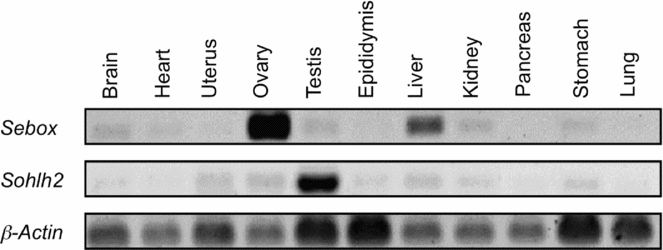
Equal amounts of total RNA from 11 different adult tissues were reverse transcribed and oligonucleotides complementary to Sebox, Sohlh2 and β-Actin were used to amplify affonding sequences by PCR. Results show that Sebox and Sohlh2 mRNAs are expressed in several adult mice tissues at low levels. However, Sebox showed a significantly higher expression in adult ovaries and, to a lesser extent, in liver, while Sohlh2 showed testis-enriched expression (Fig. 1).
RNA expression of Sebox and Sohlh2 in adult mouse tissues. Total RNA from adult tissues was isolated and then amplified with Sohlh2 or Sebox-specific primers. Amplification using β-Actin-specific primers was used as a control for equivalent RNA levels.

Sebox RNA expression in fetal and newborn mouse ovaries
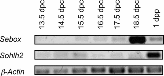
Expression of Sebox and Sohlh2 in 13.5–18.5 dpc ovaries and in newborn mice (1 dpp) is shown in Fig. 2. Expression of Sebox was low in 15.5 to 17.5 dpc ovaries, showed a peak at 18.5 dpc, and its expression decreased dramatically in newborn ovaries, while expression of Sohlh2 was low in fetal ovaries, increasing significantly in newborn ovaries.
Sebox and Sohlh2 RNA expression in fetal and newborn mouse ovaries. RNA from 13.5–18.5 days post coitum (dpc) ovaries and 1-day post- partum (dpp) ovaries were reverse transcribed and oligonucleotides complementary to Sebox and Sohlh2 were used to amplify corresponding sequences by PCR. Amplification using β-Actin-specific primers was used as a control for equivalent RNA levels.
Expression of Sebox mRNA in fetal ovary explants in culture
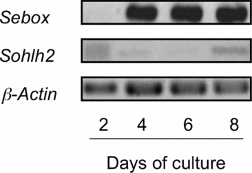
Expression of Sebox and Sohlh2 mRNAs in fetal ovaries cultured in vitro is shown in Fig. 3. Sebox was highly expressed after day 4 of culture and continued to be highly expressed through to day 8 of culture. Conversely, Sohlh2 RNA was present from day 2 to day 8 of culture, but at low levels.
Expression of Sebox and Sohlh2 mRNA in mouse fetal ovary explants cultured in vitro. RNA from fetal ovary explants at 2, 4, 6 and 8 days of culture was reverse transcribed and oligonucleotides complementary to Sebox and Sohlh2 were used to amplify corresponding sequences by PCR. β-Actin amplification was used as a control for equivalent RNA levels.
Transfection efficiency in mouse fetal ovary explants
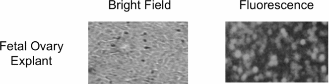
Transfection conditions were optimised using cellular internalization of a fluorescently labelled short RNAi (BLOCK-iT Fluorescent Oligo) as an indicator of transfection efficiency. Transfection efficiency was determined by counting the total number of cells per field versus the number of corresponding cells with visible fluorescence uptake and averaging across different fields (300 cells). The transfection efficiency for fetal ovary cells was routinely observed to be >80% (Fig. 4).
Transfection efficiency. Mouse fetal ovary explants in culture 16–18 h after transfection with BLOCK-iT Fluorescent Oligo.
Targeting Sebox in mouse fetal ovaries in culture
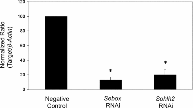
Stealth RNAi duplexes targeting Sebox and the positive control Sohlh2 inhibited their expression by ≥80% relative to the Stealth RNAi negative control after 24 h in fetal ovary explants in culture (Fig. 5).
Targeting Sebox in mouse fetal ovaries in culture. Sebox-specific RNAi and corresponding controls (RNAi Negative Control and Sohlh2-specific RNAi, used as positive control) were transfected into mouse fetal ovary explants on day 3 of culture using Lipofectamine 2000. At 24 h post-transfection, expression of target and internal control genes was analysed by RT-semiquantitative PCR. Expression of Sebox and Sohlh2 is normalized to β-Actin and is presented as the fold change in expression relative to negative control. Experiments were conducted in triplicate, and the data are expressed as the mean ± standard error of the mean (SEM). *Statistically significant changes in expression relative to the negative control (P < 0.05).
Effect of Sebox knockdown on mouse fetal ovaries cultured in vitro
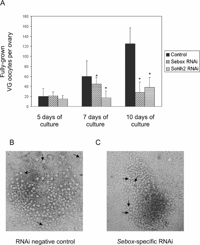
A Sebox-specific RNAi was used to determine whether Sebox plays an essential role in the early oogenesis in mouse in vitro. Sohlh2-specific RNAi was used as a positive control. In fetal ovaries transfected with Sebox-specific RNAi, the number of oocytes at GV stage and that showed a diameter of 40–70 μm was decreased to 75% after 7 days of culture relative to the negative control, and to 22.4% after 10 days of culture (Fig. 6). A similar effect on oocytes growth was observed with Sohlh2-specific RNAi.
Effect of Sebox knockdown on mouse fetal ovary explants cultured in vitro. On day 3 of culture, fetal ovary explants were transfected with RNAi negative control, Sebox-specific RNAi or Sohlh2-specific RNAi (used as a positive control). (A) The number of oocytes at the germinal vesicle (GV) stage and showing a diameter of 40–70 μm grown out of each fetal ovary was analysed on days 5, 7 and 10 of culture. Experiments were repeated three times and data are presented as the mean ± SEM. *When compared with the paired RNAi negative controls, P< 0.05. (B) Representative fetal ovary explant transfected with RNAi negative control. (C) Representative fetal ovary explant transfected with Sebox-specific RNAi. Arrows show fully grown GV oocytes.
Discussion
Oocyte development must be, in part, dictated by networks of genes preferentially expressed in the ovary, which in turn are regulated by ovary-specific transcriptional regulators (Jagarlamudi & Rajkovic, 2011). In the present study, the role of Sebox, which encodes a transcription factor with ovary-restricted expression, was analysed in vitro during the early oogenesis in mice.
Sebox expression could be barely detected in 15.5 to 17.5 dpc fetal ovaries explants, increased dramatically at 18.5 dpc, and then decreased significantly in newborn ovaries (Fig. 2). This developmentally regulated and stage-specific expression pattern was also observed for Sohlh2, the positive control used in this study, and is a hallmark of primary regulators of germ cell development (Hong et al., Reference Hong, Choi, Woo, Oh, Kim, Choi, Kim, Jung, Kim, Sun, Yi, Eddy and Cho2005; Bonilla & Xu, Reference Bonilla and Xu2008).
Transfection efficiency in fetal ovaries in culture, as evaluated by cellular internalization of a fluorescently labelled short RNAi, was observed to be >80% (Fig. 4). This result is similar to that reported by Xu et al. (Reference Xu, Hua, Zhang, Jiang, Zhang, Ma, Zheng, Sun, Shen, Sha, Cooke and Shi2011). In their study, whole mouse fetal ovaries were cultured and transfected with cyanine 3 (Cy3)-labelled RNAi targeting luciferase GL2. At 96 h after transfection, 92% of oocytes and somatic cells showed intense Cy3 fluorescence. These results show that RNAi could be delivered efficiently into mouse ovarian cells from whole fetal ovaries (Xu et al., Reference Xu, Hua, Zhang, Jiang, Zhang, Ma, Zheng, Sun, Shen, Sha, Cooke and Shi2011) or fetal ovary explants in culture (present study) under the described experimental conditions.
In fetal ovary explants transfected with Sebox- and Sohlh2-specific RNAi, the corresponding mRNA levels were significantly reduced by ≥80% at 24 h after transfection in comparison with negative control fetal ovary explants (Fig. 5). Xu et al. (Reference Xu, Hua, Zhang, Jiang, Zhang, Ma, Zheng, Sun, Shen, Sha, Cooke and Shi2011) found that proliferating cell nuclear antigen (PCNA) mRNAs were significantly reduced to 38% of the control level, at 96 h after transfection with RNAi targeting PCNA in mouse whole fetal ovaries in culture. These results show that efficient down-regulation of targeted genes by RNAi technology can be accomplished in fetal ovary explants and whole fetal ovaries in culture.
In a previous study, it was observed that microinjection of Sebox RNAi into mouse GV oocytes resulted in a marked decrease in Sebox RNA and protein expression, but oocyte maturation rate was not affected. In contrast, microinjection of Sebox RNAi into pronuclear-stage embryos resulted in holding embryo development at the 2-cell stage (Kim et al., Reference Kim, Kim and Lee2008). In the present study, the number of fully grown oocytes in GV stage was significantly reduced in fetal ovary explants transfected with Sebox-specific RNAi, in comparison to fetal ovaries transfected with the negative control RNAi (Fig. 6), thus indicating that Sebox plays a pivotal role in the early oogenesis in mice. Similar results were obtained in fetal ovary explants transfected with Sohlh2-specific RNAi. The role of Sohlh2 has been studied in vivo, by generating Sohlh2-knockout mice (Choi et al., Reference Choi, Yuan and Rajkovic2008). It was found that Sohlh2-deficient ovaries can form primordial follicles but they do not differentiate surrounding granulosa cells into cuboidal and multilayered structures. In addition, the formed primordial follicles were abnormal at the molecular level as they misexpressed several germ cell- and oocyte-specific genes, including Sohlh1, Nobox, Figla, Gdf9, Zp1, Zp3, Kit (kit oncogene) and stra8 (stimulated by retinoic acid gene 8). As a consequence, oocytes are rapidly lost in Sohlh2-deficient ovaries, and only few are present by postnatal day 14. Further studies are necessary to determine the germ cell- and oocyte-specific genes regulated by Sebox in order to clarify the molecular mechanisms by which Sebox participates in the maintenance and differentiation of the oocyte during the early oogenesis in mammals.