INTRODUCTION
Cardiomyopathy is an important manifestation of the American trypanosomiasis or Chagas' disease, caused by the protozoan Trypanosoma cruzi, which leads to a considerable morbidity and mortality. Epidemiological surveys estimate that 16–18 million individuals are infected in the American continent (WHO, 2000). About 10–30% of chronic cases develop symptoms and signs of chronic chagasic myocarditis, leading to myocardium dysfunction and heart failure, and less frequently to megacolon and megaesophagus (Elizari, 1999, 2002; Higuchi et al. 2003). Currently, there is no reliable therapy for chronic infection (Coura and De Castro, 2002) and novel targets for chemotherapy are under investigation (reviewed by Urbina, 1999; Do Campo, 2001; Rodriguez, 2001). Since the cardiomyocyte is an important target of this infection we investigated the biochemical and molecular interactions between the parasite and the cardiomyocyte.
In order to perform their biological function heart cells require a mechanical binding to the extracellular matrix, which provides not only structural and mechanical support but also transmits signals that trigger many biological processes, including regulation of gene expression, cell proliferation, differentiation, and cell death (Buck and Horwitz, 1987; Fassler et al. 1996; Lin et al. 1997; Babbitt et al. 2002). Cytoskeleton proteins play a key role in many of these events and changes in the cellular cytoarchitecture have been reported to generate cardiac dysfunction. It has been proposed that cardiac hypertrophy is a consequence of sarcomeric protein alterations (Olson et al. 2001a; Harada and Morimoto, 2004) and dilated cardiomyopathy may be caused by mutation or disturbance of cytoskeletal proteins (Olson et al. 1998, 2001b; Kamisago et al. 2000).
In T. cruzi infection, remarkable changes have also been demonstrated in the cardiomyocyte structure and physiology, including disturbance in the calcium (Ca2+) homeostasis (Garzoni et al. 2003), conduction system (Andrade et al. 1978; Campos de Carvalho et al. 1992) and cytoskeletal organization (Pereira et al. 1993; Taniwaki et al. 2005). One of the most striking observations in the study of the T. cruzi-induced alterations in cytoskeleton architecture has been the disruption of actin filaments (Pereira et al. 1993; Taniwaki et al. 2005) and costameres (Melo et al. 2004), which may contribute to the abnormalities in the contraction force evidenced in Chagas' disease cardiomyopathy.
Alpha-actinin, an anti-parallel homodimer that displays actin-binding domains, is a member of the spectrin superfamily (Dubreuil, 1991). The phylogenic analysis of α-actinin has demonstrated that 4 distinct α-actinin isoforms are present in vertebrates, while invertebrates and protista display only 1 isoform (Virel and Backman, 2004). In vertebrates, α-actinin is classified into non-muscle isoforms, α-actinin 1 and 4, and muscle isoforms, α-actinin 2 and 3. Alpha-actinin, a key protein that orchestrates the link between actin filament and integrin receptors within focal adhesions (Pavalko, 1991; Critchley et al. 1999; Mills et al. 2001; Brakebusch and Fässler, 2003), contributes to the formation and maintenance of Z-bands (Beggs et al. 1992; Wang et al. 2005) and in the force transduction between myofibrils (Shah and Lieber, 2003). Therefore, we investigated the expression and distribution of α-actinin in cardiomyocytes infected by T. cruzi in vitro.
In this paper we report that T. cruzi infection alters Z-line distribution of α-actinin, but not its association to focal adhesion contacts, which may be related to the disturbance of transmission force within myofibrils rather than cell–cell adhesion sites.
MATERIALS AND METHODS
Cell culture
Primary cultures of cardiomyocytes were obtained from 18-day-old mouse embryos as previously described (Meirelles et al. 1986). Briefly, cardiac fragments were dissociated in phosphate-buffered saline (PBS, pH 7·2) containing 0·025% trypsin (Sigma; Missouri, USA) plus 0·01% collagenase (Worthington, New Jersey, USA) and plated (1×10⁵ cells/ml) into 24-wells culture dishes containing glass cover-slips previously coated with 0·01% gelatin (Sigma). For biochemical analysis, the isolated cells were plated at a density of 2·5×106 cells in 60 mm culture dishes. The cells were cultivated in Dulbecco's modified Eagle medium (DMEM; Sigma) supplemented with 10% horse serum (Sigma), 5% fetal bovine serum (FBS; Sigma), 2·5 mM CaCl2, 1 mM L-glutamine (Sigma), 2% chicken embryo extract, and maintained at 37 °C in a 5% CO2 atmosphere.
Parasites and cell culture infection
Bloodstream trypomastigote forms of Trypanosoma cruzi, Y strain, were obtained from Swiss mice at the peak of the parasitaemia as described (Meirelles et al. 1984). Cardiac cells were infected after 24 h of culture at a multiplicity of 10 parasites per host cell. After 24 h of interaction, free trypomastigotes were removed by washing the cultures with Ringer's solution (154 mM NaCl, 3·35 mM KCl, 1·7 mM CaCl2, pH 7·0). The time-course of infection was interrupted after 24 and 72 h.
Indirect immunofluorescence
Uninfected and T. cruzi-infected cardiomyocytes were fixed for 5 min at −20 °C with acetone (Merck). Later, the cells were washed and permeabilized with PBS containing 0·5% Triton X-100 (Sigma). The cultures were then washed with PBS containing 4% bovine serum albumin (BSA; Sigma) to block unspecific reaction and incubated overnight at 4 °C with anti-α-actinin antibody (Santa Cruz) diluted 1[ratio ]100 in PBS. The α-actinin antibody recognizes the amino acids 593–892 mapping at the carboxy terminus of α-actinin-1 of human origin and reacts with alpha-actinin-1, -2, -3 and -4 isoforms. The antigen-antibody complex was revealed by incubation for 1h at 37 °C with FITC-conjugated anti-rabbit IgG (1[ratio ]100; Sigma). Controls were performed by incubation with pre-immune serum or by omission of the primary antibody. DNA was detected by using 10 μg/ml 4,6-diamidino-2-phenilyndole (DAPI; Sigma). The cells were then mounted in 2·5% 1,4-diazabicyclo-(2,2,2)-octane (DABCO; Sigma) in PBS containing 50% glycerol, pH 7·2, and the fluorescence images were obtained using either an Axioplan 2 microscope equipped with epifluorescence (Carl Zeiss, Oberkochen, Germany). A total of 5 complete experiments were carried out by indirect immunofluorescence.
Protein extraction and immunoblotting assay
Total proteins were extracted using a lysis solution (50 mM Tris-HCl containing 1% Triton X-100 plus the protease inhibitors, 10 μM E-64 (Sigma), 1 mM phenylmethylsulfonyl fluoride (Sigma) and 1 μM pepstatin (Sigma), pH 8·0). The amount of protein was determined by the Bradford method (Bradford, 1976) and a total of 20 μg of protein was eletrophoretically separated in a 12% polyacrylamide gel, by SDS-PAGE. The proteins were then transferred at 30V/60 mA overnight at 4 °C to a polyvinylidene difluoride (PVDF) membrane (Bio-Rad, California, USA) in transfer buffer (25 mM Tris, 192 mM glycine and 10% methanol, pH 8·3). After protein transference, the membrane was blocked with 0·5% I-Block (Sigma), 0·1% Tween 20 (Sigma) in PBS and incubated overnight at room temperature with anti-alpha-actinin antibody diluted 1[ratio ]2000 in blocking solution. After several washes, the membrane was incubated with anti-rabbit antibody conjugated to alkaline phosphatase (1[ratio ]3000; Sigma). The enzyme activity was revealed with 0·17 mg/ml 5-bromo-4-chloro-3 indolyl phosphatase (BCIP; Sigma) and 0·34 mg/ml nitro blue tetrazolium (NBT; Sigma) in substrate buffer (0·1 M Tris, 100 mM NaCl, 50 mM MgCl2, pH 9·5). The densitometry analysis was performed using the image-Pro Plus software. The immunoblotting assays were performed independently at least 3 times, beginning with new cultures in each experiment.
Statistical analysis
Student's t-test was used to determine the significance of differences between means in Western blot assays; a P value [les ]0·05 was considered significant.
RESULTS
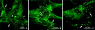
Primary cultures of cardiomyocytes have been used as an important tool to investigate the T. cruzi-cardiomyocyte interaction. Our group developed a suitable embryonic cardiomyocyte-in vitro system that displays most of the isolated cells coupled and spontaneously beating after 24 h of culture. The cultured cardiomyocytes contained prominent myofibrils, showing α-actinin immunofluorescent staining at the Z line of sarcomeres, focal adhesion sites and also at cell–cell junctions (Fig. 1A and B), while non-differentiated cells, myoblasts and fibroblasts, displayed strong labelling of α-actinin at the focal adhesion contacts (Fig. 1C). In contrast to non-differentiated cells, focal adhesion sites were less evident in myocytes probably as a result of the strong reaction with Z lines of myofibrils (Fig. 1A). In addition, an intense labelling was observed at the adherens junctions (AJs), maintaining the association between groups of adjacent cells (Fig. 1B).

Fig. 1. Immunofluorescence localization of α-actinin in cardiac muscle cell cultures. Alpha-actinin reactivity was visualized in Z-line of sarcomeres (arrowheads), focal adhesion sites (arrows) and cell–cell junctions (long arrow) in cardiomyocytes (A, B). Intense labelling was observed in focal adhesion contacts of non-differentiated cells (arrow; C).
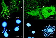
Previously, we demonstrated a disarray of sarcomeric actin filaments and costameres in T. cruzi-infected cardiomyocytes. Therefore, we now addressed the question whether this disassembly represents a specific disturbance in the distribution of these proteins or a drastic disruption of myofibrils. The distribution of α-actinin was evaluated at early (24 h) and later (72 h) times of T. cruzi-cardiomyocyte infection. After 24 h of infection the distribution of α-actinin was similar to control cells, showing the characteristic periodicity in the Z line of cardiac myofibrils (Fig. 2A). In contrast, at a later time-point (72 h post-infection) we observed a dramatic change in the sarcomeric distribution of α-actinin in the host cell, which was identified by the lack of periodic immunoreactivity (Fig. 2C). At this time-point α-actinin was mainly detected at focal contacts (Fig. 2C). Interesting, a strong positive reaction was detected throughout the cytoplasm of the intracellular parasites (Fig. 2C).

Fig. 2. Double labelling of Trypanosoma cruzi-infected cardiomyocytes with α-actinin and DAPI (A–D). In early times of infection (24 h; A), α-actinin was visualized in focal adhesion sites (arrows) and Z-line of myofibrils (arrowhead). After 72 h of infection (C), α-actinin was mostly detected at focal contacts (arrows) of highly infected cells, showing no immunoreaction at the Z-line of myofibrils. Intracellular parasites (double arrows) were also stained with anti-α-actinin antibody. DAPI stained the host cell nucleus and the parasite nucleus and kinetoplast (B, D).
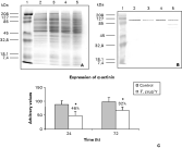
One key question was whether T. cruzi infection also affects the expression of α-actinin. To evaluate this point, total protein extract was obtained from uninfected and T. cruzi-infected cardiomyocytes and the expression of α-actinin was analysed by immunoblotting assay. Our results indicate a decline in the α-actinin expression after early and late times of infection. The quantitative data, obtained by densitometric analysis, demonstrated a reduction of 46% and 32% after 24 h and 72 h post-infection, respectively (Fig. 3).

Fig. 3. Total protein extract of uninfected and Trypanosoma cruzi-infected cardiomyocytes stained by Coomassie Blue (A). Immunoblot detection of α-actinin in uninfected and T. cruzi-infected cardiomyocyte cultures (B). Line 1, molecular weight markers, uninfected cardiomyocytes after 48 h (lane 2) and 96 h (lane 4) of culture and T. cruzi infected-cardiomyocyte after 24 h (lane 3) and 72 h (lane 5) of infection. The densitometric analysis revealed a reduction of 46% and 32% of α-actinin expression after 24 h and 72 h of T. cruzi infection, respectively (C). Student's t test: * statistically significant P<0·05.
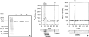
Based on the fact that some protists present α-actinin in their cytoskeletal organization, we next evaluated the immunoreactivity of T. cruzi with anti-α-actinin antibody by Western blot analysis. Total protein extract of different life-stage forms of T. cruzi (epimastigotes, trypomastigotes and amastigotes) was separated by SDS-polyacrylamide gel electrophoresis, followed by Western blotting using anti-α-actinin antibody. There was a protein that migrated with a molecular mass of 78 kDa (Fig. 4), which did not match with the protein mobility of 100 kDa detected in the cardiac muscle samples (Fig. 3). The 78 kDa protein was also detected in the total protein extract of 72h-infected cardiomyocytes (Fig. 3, lane 5). This may correspond to the protein of the intracellular parasites. This band was not visualized in 24 h-infected cells, probably due to the low infection ratio (20–22%) at this time of infection, which was increased to 50–60% after 72 h of infection (Meirelles et al. 1986).

Fig. 4. Immunoblot detection of α-actinin-like protein in Trypanosoma cruzi (A). Line 1, molecular weight standards; line 2, amastigote forms; line 3, epimastigote forms and line 4, trypomastigote forms. The Western blotting assays revealed an immunoreactive band in all three evolutive forms of T. cruzi. The densitometry analysis revealed a protein with molecular mass of 78 kDa (B).
DISCUSSION
Structural and functional changes of cytoskeletal proteins such as sarcomeric disorganization and contractile proteins are thought to be the basis of the myocardial dysfunction that accompanies cardiomyopathy (Hein et al. 1994; Muraishi et al. 1999). Loss of myofibrils is one striking cytoskeleton alteration observed in T. cruzi-infected cardiomyocytes (Pereira et al. 1993). However, it was unclear if this event corresponds to complete degeneration of the myofibrils or to a specific disruption of sarcomeric actin. Therefore, in this study we evaluated the distribution of α-actinin, an actin-binding and cross-linking protein, during T. cruzi-cardiomyocyte interaction in vitro.
As expected, fibroblasts and non-differentiated muscle cells (myoblasts) demonstrated α-actinin localized at focal adhesion sites. Further studies have provided evidence that α-actinin presents a high-affinity binding site on the integrin cytoplasmic domain (Otey et al. 1990; Kelly and Taylor, 2005), residues F-743-A753, playing a key role in the maintenance of the linkage between stress fibres and extracellular matrix (Rajfur et al. 2002) and also may participate in the formation of focal adhesion in vinculin-deficient cells (Volberg et al. 1995). Besides cytoskeleton remodelling, the interaction of α-actinin with integrins (Brakebusch and Fässler, 2003) or syndecan-4 (Greene et al. 2003) transduces signals that control many cellular functions, including cell cycle, differentiation, migration and focal adhesion formation.
In myocytes, the immunolabelling of α-actinin showed a typical pattern of cross-striation, which corresponds to the Z bands of myofibrils, and was also localized at intercellular junctions and focal adhesion sites. The distribution of α-actinin in adherent junctions has been previously reported due to its co-localization with N-cadherin, transmembrane glycoproteins that mediate cell–cell adhesion, in several cells types, including cardiomyocytes, fibroblasts and epithelial cells (Knudsen et al. 1995; Wu et al. 1996).
Our data on T. cruzi-cardiomyocyte interaction revealed that the distribution of α-actinin remains similar to control cells at early times of infection (24 h). In contrast, no sarcomeric distribution of α-actinin was detected in cardiomyocytes at 72 h post-infection, while this protein remained associated with focal adhesions, indicating a tight association of this protein with transmembrane receptors and the extracellular matrix. Although a drastic effect on myofibril organization has been noted only at later times of infection, the biochemical assays revealed a decrease in the α-actinin expression after only 24 h of infection, when most of the cells displayed only few intracellular parasites. These results corroborate our previous data that demonstrated a severe disorganization of α-cardiac actin within myofibrils (Pereira et al. 1993) and a down-regulation of α-cardiac mRNA levels (Pereira et al. 2000) after T. cruzi-cardiomyocyte interaction. In addition, the disruption of muscle cell cytoarchitecture has been recently demonstrated in Calomys callosus chronically infected with T. cruzi and immunosupressed with cyclophosphamide, suggesting that the reactivation of the infection intensifies the process of myofibril disorganization (Taniwaki et al. 2005). Moreover, several cytoskeleton alterations have also been reported in hamsters chronically infected with T. cruzi, showing disorganization and myofibril lysis in some areas of the cardiac tissue while other regions displayed an enlargement of sarcomeres and reduction of myofibril thickness (Colmanetti et al. 2005). Depletion of microfilaments was also demonstrated in human placenta syncytiotrophoblast (Sartori et al. 2003). Recently, we reported alterations on the costameric distribution of vinculin in T. cruzi-infected cardiomyocytes (Melo et al. 2004), which may lead to a reduction of transmission of cardiac contraction force, contributing to the cardiac dysfunction reported in chronic chagasic cardiomyopathy.
Recent studies have provided some details elucidating the host cellular response induced by T. cruzi infection. It has been demonstrated that this parasite has the ability to modulate many signal transduction systems and transcription levels of various host genes (Garg et al. 2003; Mukherjee et al. 2003; Moore-Lai and Rowland, 2004). Changes in the host cell transcripts have been demonstrated as an immediate response of many intracellular pathogens and may facilitate the success of the establishment of the infection (Yowe et al. 2001; Greenwell-Wild et al. 2002; Sexton et al. 2004). Likewise, T. cruzi induces an immediate global transcription response in cardiomyocyte at an early stage (1–4 h) of infection, including genes involved in the immune response, cytoskeleton organization and cell cycle (Pereira et al. 2002; Probst et al. 2003).
Interestingly, the intracellular amastigotes reacted with anti-α-actinin antibody by indirect immunofluorescence assay. This observation led us to speculate that the reaction could be due to acquisition of this protein from the host cell. Therefore, we performed the Western blotting assay with epimastigotes, trypomastigotes and amastigotes. Surprisingly, a protein with 78 kDa was detected in all life-stage forms, suggesting that it may be a constitutive protein of the parasite.
Alpha-actinin has been identified in most eukaryotic organisms, from human (Beggs et al. 1992) to worms (Fyrberg et al. 1990; Barstead et al. 1991), including the protozoan Trichomonas vaginalis (Virel and Backman, 2004). Although no α-actinin gene has yet been described in the T. cruzi genome, blast search revealed that this parasites display a putative protein related to actin (Accession no. AAX80912), actin-like protein (Accession no. AAX79472) and also actin-associated proteins, such as G-actin binding protein (Accession no. AAX79053) and cofilin (Accession no. AAX81027). When a more detailed Blast analysis was performed in the genomic data base of T. cruzi using the C-terminal sequence of α-actinin (residues 593-892), which correspond to the amino acid sequence recognized by the anti-α-actinin antibody, approximately 20% identity was observed with the family of Ca2+ binding proteins, such as calmodulin and centrin, and many hypothetical proteins. In addition, some homology with the N-terminal sequence of α-actinin, an actin-binding domain, was also observed with few hypothetical proteins of T. cruzi when the complete sequence of human α-actinin 1 was analysed. Therefore, even though we can not discard the possibility of cross-reaction with other T. cruzi cytoplasmic proteins unrelated with cytoskeletal proteins, it is possible that T. cruzi display an α-actinin-like protein with similar domains within the N and C-terminal of α-actinin as occurs with Trichomonas vaginalis, showing an atypical amino acid sequence in the rod domain (Virel and Backman, 2004).
In summary, our present results demonstrate that T. cruzi affects α-actinin sarcomeric distribution, leading to a disassembly of the contraction apparatus in cardiomyocytes. The degeneration of myofibrils may contribute to pathogenesis of chagasic cardiomyopathy.
The authors thank Dr Maurilio José Soares for critical reviews of the manuscript and Bruno Ávila and Genilton J. B. Vieira for his skillful computer imaging support. This work was supported by grants from Fundação Oswaldo Cruz (FIOCRUZ), Fundação de Amparo à Pesquisa do Estado do Rio de Janeiro (FAPERJ), Conselho Nacional de Desenvolvimento Científico e Tecnológico (CNPq) and Programa Estratégico de Apoio à Pesquisa em Saúde (PAPES iv).