Introduction
Acclimation from freshwater to seawater environments is a species-specific feature among fish and varies among developmental stages (Watanabe et al., Reference Watanabe, Kuo and Huang1985; Villegas, Reference Villegas1990; Suresh and Lin, Reference Suresh and Lin1992). The yolk-sac stage is especially important because larvae are more susceptible to infections and other external influences that can lead to developmental alterations, disease, and mortality (Fridman et al., Reference Fridman, Bron and Rana2012a,b). As a consequence, low salinity treatments have been used as a prophylactic measure in larviculture to prevent environmental contamination and microbiological infections (Altinok and Grizzle, Reference Altinok and Grizzle2001; Garcia et al., Reference Garcia, Becker, Copatti and Baldisserotto2007; Souza-Bastos and Freire, Reference Souza-Bastos and Freire2009). Furthermore, several studies have shown that low salinity environments provide higher survival rates during larviculture than freshwater (Likongue et al., Reference Likongue, Stecko, Stauffer and Carline1996; Wang et al., Reference Wang, Lui, Po and Fan1996; Borode et al., Reference Borode, Balogun and Omoyeni2002; Beux and Zaniboni-Filho, Reference Beux and Zaniboni-Filho2007).
Skeletal muscle is one of the most interesting biological systems in fish due to its phenotypic plasticity in response to external agents (Johnston, Reference Johnston2006). Muscular injuries, including morphological alterations, frequently found in fish, can be the result of ionic imbalance due to physiological or environmental conditions (Rowlerson et al., Reference Rowlerson, Radaelli, Mascarello and Veggetti1997; Betancor et al., Reference Betancor, Caballero, Benítez-Santana, Saleh, Atalah and Izquierdo2013). Nonetheless, few studies have addressed the effects that salinity has on muscle development in larvae of freshwater fish species (Martins et al., Reference Martins, Melo, Campo-Junior, Santos, Luz, Rizzo and Bazzoli2014). The resulting variation in muscle growth and associated changes in interstitial tissue are known to be determinants of texture and other flesh quality characteristics (Johnston, Reference Johnston1999).
Tilapias are the second most important group of cultured fish worldwide with the Nile tilapia, Oreochromis niloticus (Linnaeus, 1758), being the most predominantly cultured species (FAO, 2016). The species has the ability to adapt to various culture systems, has a fast growth rate, and is resistant to disease and infection (Bhujel, Reference Bhujel2000; Little and Hulata, Reference Little, Hulata, Beveridge and McAndrew2000). Even though wild strains of O. niloticus inhabit freshwater environments, it has been successfully cultured in brackish and seawater due to its adaptability and ease of culture (Likongue et al., Reference Likongue, Stecko, Stauffer and Carline1996; Suresh and Lin, Reference Suresh and Lin1992; Jun et al., Reference Jun, Pao, Haizhen, Ruiwei and Hui2012). Moreover, O. niloticus has been acclimated to saltwater environments due to the crisis facing inland water resources worldwide (Fridman et al., Reference Fridman, Bron and Rana2012a,b).
The influence of salinity on zootechnical parameters of O. niloticus larvae have been previously studied and have revealed significantly lower survival rates when newly hatched larvae are transferred from freshwater to saline environments at 15 parts per thousand (ppt) and above (Watanabe et al., Reference Watanabe, Kuo and Huang1985). Higher mortality, slower development, and higher malformation rates have also been detected among O. niloticus larvae cultured in saline environments, and the physiological changes that they exhibit are dependent on the time of transfer after hatching (Fridman et al., Reference Fridman, Bron and Rana2012a,b). In addition, the number and size of chloride cells have been shown to be significantly altered when O. niloticus larvae are moved from freshwater to seawater (Fridman et al., Reference Fridman, Rana and Bron2013). It has been well established that these mitochondria-rich cells are primary osmoregulatory sites for the trans-epithelial transport of ions in teleosts (Alderdice, Reference Alderdice, Hoar and Randall1988; Fridman et al., Reference Fridman, Bron and Rana2011).
In addition to morphological features, information regarding molecular parameters can provide relevant knowledge about the regulatory mechanisms operating during early development of teleosts under natural and experimental conditions. In this regard, tissue development is dependent on an intricate balance between cell proliferation and apoptosis, which regulates cell number and homeostasis in embryos and larvae (Fuchs and Steller, Reference Fuchs and Steller2011). Proliferating cell nuclear antigen (PCNA) plays an essential role in DNA metabolism as a component of replication and repair machinery (Kelman, Reference Kelman1997). Caspase-3 is an effector molecule of apoptosis responsible for the destruction of damaged cells via the cleavage of many vital proteins (Takle and Andersen, Reference Takle and Andersen2007). These molecules have been frequently used as biomarkers of cell proliferation and apoptosis in developmental studies.
The goal of the present study was to investigate how low salinity environments affected early larval development of O. niloticus. Towards this aim, yolk-sac surface, white muscle, larval trunk area, and protein expression of PCNA and caspase-3 in yolk-sac larvae were assessed.
Materials and Methods
Experimental design and fish sampling
The present study was carried out at the Laboratório de Aquacultura da Universidade Federal de Minas Gerais (LAQUA-UFMG) in the municipality of Belo Horizonte, Southeast Brazil. All procedures were approved and certified by the Ethics Committee on Animal Use (CEUA/UFMG, 396/2012). For breeding, specimens of O. niloticus (87 ± 28.8 g) were maintained in 500-l freshwater tanks at a stocking density of about one fish per 60 l, and maintained at 28 ± 1°C with dissolved oxygen (DO) concentration of at least 5 mg l−1 and a photoperiod of 12 h light: 12 h dark. Inspections were conducted daily when eggs present in the mouth of females were removed and incubated at 28 ± 1°C until larvae hatching.
Newly hatched larvae (up to 24 h post-hatching) were submitted to four salinity treatments, in duplicate: (S0) freshwater/control, (S2) 2 g l−1, (S4) 4 g l−1, and (S6) 6 g l−1 NaCl. Each treatment comprised about 200 larvae stocked in artificially aerated 20-l plastic buckets containing a plastic strainer (1 mm mesh net) to facilitate the handling of larvae. Water parameters were monitored daily (mean ± standard deviation (SD)) and measured: 28.5 ± 0.8°C, dissolved oxygen 5.8 ± 0.3 mg l−1 and pH 7.3 ± 0.2. Fish were sampled after 48 or 72 h of exposure to assess the acute effects of salinity on early development. The salinities used in this study resulted in high larval survival (above 90%) and larvae with total length ranging 8.13 to 10.25 mm after 72 h of treatment. Larvae were euthanized with an overdose of Eugenol (285 mg/l), in agreement with ethics rules and procedures of the Conselho Nacional de Controle de Experimentação Animal (National Council for Control of Animal Experimentation) (CONCEA) and sampled for posterior analyses by histology, electron microscopy and ELISA assay.
Histology
For histological analyses, 12 larvae of each treatment sampled at 72 h exposure were fixed in Bouin’s fluid for 12 h, dehydrated through a graded series of ethanol concentrations followed by xylene, and then embedded in paraffin. Cross-sections (5 μm) of the larvae body were made caudal to the yolk sac, which were stained with haematoxylin–eosin and examined using an Axioplan 2 Zeiss light microscope.
Scanning and transmission electron microscopy
Samples of larvae for electron microscopy were fixed in Karnovsky solution (2.5% glutaraldehyde and 2% paraformaldehyde) for 12 h at 4°C and then transferred to 0.1 M phosphate buffer pH 7.3 at 4°C. For analysis of the yolk-sac surface, samples were post-fixed in 1% osmium tetroxide for 2 h at room temperature, then 1% tannic acid for 20 min, and finally 1% osmium tetroxide for another 2 h. Samples were then dehydrated, submitted to critical point drying with CO2, mounted on stubs, metalized with gold for 1–2 min at 15 mV, and examined using a Zeiss DSM-950 scanning electron microscope. For the ultrastructural analysis of white fibres (WFs), lateral skeletal muscle samples were post-fixed with 1% osmium tetroxide reduced with 1.5% potassium ferrocyanide for 2 h, dehydrated through a series of increasing concentrations of ethanol, and embedded in Epon/araldite plastic resin. Ultrathin sections were contrasted with uranyl acetate and lead citrate, and examined using a FEI Tecnai G2–12 transmission electron microscope.
ELISA assay
Four samples of 100 to 150 larvae of each salinity treatment were taken at 48 and 72 h of exposure and stored at −80°C for molecular analysis of cell proliferation and apoptosis. Indirect ELISA assay was performed for detection of caspase-3 and PCNA using the primary antibodies anti-active caspase-3 (c-8587; Sigma, USA) and anti-PCNA, clone PC-10 (sc-56; Santa Cruz, USA). The samples were homogenized in extraction buffer (50 mM Tris–HCl pH 8.0 with 0.02% aprotinin and 1 mM phenylmethylsulfonyl fluoride) using a Potter S homogenizer (Thomas Scientific, USA) at a 1:2 ratio of tissue weight:buffer volume and held in an ice-water bath. The extracts were then centrifuged at 15,000 g for 60 min at 4°C, the pellet removed, and the supernatant stored in aliquots at −80°C until analysis. Total soluble protein of the larval homogenates was determined by the Bradford method using bovine serum albumin (BSA) as a standard. Duplicate samples of 250 μg ml−1 were incubated for 1 h at 37 ºC in a 96-well microplate (Nunc, Denmark) and blocked with 2% BSA for 1 h at room temperature. Incubation with primary antibody (1:1000 dilution) was then performed at 37°C for 1 h for caspase-3 and 4°C overnight for PCNA. The samples were then washed with PBS–Tween 0.05%, and then incubated with secondary antibody anti-IgG (1:1500 Sigma) conjugated with horseradish peroxidase for 2 h at 37°C. The samples were then washed again and the subsequent reaction revealed with 100 μl of 0.04% O-phenylenediamine dihydrochloride (Sigma, USA) in 0.05 M phosphate-citrate buffer solution with 0.0025% hydrogen peroxide. The reaction was stopped by adding 50 μl of 5% H2SO4 to each well, and the absorbance measured at 490 nm using an ELX800 Microplate Reader (Biotek, USA).
Morphometry
The transverse area of the larval trunk was assessed at 72 h exposure. Histological cross-sections of the body of 12 larvae per treatment were obtained caudal to the yolk sac and measured from digital images using the Zeiss AxioVision image analysis system coupled to a Zeiss Axioplan 2 microscope. Muscle tissue was analysed regarding the presence of the following structural alterations in the WF: cytoplasmic vacuolization, swollen fibres, irregular fibres, presence of sarcoplasmic halo, ruptured fibres, and presence of oedema and mononuclear infiltrate (Betancor et al., Reference Betancor, Caballero, Benítez-Santana, Saleh, Atalah and Izquierdo2013). The percentages of normal and abnormal WF showing structural alterations were determined per treatment. The analyses were carried out for the inner muscle compartment located to the right side of the notochord and above the transversal septa.
Statistics
Statistical analysis was performed using GraphPad InStat software. Data were expressed as mean ± SD, and results were considered significant at P-values < 0.05. Data were transformed
 $ ( {\root 4 \of x } ) $
prior to statistical analyses. One-way analysis of variance (ANOVA) followed by Tukey’s post hoc test were used to compare larvae trunk area, normal and abnormal WF in muscle tissue, and expression levels of caspase-3 and PCNA among salinity treatments. Student’s t-test was used to analyse molecular data at 48 and 72 h exposures.
Results
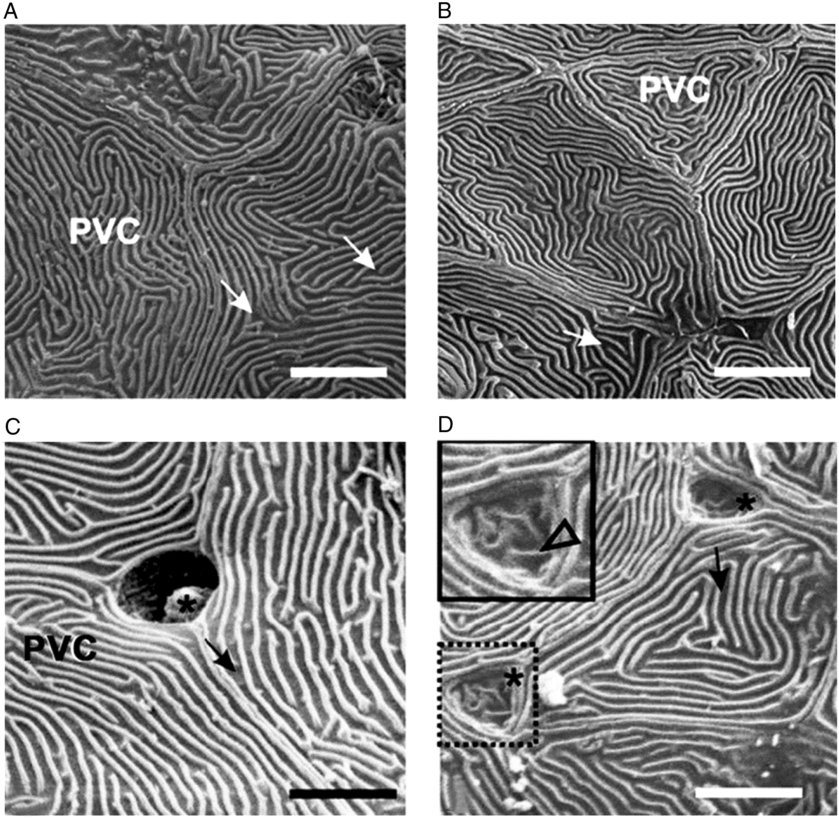
To determine if acute increase in salinity has morphological effects on O. niloticus larvae, observations were first made of the yolk-sac surface. Scanning electron microscopy revealed pavement cells (PVCs) interspaced by chloride cells (CCs), which were responsible for osmoregulation in the yolk-sac larvae (Fig. 1). In freshwater (S0) the CCs were morphologically distinct because of their rounded apical opening and the occasional presence of mucous material in their crypts. PVCs were large and possessed a great number of microridges (Fig. 1 A, C).
Scanning electron micrographs of the yolk-sac surface in O. niloticus larvae at 72 h exposure to different salinity treatments: (S0) freshwater and (S6) 6 g l−1 NaCl. (A, B) Pavement cells (PVC) at S0 (A) and S6 (B). (C, D) Chloride cells (*) at S0 (C) and S6 (D). Insert: detail of altered chloride cell; arrows: microridges; arrowhead: microvilli. Scale bar: 7 μm (A, B) and 5 μm (C, D).

Morphological alterations to the surface of the yolk sac were noticed with increasing salinity (Fig. 1 B, D). The apical opening and inner space of crypts of the CCs were smaller in larvae exposed to saline treatments (S2, S4, and S6) than in those cultured in freshwater (S0). Furthermore, these structures displayed a triangular form and shallower crypts with microvilli on their apical epithelium (Fig. 1 D). The PVCs were smaller in size in saline treatments than in freshwater and displayed a polygonal form and a discrete concave surface (Fig. 1 B). These cellular alterations to PVCs were more commonly observed in larvae of the S6 treatment.
Muscle morphology and larvae trunk area
Histological cross-sections of O. niloticus larvae at 72 h exposure revealed the morphological organization of the lateral skeletal muscle, which was composed of myotomal blocks radially structured along the trunk of larvae. Each block was composed of a single layer of red fibres located next to the skin and connective septa surrounding the inner muscle compartment. The deep compartment was composed mainly of WF, which occupied the majority of the larvae trunk.
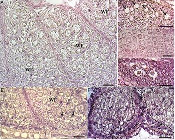
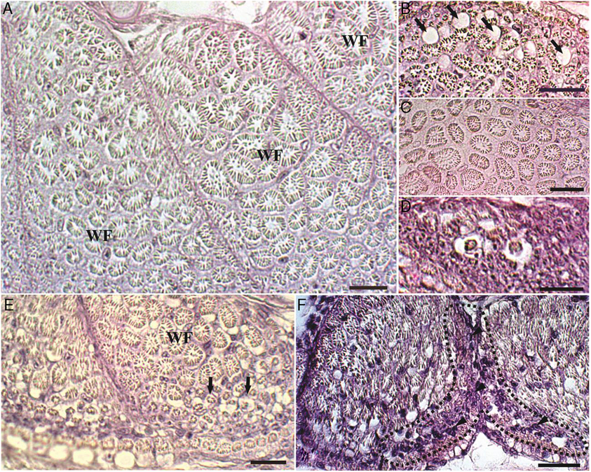
Histologically, cross-sections of the inner compartment of the skeletal muscle revealed the regular organization of WF in larvae of S0 and S2 (Fig. 2 A). In larvae of S4 and S6, however, the muscle tissue had areas with irregular fibres, as well as tissue discontinuity marked by damaged areas. The damaged areas contained vacuolated fibres, ruptured fibres, concentrations of undifferentiated cells, sarcoplasmic halo WF and swollen WF (Fig. 2 B–E). The sarcoplasmic halo around myofibrils was the most commonly found injury in white muscle. Oedema and mononuclear infiltrate were also recorded in damaged areas of larvae of the S4 and S6 treatments (Fig. 2 F).
Histological cross-sections of the skeletal muscle in yolk-sac larvae of O. niloticus at 72 h exposure to different salinity treatments: (S0) freshwater, (S2) 2 g l−1, (S4) 4 g l−1 and (S6) 6 g l−1 NaCl. Sections were stained with haematoxylin and eosin. (A) Structural organization of the skeletal muscle showing normal white fibres (WF) with acidophilic myofibrils and peripheral nuclei found mainly in fish from S0 and S2. (B–F) Histological alterations in the muscle tissue found mainly in fish from S4 and S6: (B) Vacuolated WF (black arrow), (C) Swollen WF, (D, E) sarcoplasmic halo WF and (F) mononuclear infiltrate (delineated area). Scale bar: 20 μm.
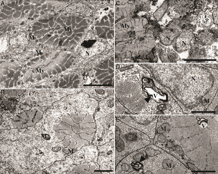
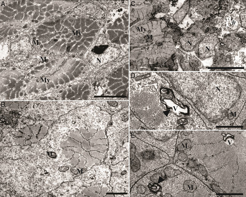
Transmission electron microscopy revealed that the sarcoplasm of the WF was almost totally occupied by myofibrils, with regular mitochondria and endoplasmic reticulum surrounding them (Fig. 3 A). Moreover, altered WF presented structural disorganization, with an electro-lucid area occupying almost all the sarcoplasm and myofibrils concentrated at the cell periphery (Fig. 3 B). The damaged areas also contained ruptured fibres and altered WF exhibiting myelin figures (Fig. 3 C–E).
Transmission electron micrographs of the lateral skeletal muscle in yolk-sac larvae of O. niloticus at 72 h exposure to different salinity treatments: (S0) freshwater, (S2) 2 g l−1, (S4) 4 g l−1 and (S6) 6 g l−1 NaCl. (A) Normal white fibres (WF) showing peripheral nucleus (N), myofibrils (My) occupying most of the sarcoplasm and mitochondria (M) at periphery in larva from S0; (B–E) WF with structural alterations: (B) swollen sarcoplasm with electron-lucid areas surrounding reduced myofibrils and altered mitochondria (M); (C) damaged fibres (*); (D, E) myelin figures (arrowhead) associated with vacuolated areas (V). Scale bar: 5 μm (A, C), 1 μm (B, D, F).
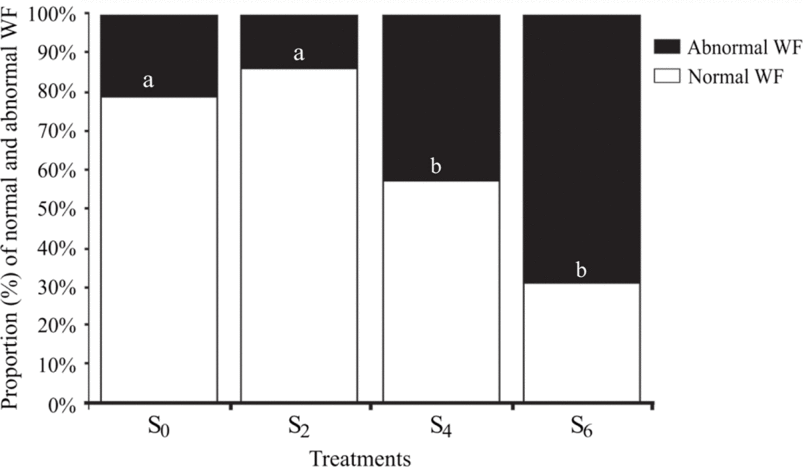
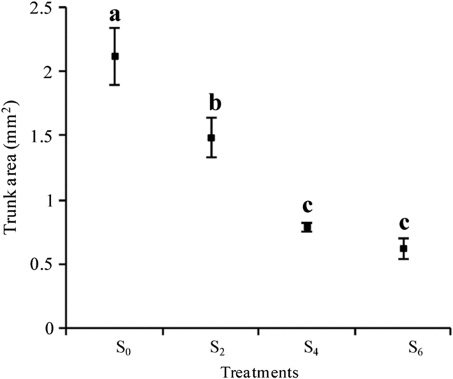
Morphometric analysis of normal and abnormal WF at 72 h exposure (Fig. 4) revealed that normal WF were predominant in S0 and S2 while abnormal WF were most frequent in S4 and S6, P < 0.05. Because morphological alterations disturb muscular development and can affect body size, the cross-sectional area of the larvae trunk in the different salinity treatments was measured. A significant smaller larvae trunk area was found for the saline treatments compared with the freshwater treatment at 72 h exposure (P < 0.05) with more drastic effect at S4 and S6 (Fig. 5).
Percentage of normal white muscle fibres (WF) and abnormal WF with histological alterations found in yolk-sac larvae of O. niloticus at 72 h exposure to different salinity treatments: (S0) freshwater, (S2) 2 g l−1, (S4) 4 g l−1 and (S6) 6 g l−1 NaCl.

Transversal trunk area in yolk-sac larvae of O. niloticus at 72 h exposure to different salinity treatments: (S0) freshwater, (S2) 2 g l−1, (S4) 4 g l−1 and (S6) 6 g l−1 NaCl. Values are expressed as mean ± standard deviation (SD). a,b,cDifferent letters indicate significant difference between treatments (P < 0.05).

Molecular biomarkers
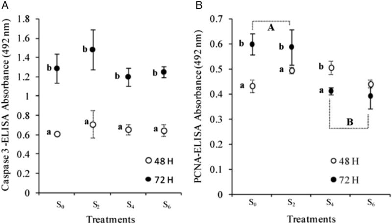
The influence of salinity on early larval development of O. niloticus larvae was also evaluated by protein expression of caspase-3 and PCNA, biomarkers of apoptosis and cell proliferation, respectively. Caspase-3 levels increased significantly from 48 to 72 h post-hatching (P < 0.05), but no significant difference was detected among salinity treatments (Fig. 6 A). Regarding cell proliferation, there was a significant increase in PCNA levels from 48 to 72 h for S0 and S2 (Fig. 6 B), while there was a decrease from 48 to 72 h for S4 and S6, with significant difference for S4 (P < 0.05). Comparing the salinity treatments, PCNA levels were significantly higher for S0 and S2 than for S4 and S6 at 72 h exposure (P < 0.05).
Indirect ELISA in yolk-sac larvae of O. niloticus exposed to different salinity treatments: (S0) freshwater, (S2) 2 g l−1, (S4) 4 g l−1 and (S6) 6 g l−1 NaCl. Protein levels (mean ± standard deviation (SD)) of caspase-3 (A) and PCNA (B) at 48 h and 72 h exposure. A,BDifferent capital letters indicate a significant difference between salinity treatments at 72 h exposure (P < 0.05). a,bDifferent small letters indicate a significant difference between exposure times in the same salinity (P < 0.05).

Discussion
Chloride cells in the epithelium of gills are the main sites for the elimination of Na+ and Cl− for ionic homeostasis in adult fish adapted to saline environments (Zadunaisky, Reference Zadunaisky1996). Gills, however, are not yet developed and functional in embryos and yolk-sac larvae, and therefore osmoregulation, is mainly performed by CCs in the yolk-sac membrane and the skin of the body (Shiraishi et al., Reference Shiraishi, Kaneko, Hasegawa and Hirano1997; Hiroi et al., Reference Hiroi, Kaneko, Uchida, Hasegawa and Tanaka1998). During early development these cells exhibit morphological and functional plasticity related to an adaptive response of the cell to environmental changes, which serves to preserve physiological homeostasis and conserve energy in fluctuating environments (Fridman et al., Reference Fridman, Bron and Rana2011). The driving force for salt transport in freshwater and saline environments is generated by Na+/K+-ATPase, which is located mostly in membranes of the tubular network of the CCs (Hirose et al., Reference Hirose, Kaneko, Naito and Takei2003).
The acclimation of euryhaline fish, such as the Nile tilapia, from freshwater to seawater occurs in two distinct phases (Zadunaisky, Reference Zadunaisky1996). The primary response is a rapid reduction in cellular permeability and the start of chloride secretion. A second adaptation takes place after days or weeks and consists of an increase in the number and size of the CCs accompanied by increased Na+/K+-ATPase activity. In O. mossambicus, CCs form multicellular complexes together with adjacent accessory cells, and these changes occur days or weeks after transfer from freshwater to seawater (Zadunaisky, Reference Zadunaisky1996; Shiraishi et al., Reference Shiraishi, Kaneko, Hasegawa and Hirano1997). Scanning electron microscopy of the present study revealed structural differences in the opening of the CCs of O. niloticus according to environmental conditions. However, the short exposure time and low salinities employed in the present study resulted in a less expressive cellular rearrangement of the yolk-sac surface than observed for the embryo and larvae of O. mossambicus.
As acclimation to saline environments resulted in morphological changes to osmoregulatory cells, the lateral skeletal muscle was examined histologically to determine if salinity had some effect on WF. Reports about damage to muscle caused by external factors in fish are scarce, and those that do exist assessed mechanical injury and feeding influence (Rowlerson et al., Reference Rowlerson, Radaelli, Mascarello and Veggetti1997; Betancor et al., Reference Betancor, Caballero, Benítez-Santana, Saleh, Atalah and Izquierdo2013). The mononuclear infiltrate detected in larvae of S4 and S6 in the present study could be related to muscular remodelling. This process involves the migration of undifferentiated myoblasts and satellite cells to areas of damaged muscle to regenerate or form new myofibres through fusion or repair mechanisms (Schultz and McCormick, Reference Schultz and McCormick1994). Although the diagnosed muscle alterations were more frequent in S4 and S6, most were seen in all the treatments, however the number of normal WF was markedly higher in larvae of S0 and S2 than in larvae of S4 and S6. Even though low salinity treatments are known to improve survival of O. niloticus larvae (Villegas, Reference Villegas1990; Fridman et al., Reference Fridman, Bron and Rana2012a), the reduction of both normal WF and trunk area reported here demonstrated that the S4 and S6 treatments may have limited the development of muscle, which may be reflected in later fish performance. To determine if the analysed salinity treatments, and their associated osmoregulatory expenditures, were reflected in the homeostasis of O. niloticus larvae, the protein expression of an executioner molecule of apoptosis, caspase-3, was analysed. Apoptosis is an essential mechanism for maintaining an appropriate number of cells in a tissue and plays a fundamental role during the development and homeostasis of multicellular organisms (Jenkins et al., Reference Jenkins, Timmons and McCall2013; Melo et al., Reference Melo, Martins, Luz, Rizzo and Bazzoli2015). Although caspase-3 is commonly used as an environmental stress biomarker (Takle and Andersen, Reference Takle and Andersen2007; Thomé et al., Reference Thomé, Domingos, Santos, Martinelli, Sato, Rizzo and Bazzoli2012), our results revealed that the larvae did not experience an apoptotic imbalance in saline environments up to 6 g l−1 NaCl, as shown by the statistically equivalent levels of caspase-3 among all treatments. However, the significant increase in caspase-3 activity from 48 to 72 h was expected because apoptosis is a cellular mechanism that induces suicide of supernumerary, misplaced, or damaged cells, and is essential for tissue renewal and repair during animal development (Meier et al., Reference Meier, Finch and Evan2000). Despite the morphological alterations to the yolk-sac surface and muscle tissue in the saline environments of S4 and S6, these treatments did not stimulate an increase in apoptosis in O. niloticus larvae.
In contrast to caspase-3, PCNA expression is a useful tool for identifying cell proliferation and DNA repair throughout the cell cycle during embryogenesis and tissue development in vertebrates (Loppion et al., Reference Loppion, Crespel, Martinez, Auvray and Sourdaine2008; Melo et al., Reference Melo, Martins, Luz, Rizzo and Bazzoli2015). In the present study, the lower levels of PCNA expression for S4 and S6 at 72 h exposure were in agreement with previous fish studies that demonstrated greater PCNA expression in better environmental or culture conditions than in adverse conditions (Brodeur et al., Reference Brodeur, Peck and Johnston2002; Thomé et al., Reference Thomé, Domingos, Santos, Martinelli, Sato, Rizzo and Bazzoli2012; Martins et al., Reference Martins, Melo, Campo-Junior, Santos, Luz, Rizzo and Bazzoli2014). The decreased levels of PCNA expression in the present study may reflect reduced muscular development in yolk-sac larvae, suggesting that the higher salinities of S4 and S6 may have limited the somatic growth of O. niloticus larvae during early development. This finding was supported by the morphometric analysis, which showed that salinities above 2 g l−1 NaCl significantly reduced the trunk area of larvae. Furthermore, lower dry weight and reduced yolk consumption has been reported for O. niloticus larvae maintained in environments with salinities greater than 15 ppt (Fridman et al., Reference Fridman, Bron and Rana2012a). Together, these data reinforced the idea that, even though low salinity environments can improve the survival of tropical freshwater fish larvae, there is a cost to somatic growth during larval development (Fridman et al., Reference Fridman, Bron and Rana2012a; Jomori et al., Reference Jomori, Luz and Portella2012; Martins et al., Reference Martins, Melo, Campo-Junior, Santos, Luz, Rizzo and Bazzoli2014).
In summary, our results suggested that yolk-sac larvae of O. niloticus may experience osmoregulatory spending in low salinities, as evidenced by alterations to the surface of CCs covering the yolk sac. At salinities of 4 and 6 g l−1 NaCl, O. niloticus larvae experienced a disturbance to cell proliferation, trunk area, and the proportion of normal WF. Furthermore, at 6 g l−1 NaCl larvae exhibited severe histological damage to muscle. Therefore, treatments of 4 and 6 g l−1 NaCl are not recommended for early larval development of O. niloticus in aquaculture.
Acknowledgements
The authors wish to thank the technicians at the Laboratory of Aquaculture (LAQUA) of the Universidade Federal de Minas Gerais (UFMG) for their assistance in handling the fish; the technicians at the Centro de Microscopia and the Centro de Aquisição e processamento de imagens (CAPI) of the UFMG for preparing the biological material for electron microscopy.
Financial support
The research was funded by Brazilian institutions: CNPq (grants 305132/2015-6 and 407778/2016-0), CAPES (Finance Code 001) and FAPEMIG (grant APQ-00898-15).
Conflicts of interest
None
Ethical standards
All procedures were approved and certified by the Ethics Committee on Animal Use (CEUA/UFMG, 396/2012).