Introduction
Anisakidae nematodes, particularly Contracaecum Railliet & Henry, 1912, are known for their wide host range and geographic distribution. These parasites commonly infect aquatic organisms worldwide (Rohde Reference Rohde2005). The life cycle of Contracaecum typically involves aquatic invertebrates and fish as intermediate and/or paratenic hosts, and piscivorous birds and mammals serve as definitive hosts (Anderson Reference Anderson2000). Third-stage Contracaecum spp. larvae have been reported in the stomach mucosa of humans causing anisakidosis, a painful condition after ingestion of raw or undercooked fish (Shamsi Reference Shamsi2019). In Argentina, only a few cases of anisakidosis have been reported so far (Degese et al. Reference Degese, Gatti and Krivokapich2019; Menghi et al. Reference Menghi, Gatta, Arias, Santoni, Nicola, Smayevsky, Degese and Krivokapich2020).
Morphological identification of Contracaecum species can be challenging due to the presence of overlapping diagnostic characters. Traditional taxonomic approaches rely on morphological features such as cephalic, buccal, and pharyngeal structures, cuticular details, and male reproductive system characteristics, such as spicules and the papillae arrangement at the posterior end (Fagerholm Reference Fagerholm1990; Hartwich Reference Hartwich1964; Mattiucci and Nascetti Reference Mattiucci and Nascetti2008). This classification scheme, originally proposed by Fagerholm (Reference Fagerholm1990), provides a framework for distinguishing the different species within the genus into four morphotypes defined by the arrangement of the male pre- and postcloacal papillae. More recently, Garbin et al. (Reference Garbin, Servián, Fuentes, Morgenthaler, Millones, Fuchs, Diaz and Navone2024) proposed a modified classification criterion of Contracaecum species into three morphotypes, also based on the arrangement of caudal papillae in males.
Additionally, taxonomists have encountered challenges in distinguishing between cryptic or sibling species, which appear morphologically similar but are genetically distinct (D’Amelio et al. Reference D’Amelio, Cavallero, Dronen, Barros and Paggi2012; Mattiucci et al. Reference Mattiucci, Paoletti, Webb, Sardella, Timi, Berland and Nascetti2008a, Reference Mattiucci, Paoletti, Consuegra-Solorzano and Nascetti2010; Shamsi et al. Reference Shamsi, Gasser and Beveridge2008). To address these issues, molecular studies have been crucial in clarifying the identification of Contracaecum species (D’Amelio et al. Reference D’Amelio, Barros, Ingrosso, Fauquier, Russo and Paggi2007; Mattiucci et al. Reference Mattiucci, Paoletti, Olivero-Verbel, Baldiris, Arroyo-Salgado, Garbin, Navone and Nascetti2008b; Shamsi Reference Shamsi2019). Techniques such as sequencing of nuclear and mitochondrial genes have been used for identifying Contracaecum species and linking the larvae parasitizing paratenic/intermediate hosts (e.g. crustaceans, squids, fishes, etc.) to adults parasitizing definitive hosts, mainly seabirds and marine mammals (Garbin et al. Reference Garbin, Mattiucci, Paoletti, Diaz, Nascetti and Navone2013; Mattiucci et al. Reference Mattiucci, Paoletti, Consuegra-Solorzano and Nascetti2010; Mattiucci and Nascetti Reference Mattiucci and Nascetti2008). Contracaecum larvae can only be identified to species level with the help of molecular markers. In all cases of anisakidosis in humans, morphological identification of larvae has only been possible to genus level (Buchmann Reference Buchmann2023; Caffara et al. Reference Caffara, Tedesco, Davidovich, Locke, Gustinelli, King, Nuytten, Nuzzo and Fioravanti2023; Shamsi Reference Shamsi2019).
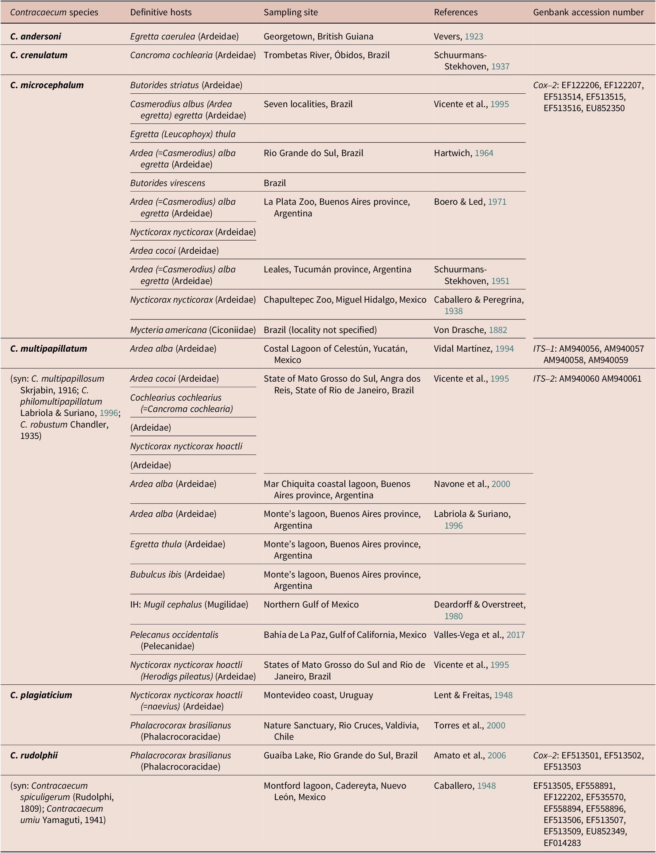
Several Contracaecum species were documented parasitizing Ardeidae in South and Central America (Table 1), with some records in Argentina (Garbin et al. Reference Garbin, Diaz, Servián, Fusaro and Navone2023). Thus, Contracaecum microcephalum (Rudolphi 1809) was found parasitizing the Great egret Ardea alba Linnaeus, 1758, the Cocoi heron Ardea cocoi Linnaeus, 1776, and the Black-Crowned Night heron Nycticorax nycticorax Linnaeus, 1758 (Boero and Led Reference Boero and Led1971; Schuurmans-Stekhoven Reference Schuurmans-Stekhoven1951). Contracaecum philomultipapillatum Labriola & Suriano, Reference Labriola and Suriano1996, was reported in A. alba, the Snowy egret Egretta thula (Molina 1782), and the Western Cattle egret Bubulcus ibis Linnaeus, 1758 (Labriola and Suriano Reference Labriola and Suriano1996). Then, Navone et al. (Reference Navone, Etchegoin and Cremonte2000) reported Contracaecum multipapillatum (von Drasche Reference Von Drasche1882) in A. alba and suggested that C. philomultipapillatum is a junior synonym of C. multipapillatum. Other records are detailed in Table 1.
Records of Contracaecum species parasitizing Ardeidae in Argentina, Central and South America

The Cocoi heron is a resident bird found in aquatic environments widely distributed in South America and considered common throughout its range, typically inhabiting shorelines and shallow waters where it hunts for fish, amphibians, rodents, crustaceans, and insects (Ducommun and Beltzer Reference Ducommun, Beltzer, Virgolini and Quiroga2010; Hayes et al. Reference Hayes, Nakamura, Hiss and Capllonch2023).
The aim of the present work was to identify two different specimens of Contracaecum parasitizing one dead Cocoi heron from the Magdalena city coast, Buenos Aires province, Argentina, using an integrative taxonomic approach involving morphological observations by optical and scanning electron microscopy, and genetic analyses of nuclear and mitochondrial markers.
Material and methods
Sampling
In September 2017, one A. cocoi specimen was found dead at the Magdalena city coast, Buenos Aires Province, Argentina (34° 58′ 59.7″ S, 57° 44′ 9.05″ W). During necropsy, 97 nematode specimens were recovered from the oesophagus and stomach and preserved in 70% ethanol for their morphological study. Thirteen specimens were preserved in 96% ethanol for molecular analysis.
Morphological study
Forty nematode specimens were cleared in lactophenol (1:9) and studied using an optical microscope (OM), Olympus BX51. Six males and six females were dehydrated in a graded ethanol series and dried in hexamethyldisilazane. Subsequently, they were mounted on stubs, coated with 40 nm of gold/palladium in a Thermo VG Scientific Polaron SC 7630, and examined in a Zeiss Supra 40 scanning electron microscope. Measurements of parasites are given in millimetres (mm), unless otherwise indicated, with mean values followed by range in parentheses. Taxonomic identification was performed based on the diagnostic features of the family Anisakidae, following the classification criteria described by Fagerholm (Reference Fagerholm1990) regarding the arrangement of male caudal papillae, and by consulting relevant bibliography (Garbin et al. Reference Garbin, Diaz and Navone2019, Reference Garbin, Servián, Fuentes, Morgenthaler, Millones, Fuchs, Diaz and Navone2024; Hartwich Reference Hartwich1964). Three morphotypes (i.e. simple, intermediate, and multiple) were established taking into account the distribution pattern on the male caudal papillae according to Garbin et al. (Reference Garbin, Servián, Fuentes, Morgenthaler, Millones, Fuchs, Diaz and Navone2024) and based on Fagerholm (Reference Fagerholm1990). Type and voucher specimens are deposited in the Helminthological Collection of the Museo de La Plata (MLP-He), La Plata, Buenos Aires province, Argentina.
DNA extraction, amplification, and sequencing
DNA extraction was performed using the Wizard® Genomic DNA Purification Kit (Promega, USA) according to the manufacturer’s instructions. All amplifications were performed with a Mastercycler thermocycler (Eppendorf) in a 50-μl reaction mixture consisting of 25 μl of PB-L master mix (PB-L Productos Bio-Lógicos, Quilmes, Argentina), 0.4 μM of each forward and reverse primer, and 4 μl of the template DNA (120–200 ng).
The cox2 mtDNA gene from both Contracaecum specimens was amplified following Mattiucci et al. (Reference Mattiucci, Paoletti, Consuegra-Solorzano and Nascetti2010), with the primers 211F (5′-TTT TCT AGT TAT ATA GAT TGR TTY AT-3′) and 210R (5′-CAC CAA CTC TTA AAA TTA TC-3′) from Nadler and Hudspeth (Reference Nadler and Hudspeth2000). Cycling parameters were as follows: an initial step at 94°C for 3 min, followed by 34 cycles at 94°C for 30 sec, 47°C for 1 min, and 72°C for 90 sec. An additional elongation step was performed for 10 min at 72°C.
The amplification of the ITS- 1 region was carried out with the primers SS1 (5′-GTTTCCGTAGGTGAACCTGCG-3′) and NC13R (5′-GCTGCGTTCTTCATCGAT-3′) and the ITS- 2 region with the primers SS2 (5′-TTGCAGACACATTGAGCACT-3′) and NC2 (5′-TTAGTTTCTTTTCCTCCGCT-3′), following Shamsi et al. (Reference Shamsi, Gasser and Beveridge2008, Reference Shamsi, Norman, Gasser and Beveridge2009). Cycling conditions for both were: 94°C for 5 min, followed by 30 cycles at 94°C for 30 sec, at 55°C for 30 sec, at 72°C for 30 sec, and a final extension at 72°C for 5 min.
The amplification of SSU rRNA gene was performed according to D’Amelio et al. (Reference D’Amelio, Barros, Ingrosso, Fauquier, Russo and Paggi2007) with the primers MH3 (5′-TTGTTCCAGAATAATCGGCTAGACTT-3′) and MH4.5 (5′TCTACTTTACTACAACTTACTCC-3′). PCR cycling conditions included 10 min at 95°C, 35 cycles of 30 sec at 95°C, 30 sec at 55°C, 30 sec at 72°C, and a final extension of 7 min at 72°C.
PCR amplicons were confirmed by 1% agarose electrophoresis gel and then purified and sequenced by Macrogen (Seoul, South Korea) with the same PCR primers. The nucleotide sequences obtained here were deposited in DDBJ/EMBL/GenBank databases.
Phylogenetic analysis
Sequences were assembled using the Geneious program. The assembled sequences were checked for the presence of pseudogenes in the cox2 mtDNA gene by translating the amino acid sequences using the invertebrate mitochondrial genetic code within Geneious. Contracaecum spp. sequences were aligned using the online version of MAFFT 7 (Katoh and Standley Reference Katoh and Standley2013) with sequences of Anisakis spp. used as outgroups. The best partitioning scheme and substitution model for each DNA partition were chosen under the Bayesian Information Criterion (Schwarz Reference Schwarz1978) using the ‘greedy’ search strategy in Partition Finder v. 1.1.1 (Lanfear et al. Reference Lanfear, Calcott, Ho and Guindon2012). Only the cox2 mtDNA fragment dataset was partitioned into first-, second-, and third-codon positions. The appropriate nucleotide substitution models implemented for each partition were HKY+I for the first and third codon positions, and HKY+G for the second codon position. For the datasets of SSU rRNA and ITS2 genes, the HKY+G was used, while K80+G model was used for the ITS1 gene.
Phylogenetic reconstruction was performed using Bayesian Inference (BI) through Mr. Bayes 3.2.3 (Ronquist and Huelsenbeck Reference Ronquist and Huelsenbeck2003). Phylogenetic trees were constructed using two parallel analyses of Metropolis-Coupled Markov Chain Monte Carlo (MCMC) for 20 million generations each, to estimate the posterior probability (PP) distribution. Topologies were sampled every 1,000 generations and the average standard deviation of split frequencies was observed to be less than 0.01 at the end of the run, as suggested by Ronquist et al. (Reference Ronquist, Teslenko, van der Mark, Ayres, Darling, Höhna, Larget, Liu, Suchard and Huelsenbeck2012). The robustness of the clades was assessed using Bayesian posterior probability (PP), where PP > 0.90 was considered strongly supported. A majority consensus tree with branch lengths was reconstructed for each run after discarding the first 25% trees sampled as ‘burn-in’.
Additionally, genetic distances among Contracaecum species were computed under the Kimura 2-parameter (K2P) substitution model (Kimura Reference Kimura1980) using MEGA v11, to evaluate inter- and intraspecific levels of divergence in the cox2, ITS1, ITS2, and SSU rRNA dataset.
Results
Contracaecum jorgei Sardella, Mancini, Salinas, Simões & Luque, Reference Sardella, Mancini, Salinas, Simões and Luque2020
Type host: Nannopterum (=Phalacrocorax) brasilianum (King, 1828) (Pelecaniformes: Phalacrocoracidae).
New host: Ardea cocoi Linnaeus, 1766 (Pelecaniformes: Ardeidae).
Type locality: Pampean shallow lakes, Unión Department, Córdoba province, Argentina (33°25′ S, 62°54′ W).
New locality: Magdalena city coast, Buenos Aires Province, Argentina (34°58′59.7″ S, 57°44′ 9.05″ W).
Infection site: Oesophagus and stomach.
Specimens deposited: 20 voucher specimens (MLP-He 8340).
Description
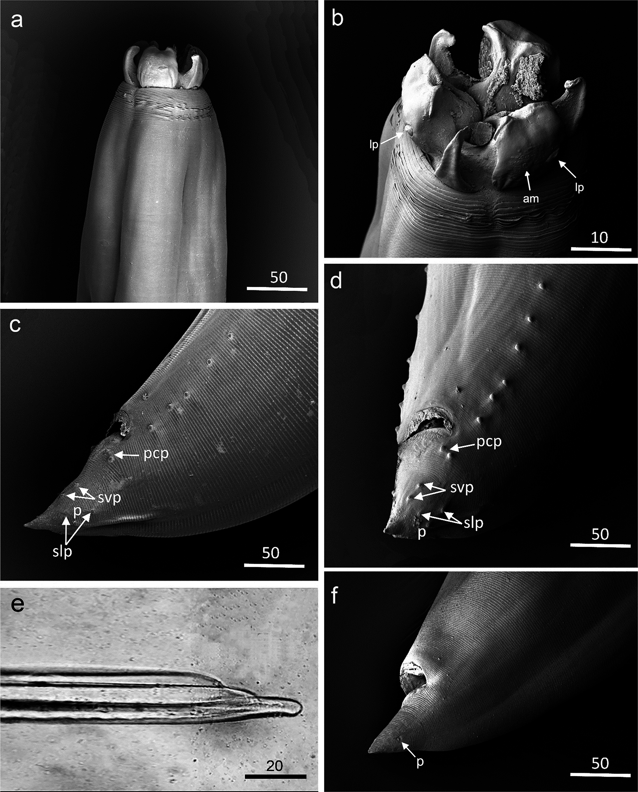
General morphology (based on 10 males and 10 females): Cuticle transversally striated throughout body (Figs. 1b, c, f). Non-conspicuous cephalic collar, V-shaped lateral region without striations (Fig. 1b). Three smooth interlabia (Figs. 1a,b). Lips barely longer than interlabia with non-visible notches (Figs. 1a,b). Lips with two non-conspicuous and lobed auricles, no apparent tips (Figs. 1a,b). Remarkable lip papillae (lp), two on the dorsal lip, and one on each ventrolateral lip, visible amphid (am) (Fig. 1b, see arrows).
Contracaecum jorgei parasitizing Ardea cocoi from the Magdalena coast, Buenos Aires province, Argentina, at the SEM. (a) Anterior end, lateral view; (b) anterior end, detail of lateroventral lip, interlabia, labial papilla (lp), lip auricles, amphid (am) (see arrows); (c) male posterior end, lateral view: precloacal papillae, paracloacal papillae (pcp), subventral postcloacal papillae (svp), sublateral postcloacal papillae (slp), phasmid (p); (d) male posterior end, sublateral view: precloacal papillae, paracloacal papillae (pcp), subventral postcloacal papillae (svp), sublateral postcloacal papillae (slp), phasmid (p); (e) spicule free-distal end, lateral view (LM photo); (f) female posterior end, phasmid (p). Scale bars expressed in micrometres.

Male: No apparent tail distal constriction (Figs. 1c,d). Short spicules approximately one-fifth the body length, and short spicule free-distal end (Fig. 1e and Table 2). Caudal papillae distribution fits into a simple morphotype (A), consisted of two paracloacal papillae pairs (pcp), two subventral papillae pairs (svp), two sublateral papillae pairs (slp), and a phasmid pair (p) (Figs. 1c,d). Morphometric measurements of features such as the body length, maximum body width, nerve ring, deirids, oesophagus length, intestinal caecum length, ventricle length, ventricular appendix length, spicule length, tail length are detailed in Table 2.
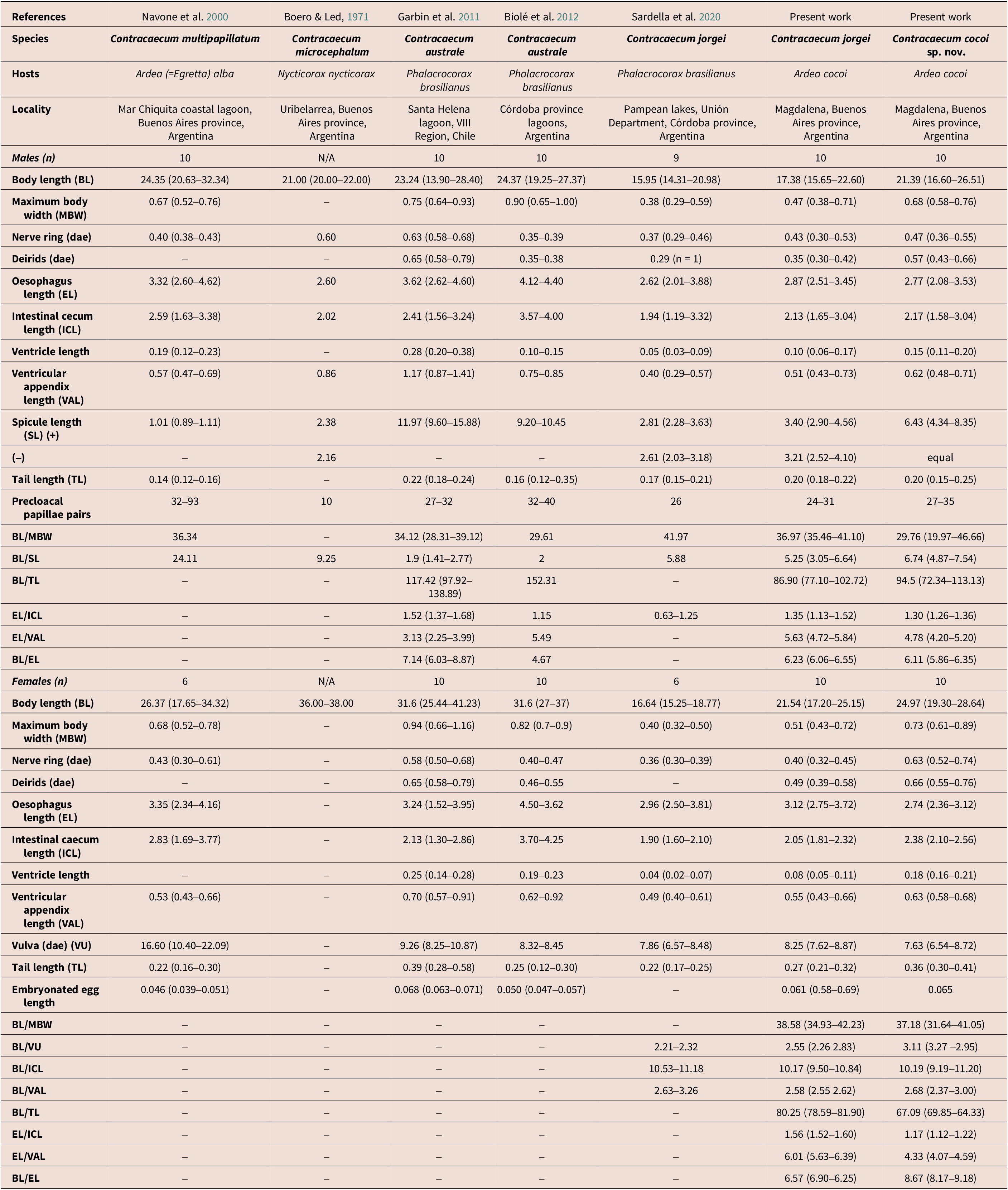
Morphometric data of Contracaecum cocoi sp. nov. and C. jorgei parasitizing Ardea cocoi from Magdalena, Buenos Aires province, and other related Contracaecum spp. descriptions

Note: All measurements are expressed in millimetres (mm). Distance to anterior end (dae), (+) Right spicule, (–) Left spicule.
Female: One pair of phasmids on the tail end (p) (Fig. 1f). Morphometric features and their measurements are detailed in Table 2.
Contracaecum cocoi sp. nov.
Type hosts: Ardea cocoi Linnaeus, 1766 (Pelecaniformes: Ardeidae).
Type locality: Magdalena city coast, Buenos Aires Province, Argentina (34°58′59.7″ S, 57°44′ 9.05″ W).
Collection date: September 2017.
Infection site: Oesophagus and stomach.
Studied host: 1 (one).
Intensity: 38 adult specimens.
Specimens deposited: one holotype (male) (MLP-He 8337), one allotype (female) (MLP-He 8338), and 18 paratypes (MLP-He 8339).
Description
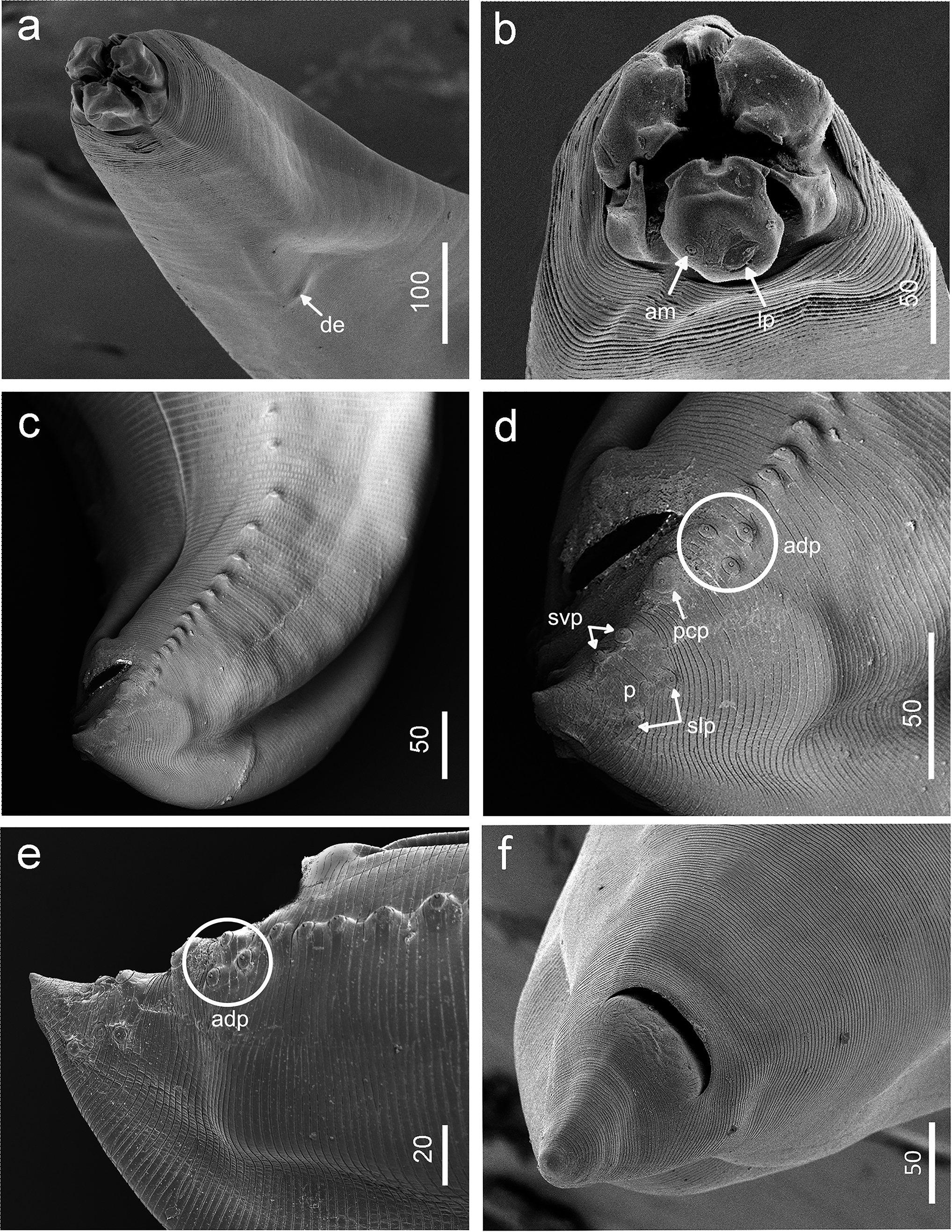
General morphology (based on 10 males and 10 females): Entire body transversally striated (Fig. 2). Conspicuous cephalic collar, with deep concentric anteriorly directed free edges and narrow V-shaped lateral region without striations (Fig. 2b). Lips with one apical shallow notch bearing two notorious auricles with a sensory pit at distal end (Fig. 2b). Large double lip papillae, two on the dorsal lip and one on each ventrolateral lip (lp), one visible amphid (am) (see arrows) (Fig. 2b). Three barely bifurcated interlabia shorter than lips (Figs. 2a,b). Deirids inconspicuous with button shaped (de) (Fig. 2a). Morphometric measurements of features such as the body length, maximum body width, nerve ring, deirids, oesophagus length, intestinal caecum length, ventricle length, ventricular appendix length, spicule length, tail length are detailed in Table 2.
Contracaecum cocoi sp. nov. parasitizing Ardea cocoi from the Magdalena coast, Buenos Aires province, Argentina, at the SEM. (a) Anterior end, subapical view; (b) anterior end, apical view: detail of lateroventral lip, lip auricles, bifid interlabia, cephalic labial papilla (lp), amphid (am) (arrows); (c) male posterior end, oblique lateral view: precloacal papillae, postcloacal papillae; (d) male posterior end, oblique lateral view: precloacal papillae, paracloacal papillae (pcp), adcloacal papillae (adp), subventral postcloacal papillae (svp), sublateral postcloacal papillae (slp), phasmid (p); (e) male posterior end, lateral view: precloacal papillae, paracloacal papillae, adcloacal papillae (adp, encircled), subventral postcloacal papillae, sublateral postcloacal papillae, phasmid; (f) female posterior end, ventral view. Scale bars expressed in micrometers.

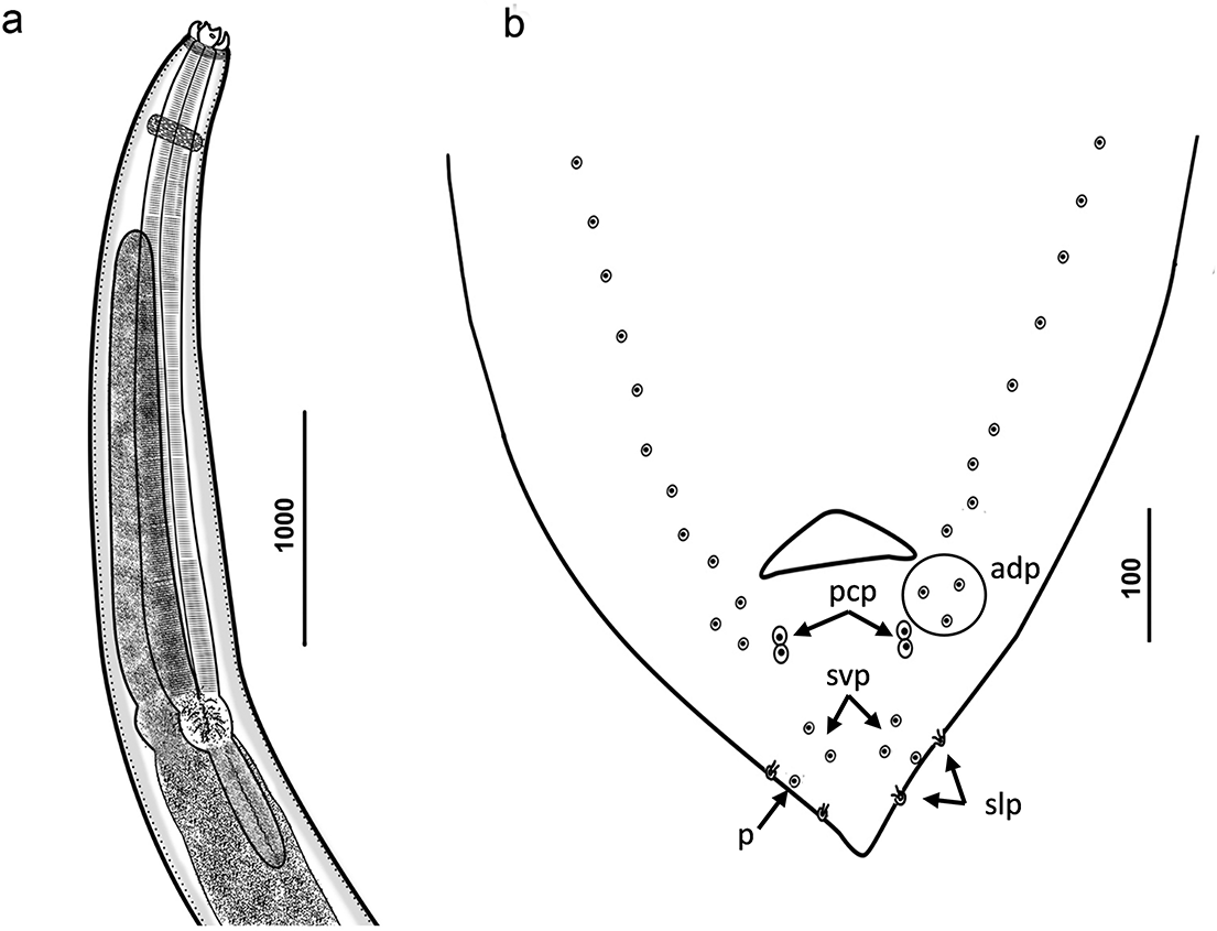
Male (holotype): Body length 22.94 mm. Maximum body width 0.73 mm. Distance from anterior end to nerve ring and deirids 0.58 and 0.66 mm, respectively. Oesophagus length 2.82 mm; intestinal caecum length 2.41 mm; ventricle length 0.18 mm; ventricular appendix length 0.60 mm (Fig. 3a). Spicules of equal length 6.82 mm reaching almost one-third of the body. Tail length 0.19 mm (Table 2). Caudal extremity conical bearing 34 pairs of precloacal papillae (Figs. 2c, 3b). Caudal papillae distribution consisted of nine pairs of postcloacal papillae, 2 large double paracloacal (pcp) pairs situated aside to cloaca in oblique position to body axis, 3 adcloacal pairs (adp) surrounding the paracloacal lateral side (encircled papillae), 2 subventral pairs (svp), 2 sublateral pairs (slp), and 1 pair of phasmids (p) among these latter ones (Figs. 2d–e, 3b). Morphometric measurements of male paratypes are detailed in Table 2.
Contracaecum cocoi sp. nov. parasitizing Ardea cocoi from the Magdalena coast, Buenos Aires province, Argentina. (a) Anterior end, lateral view: nerve ring, oesophagus, intestinal caecum, ventricle, ventricular appendix; (b) male posterior end, ventral view: precloacal papillae, paracloacal papillae (pcp), adcloacal papillae (adp), subventral postcloacal papillae (svp), sublateral postcloacal papillae (slp), phasmid (p). Scale bars expressed in micrometers.

Female (allotype): Body length 26.23 mm. Maximum body width 0.79 mm. Distance from anterior end to nerve ring and deirids 0.54 and 0.63 mm, respectively. Oesophagus length 3.12 mm; intestinal caecum length 2.42 mm; ventricle length 0.19 mm; ventricular appendix length 0.66 mm (Fig. 3a). Vulva in anterior half of body. Distance from anterior end to vulva 8.72 mm. Tail length 0.40 mm (Fig. 2f). Embryonated egg (n = 10) diameter 0.065 mm (0.061–0.074 mm). Morphometric measurements of female paratypes are detailed in Table 2.
Remarks
Currently, four species of Contracaecum are known to parasitize members of the Ardeidae family: C. multipapillatum, Contracaecum plagiaticium Lent & Freitas, Reference Lent and Freitas1948, C. microcephalum, and C. jorgei. The new species C. cocoi sp. nov. is morphologically distinct from all other species. Contracaecum cocoi sp. nov. is similar to C. multipapillatum which parasitizes A. alba and Pelecanus conspicillatus Temminck, but differs in having bifurcated interlabia (Figs. 2a,b), fewer precloacal and postcloacal (adcloacal) papillae pairs in males (Figs. 2c–f, 3b, Table 2), and much longer spicules (Table 2) (Navone et al. Reference Navone, Etchegoin and Cremonte2000; Shamsi et al. Reference Shamsi, Gasser and Beveridge2008). Contrasting with C. plagiaticium parasitizing Nycticorax nycticorax hoactli (Gmelin), the new species has double-sized spicules (4.34–8.35 vs. 2.32–3.49 mm), one additional postcloacal papillae pair (9 vs. 8), and bifid interlabia (Lent and Freitas Reference Lent and Freitas1948). Moreover, these three aforementioned species belong to the intermediate morphotype B (Garbin et al. Reference Garbin, Servián, Fuentes, Morgenthaler, Millones, Fuchs, Diaz and Navone2024). Contracaecum microcephalum, which parasitizes some species of Ardeidae (Hartwich Reference Hartwich1964; Von Drasche Reference Von Drasche1882) and belongs to the simple morphotype A (Garbin et al. Reference Garbin, Servián, Fuentes, Morgenthaler, Millones, Fuchs, Diaz and Navone2024), is distinguished from C. cocoi sp. nov. by having fewer than half the number of precloacal papillae (10 vs. 27–35), three fewer adcloacal papillae pairs (6 vs. 9), and significantly shorter spicules (2.16–2.38 vs. 4.34–8.35 mm) (Table 2). In relation to its congener C. jorgei, in the mixed infection parasitizing the same host, C. cocoi sp. nov. males possess three additional adcloacal papillae pairs as morphotype B (Garbin et al. Reference Garbin, Servián, Fuentes, Morgenthaler, Millones, Fuchs, Diaz and Navone2024), double-sized spicules, and bifid interlabia (Sardella et al. Reference Sardella, Mancini, Salinas, Simões and Luque2020) (Figs. 2, 3, Table 2).
Regarding the Contracaecum species parasitizing other birds from southern South America, the new species differs from Contracaecum australe Garbin, Mattiucci, Paoletti, González-Acuña & Nascetti, Reference Garbin, Mattiucci, Paoletti, González-Acuña and Nascetti2011, which is parasitic in the Neotropic cormorant Nannopterum (=Phalacrocorax) brasilianum (Gmelin) by having half-sized spicules (4.34–8.35 vs. 9.60–15.88 mm), three more adcloacal papillae pairs (Figs. 2, 3, Table 2), and a non-constricted tail (Biolé et al. Reference Biolé, Guagliardo, Mancini, Tanzola, Salinas and Morra2012; Garbin et al. Reference Garbin, Mattiucci, Paoletti, González-Acuña and Nascetti2011). Contracaecum caballeroi Bravo-Hollis, Reference Bravo-Hollis1939, parasitic in N. brasilianum, can be distinguished from the new species by lacking bifid interlabia, much shorter spicules (0.90–1.09 vs. 4.34–8.35 mm), and no adcloacal papillae pairs (Figs. 2, 3, Table 2) (Lent and Freitas Reference Lent and Freitas1948).
The following species resemble C. cocoi sp. nov. but they have a simple papillae arrangement on the male tail corresponding to the morphotype A, which lacks adcloacal papillae (Fig. 2d, e, 3b) (Garbin et al. Reference Garbin, Servián, Fuentes, Morgenthaler, Millones, Fuchs, Diaz and Navone2024). These include Contracaecum travassosi Gutiérrez, 1943, parasitic in Phalacrocorax atriceps (=albiventer) King (Gutierrez 1943); Contracaecum rudolphii Hartwich, Reference Hartwich1964 sensu lato from Phalacrocorax carbo (Linnaeus), Gulosus (=Phalacrocorax) aristotelis (Linnaeus), and Sula leucogaster (Boddaert) (Abollo et al. Reference Abollo, Gestal and Pascual2001; Amato et al. Reference Amato, Monteiro and Amato2006; Barus et al. Reference Barus, Tenora and Prokes2000; Hartwich Reference Hartwich1964); Contracaecum pelagicum Johnston & Mawson, 1942, parasitizing Spheniscus magellanicus (Forster) (Garbin et al. Reference Garbin, Navone, Diaz and Cremonte2007); Contracaecum magnipapillatum (= C. magnicollare Johnston & Mawson, 1941) from Anous minutus Chapin (Laridae); and Contracaecum septentrionale Kreis, Reference Kreis1955, from Phalacrocorax aristotelis (Linnaeus) (Phalacrocoracidae) (Fagerholm et al. Reference Fagerholm, Overstreet and Humphery-Smith1996; Hartwich Reference Hartwich1964; Kreis Reference Kreis1955).
When compared to its congeners parasitizing sea mammals in the southern Atlantic Ocean, Contracaecum osculatum (Rudolphi, 1802), Contracaecum radiatum (Linstow, 1907), and Contracaecum mirounga Nikolskii, 1974, are quite similar to C. cocoi sp. nov. but differ by having double rows of precloacal papillae and postcloacal papillae belonging to the morphotype C (Fagerholm Reference Fagerholm1990; Garbin et al. Reference Garbin, Servián, Fuentes, Morgenthaler, Millones, Fuchs, Diaz and Navone2024). On the other hand, Contracaecum ogmorhini sensu stricto Johnston & Mawson, 1941, and Contracaecum margolisi Mattiuci, Cianchi, Paggi, Sardella, Timi, Webb, Bastida, Rodriguez & Bullini, 2003, belong to the morphotype A (Fagerholm Reference Fagerholm1988; Fagerholm et al. Reference Fagerholm, Overstreet and Humphery-Smith1996; Garbin et al. Reference Garbin, Servián, Fuentes, Morgenthaler, Millones, Fuchs, Diaz and Navone2024; Mattiucci et al. Reference Mattiucci, Cianchi, Nascetti, Paggi, Sardella, Timi, Webb, Bastida, Rodríguez and Bullini2003).
With regard to females, C. cocoi sp. nov. exhibits a distinctive morphometric profile. It can be distinguished from C. jorgei (sharing the same host, A. cocoi) by its significantly larger body size (24.97 vs. 21.54 mm), greater maximum body width (0.73 vs. 0.51 mm), longer distance of nerve ring to the anterior end (dae) (0.63 vs. 0.40 mm), and a substantially longer tail (0.36 vs. 0.27 mm). The new species also possesses a markedly longer ventricle than C. jorgei (0.18 vs. 0.04 mm). These differences are further supported by distinct ratios, particularly a lower BL/TL ratio (67.09 vs. 80.25) and a higher BL/EL ratio (8.67 vs. 6.57) in the new species (Table 2) (Sardella et al. Reference Sardella, Mancini, Salinas, Simões and Luque2020).
Furthermore, C. cocoi sp. nov. differs from C. multipapillatum by possessing a shorter distance from the vulva to the anterior end (7.63 vs. 16.60 mm), and a longer tail (0.36 vs. 0.22 mm) (Navone et al. Reference Navone, Etchegoin and Cremonte2000). It is separated from C. australe by its shorter oesophagus length (2.74–3.24 mm vs. 3.62–4.50 mm) and a shorter ventricular appendix (0.63–0.70 mm vs. 0.62–0.92 mm) (Table 2) (Biolé et al. Reference Biolé, Guagliardo, Mancini, Tanzola, Salinas and Morra2012; Garbin et al. Reference Garbin, Mattiucci, Paoletti, González-Acuña and Nascetti2011).
The morphometric ratios provide robust diagnostic criteria. The EL/VAL ratio of C. cocoi sp. nov. (4.33) is considerably lower than that of C. jorgei (6.01) and C. multipapillatum (implied to be ~6.32), indicating a proportionally longer ventricular appendix (Table 2). Similarly, the EL/ICL ratio (1.17) is distinct from that of C. jorgei (1.56). The BL/VU ratio (3.11) is higher than that of all compared species for which data are available (C. australe, C. jorgei), indicating a proportionally more posterior position of the vulva (Table 2).
Molecular characterization and phylogenetic analysis
Four nucleotide sequences of cox2 mtDNA were obtained: three from C. jorgei (555–591 bp) and one from the new species (578 bp). In addition, two of SSU rRNA (529 bp for C. jorgei and 478 bp for C. cocoi n. sp.), two of ITS1 (494 bp for C. jorgei and 504 bp for C. cocoi n. sp.), and two of ITS2 (367 bp for C. jorgei and 397 bp for C. cocoi n. sp.). The constructed matrix consisted of 59 terminals with lengths of 591 bp (cox2 mtDNA), 12 and 460 bp (SSU rRNA), 54 and 384 bp (ITS1), 49 and 214 bp (ITS2).
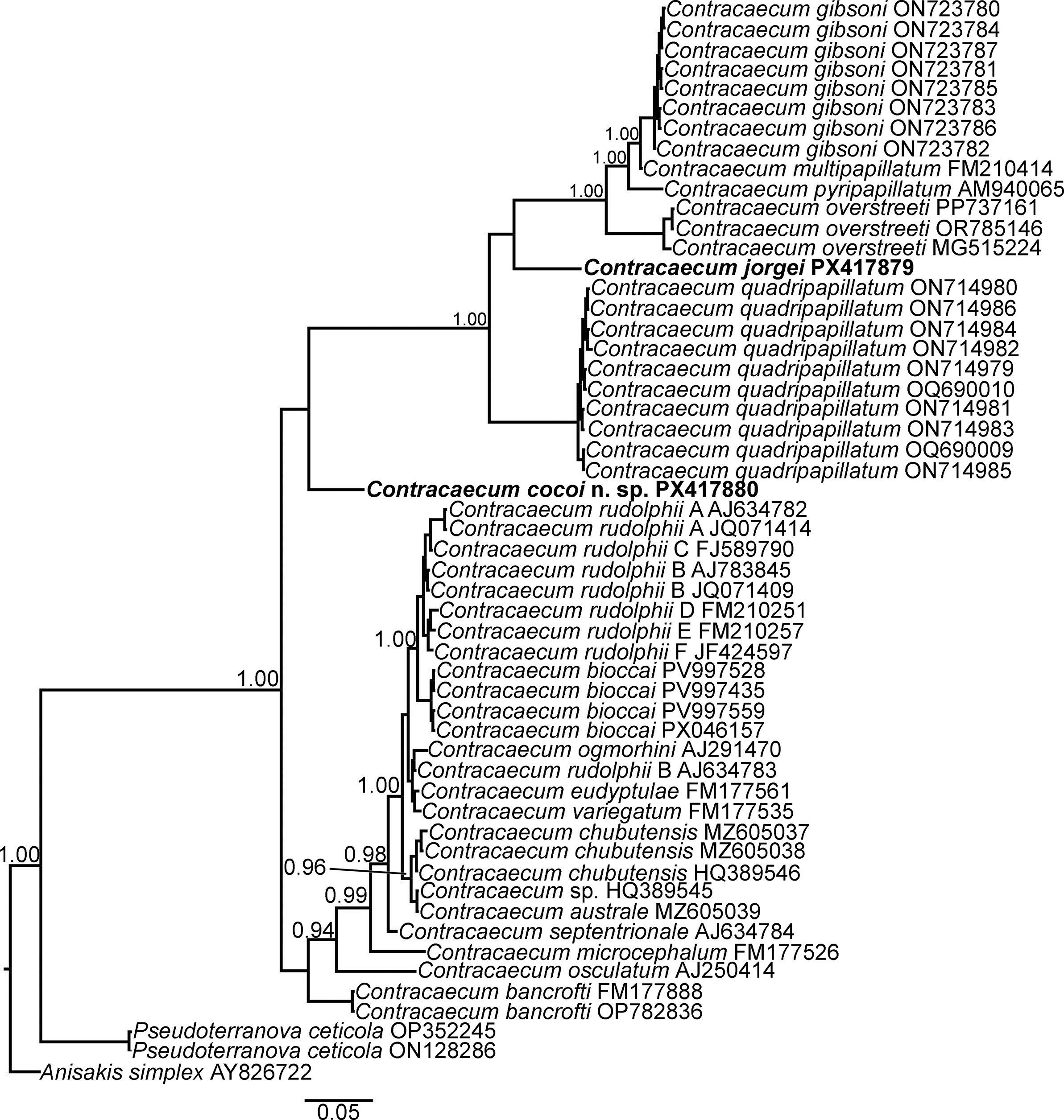
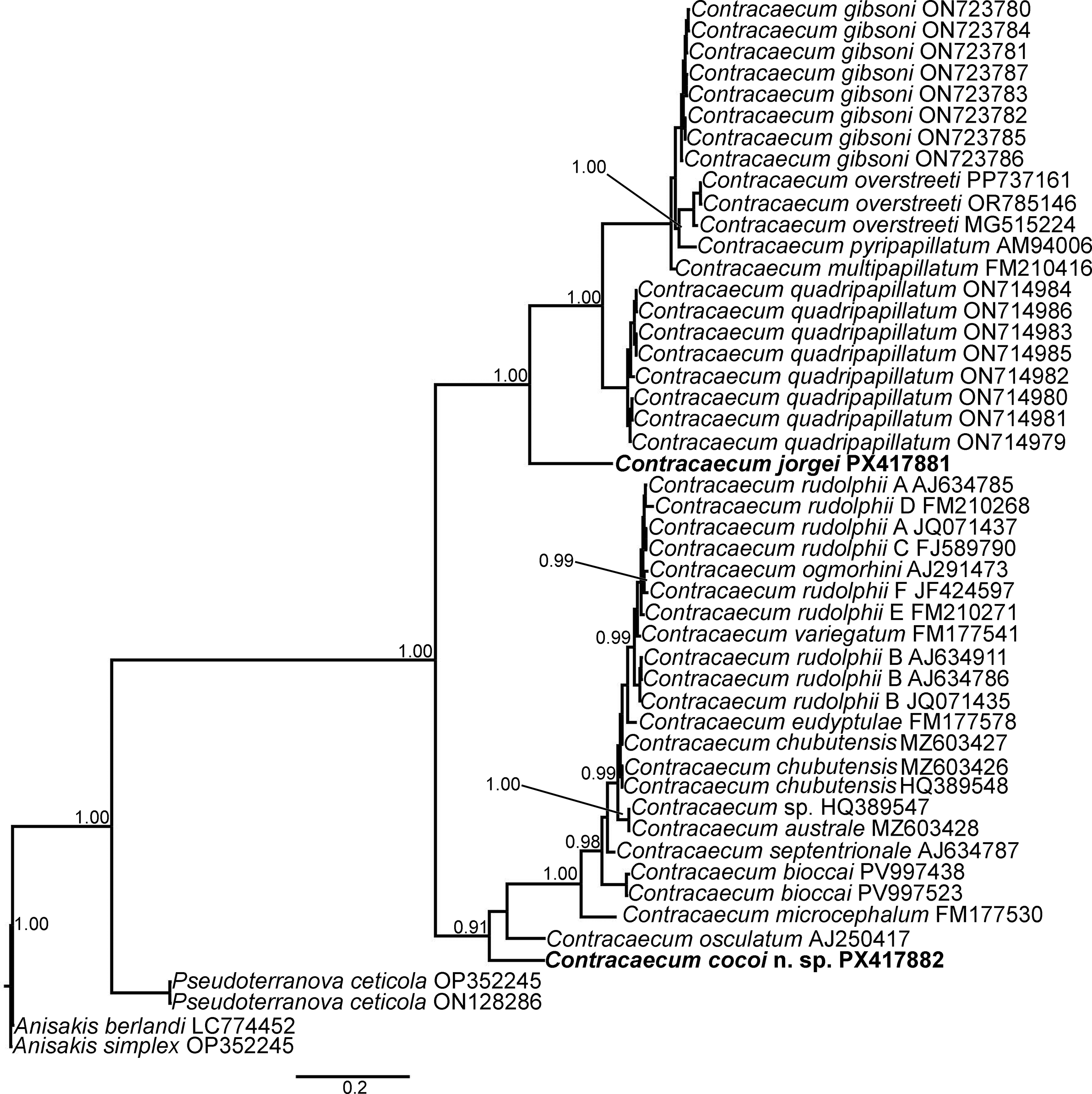
The constructed phylogenetic Bayesian Inference trees using these sequences revealed variable associations with those sequences archived in the GenBank. For instance, the ITS1 sequence (PX417880) from C. cocoi sp. nov. established as a single node positioning the species quite distant genetically from the node consisting of Contracaecum quadripapillatum Saad, Younis & Rabei, 2018, the sequence of the present studied specimens (PES) of C. jorgei (PX417879), Contracaecum overstreeti Mattiucci, Paoletti, Consuegra-Solorzano & Nascetti, Reference Mattiucci, Paoletti, Consuegra-Solorzano and Nascetti2010, Contracaecum pyripapillatum Shamsi, Gasser & Beveridge, Reference Shamsi, Gasser and Beveridge2008, C. multipapillatum, and Contracaecum gibsoni Mattiucci, Paoletti, Consuegra-Solorzano & Nascetti, Reference Mattiucci, Paoletti, Consuegra-Solorzano and Nascetti2010, well-supported on the BI trees (PP = 1.00) (Fig. 4). On the other hand, the ITS2 sequence (PX417882) displayed a different topology, clustering with the species C. osculatum, C. microcephalum, Contracaecum bioccai Mattiucci, Paoletti, Olivero-Verbel, Baldiris, Arroyo-Salgado, Garbin, Navone, & Nascetti, 2008, C. septentrionale, C. australe, Contracaecum chubutensis Garbin, Diaz, Cremonte & Navone, 2008, Contracaecum eudyptulae Johnston & Mawson, 1942, and the C. rudolphii complex (A, B, C, D, E, and F) forming a well-supported clade (PP = 0.91–1.00) (Fig. 5).
Phylogenetic trees constructed by Bayesian Inference methods based on Contracaecum spp. sequences of concatenated ITS1 markers under the substitution model HKY + G. Sequences are identified by taxon name, country, and GenBank accession number.

Phylogenetic trees constructed by Bayesian Inference methods based on Contracaecum spp. sequences of concatenated ITS2 markers under the substitution model HKY + G. Sequences are identified by taxon name, country, and GenBank accession number.

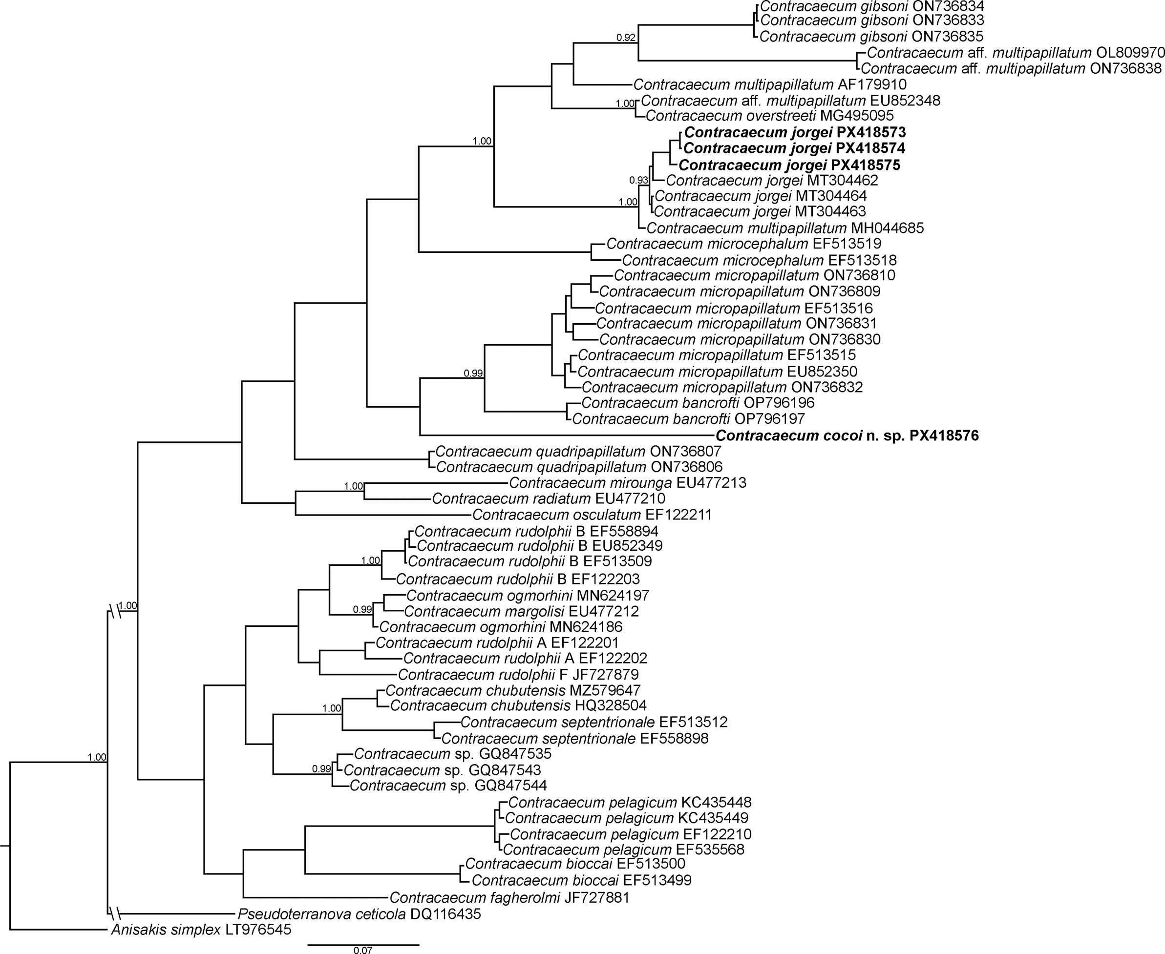
The cox2 mtDNA isolate (PX418576) exhibited C. cocoi sp. nov. as a well-supported, independent lineage (PP = 0.99) that forms the sister group to the clade composed of Contracaecum micropapillatum (Stossich, 1890) Baylis, 1920, and Contracaecum bancrofti Johnston & Mawson, 1941. The other three cox2 mtDNA analyzed sequences (PX418573-75) showed a great concordance with C. jorgei described by Sardella et al. (Reference Sardella, Mancini, Salinas, Simões and Luque2020) reflected in the strongly supported node on the BI tree (PP = 0.93) (Fig. 6). Finally, the SSU rRNA sequences showed Contracaecum cocoi sp. nov. (PX417878) and C. jorgei (PX417877) aligned with C. microcephalum in the constructed BI tree showing a weakly supported node (Fig. 7, supp. mat.). However, this analysis provides limited information due to the low number of SSU rRNA sequences available in Genbank for a more contrasting analysis.
Phylogenetic trees constructed by Bayesian Inference methods based on Contracaecum spp. sequences of a partial region of cox2 mtDNA gene under the substitution model TIM + F + I. Sequences are identified by taxon name, country, and GenBank accession number.

The shortest K2P distances calculated for the ITS1 gene matrix of Contracaecum cocoi sp. nov. displayed a distance of 0.22 with C. quadripapillatum, 0.21 with PES of C. jorgei, 0.24 with C. overstreeti, 0.27 with C. pyripapillatum, and 0.26 with both C. multipapillatum and C. gibsoni (Table 3, supp. mat.). For ITS2, the new species Contracaecum cocoi sp. nov. exhibited K2P distances of 0.21 with C. osculatum, 0.28 with C. microcephalum, 0.33 with C. bioccai, 0.32 with C. septentrionale, 0.33 with C. australe, 0.32 with C. chubutensis, 0.35 with C. eudyptulae, and 0.34–0.37 with the C. rudolphii complex (A, B, C, D, E, and F) (Table 4, supp. mat.). The cox2 mtDNA isolates of C. cocoi sp. nov. displayed a K2P distance of 0.12 with C. bancrofti, 0.13 with C. micropapillatum, and 0.16 with PES of C. jorgei (Table 5, supp. mat.). On the other hand, a K2P distance value of 0.01 was observed among the PES of C. jorgei sequences and those from the original description (Sardella et al. Reference Sardella, Mancini, Salinas, Simões and Luque2020) (Table 5, supp. mat.). Finally, K2P distance calculated for the SSU rRNA gene matrix displayed a distance of 0.15 among C. microcephalum compared to C. jorgei and C. cocoi sp. nov., respectively (Table 6, supp. mat.).
Discussion
Contracaecum cocoi sp. nov. is proposed as a new taxon clearly supported by both phylogenetic analysis and distinctive morphological features that distinguish it from its congeners. In relation to the ITS1-ITS2 Bayesian Inference trees (Figs. 4 and 5), the new species is separated from its congeners within a paraphyletic group, showing a great similarity regarding the papillae arrangement on the male tail (PAMT) with C. multipapillatum parasitizing Ardeidae and Pelecanidae (Navone et al. Reference Navone, Etchegoin and Cremonte2000; Shamsi et al. Reference Shamsi, Gasser and Beveridge2008), both C. gibsoni and C. overstreeti parasitizing Pelecanus crispus Bruch (Caffara et al. Reference Caffara, Tedesco, Davidovich, Locke, Gustinelli, King, Nuytten, Nuzzo and Fioravanti2023; Mattiucci et al. Reference Mattiucci, Paoletti, Consuegra-Solorzano and Nascetti2010), and C. pyripapillatum parasitizing P. conspicillatus (Shamsi et al. Reference Shamsi, Gasser and Beveridge2008). However, C. cocoi sp. nov. is characterized by possessing three adcloaclal pairs - one or two fewer adcloacal papillae pairs than the other species - and the spicules are twice the size. It is possible that these species, which possess an intermediate PAMT – morphotype B (Garbin et al. Reference Garbin, Servián, Fuentes, Morgenthaler, Millones, Fuchs, Diaz and Navone2024) – would tend to parasitize these two families of Pelecaniformes birds. Contracaecum quadripapillatum and C. jorgei (Saad et al. 2018; Sardella et al. Reference Sardella, Mancini, Salinas, Simões and Luque2020) would be included in a simple PAMT, morphotype A (Garbin et al. Reference Garbin, Servián, Fuentes, Morgenthaler, Millones, Fuchs, Diaz and Navone2024).
The phylogenetic analysis based on the mitochondrial cox2 mtDNA gene placed the new species as a well-supported, independent lineage sister to the clade formed by C. micropapillatum and C. bancrofti (Fig. 6). These two taxa are typically parasites of pelecanid hosts: C. micropapillatum parasitizes Pelecanus onocrotalus Linnaeus, 1758, and C. bancrofti parasitizes P. conspicillatus (Kinsella et al. Reference Kinsella, Spalding and Forrester2004; Caffara et al. Reference Caffara, Tedesco, Davidovich, Locke, Gustinelli, King, Nuytten, Nuzzo and Fioravanti2023). Furthermore, C. micropapillatum is known to parasitize not only pelecanids but also ardeids, such as Butorides striata Linnaeus, 1758 (Ezzat & Tadros, Reference Ezzat and Tadros1958) and Nycticorax nycticorax (Chow, Reference Chow1939). Whereas C. cocoi sp. nov. was found in the ardeid A. cocoi. This relationship suggests that C. cocoi sp. nov. diverged from a common ancestor with the C. micropapillatum–C. bancrofti lineage, following a host shift and subsequent specialization within Ardeidae in the South American region. The observed interspecific genetic divergence (0.12–0.13 K2P) between C. cocoi sp. nov. and its nearest relatives falls within the range reported for valid Contracaecum species, further confirming its distinctiveness. On the other hand, C. cocoi sp. nov. differs from both C. micropapillatum and C. bancrofti by possessing an intermediate PAMT (morphotype B), while the other two species show a simple PAMT, morphotype A (Garbin et al. Reference Garbin, Servián, Fuentes, Morgenthaler, Millones, Fuchs, Diaz and Navone2024). Notably, the cox2 mtDNA sequence MH044685 (unpublished data), identified as C. multipapillatum in GenBank, groups in the same branch with C. jorgei (Fig. 6). According to the information, this sequence belongs to a Contracaecum larvae specimen found parasitizing a catfish (Rhamdia sp.) in Costa Rica and Guatemala (not specified locality). It is not correct to determine species based on larval specimens since they do not develop secondary characters such as spicules and pre- and postcloacal papillae. Thus, our results suggest that this larva specimen identified as C. multipapillatum actually belongs to C. jorgei. The genetic proximity between C. jorgei and the other sequences identified as C. multipapillatum (see Fig. 6) can be explained from a parasite–host physiological point of view. Both Contracaecum species parasitize Ardeidae, specifically A. cocoi and A. alba. These birds are distributed sympatrically and have a similar diet in terms of prey items. A similar pattern could apply to C. microcephalum, which also parasitizes Ardeidae in Argentina (Table 1). The phylogenetic tree reflects these similarities, showing that Contracaecum species closely related to C. jorgei use Ardeidae birds as definitive hosts.
Regarding the SSU rRNA BI tree, both specimens studied here clustered in a monophyletic clade together with C. microcephallum. However, the PAMT of Contracaecum cocoi sp. nov. exhibits an intermediate pattern (morphotype B), whereas that of the other two species – Contracaecum jorgei and C. microcephallum – is simple (morphotype A) (Figs. 2d,e, 3b) (Garbin et al. Reference Garbin, Servián, Fuentes, Morgenthaler, Millones, Fuchs, Diaz and Navone2024). The SSU rRNA is a highly conserved gene and would not be useful for differentiating species, or at least not those that are closely related to each other, as seems to be the case here. Further SSU rRNA gene sequences are needed to determine the grouping of Contracaecum species linked to the PAMT. On the other hand, the possession of three adacloacal papillae pairs is a diagnostic feature separating this C. cocoi sp. nov. from the rest of the Contracaecum species (Figs. 2d,e, 3b).
A notable finding of this study is the sympatric and syntopic occurrence of C. cocoi sp. nov. and C. jorgei in A. cocoi. Both species were recovered from the same host individual and infection site, indicating that at least two distinct Contracaecum species can coexist within the digestive tract of a single ardeid host. Despite sharing host species and microhabitat, these taxa are genetically and morphologically well differentiated. Contracaecum jorgei exhibits a simple PAMT (morphotype A) and shorter spicules, while C. cocoi sp. nov. presents an intermediate PAMT (morphotype B) with a higher number of caudal papillae pairs (Figs. 2d,e, 3b). Their syntopic presence thus illustrates an exceptional case of niche overlap among congeners, possibly facilitated by their shared life cycle involving similar intermediate fish hosts and the generalist feeding behaviour of A. cocoi. From an evolutionary perspective, the sympatric distribution of C. cocoi sp. nov. and C. jorgei within the same host species and locality may reflect secondary contact between distinct lineages of Contracaecum specialized in Ardeidae. The occurrence of both species in A. cocoi broadens the known host range of C. jorgei – previously described from cormorants – and provides evidence of host-switching events within the genus. These findings highlight the complexity of host–parasite associations in aquatic bird systems, where overlapping trophic niches and migratory behaviour may promote parasite co-occurrence and diversification.
This work represents the second report of C. jorgei in Argentina, found parasitizing a new host, A. cocoi, in a new locality (Buenos Aires Province), located approximately 600 km south of the type locality (Córdoba Province). Additionally, considering that the specimen corresponding to sequence MH044685 originates from Costa Rica, in Central America, the geographical distribution of C. jorgei appears to be broader than previously estimated.
With regard to the host, A. cocoi has a broad distribution ranging from eastern Panama covering most of South America (Martínez-Vilalta et al. Reference Martínez-Vilalta, Motis, Kirwan, Arias, Boesman, Bosarreyes, Galluppi, Marqués Ferri, Olsen, Pyle, Rodríguez and Sheehan2025), and in Argentina it has been recorded hosting other anisakid species, such as C. microcephalum (Boero and Led Reference Boero and Led1971; Schuurmans-Stekhoven Reference Schuurmans-Stekhoven1951), as well as C. cocoi sp. nov. and C. jorgei in the present study. Infections by more than one anisakid species may be favoured by the combination of low host specificity in some Contracaecum species and the wide geographical distribution of their hosts, particularly in the case of migratory birds, as observed in this study.
Moreover, it is possible that in some records of anisakids parasitizing birds, species-level identifications may be inaccurate, as key characteristics such as the papillae arrangement on the male tail – now commonly used – were not examined. For this reason, integrative studies that include both morphological and molecular tools are essential to expand knowledge of the host and geographical distribution of parasite diversity, establish specific correspondences, and determine phylogenetic relationships within Contracaecum species. In this context, the discovery of Contracaecum cocoi sp. nov. in the Cocoi heron represents a significant contribution to the knowledge of anisakid diversity in South America. This new taxon, supported by both morphological and molecular evidence, adds to the growing recognition of cryptic speciation within Contracaecum and reveals the complexity of host–parasite relationships among aquatic birds. The occurrence of C. cocoi sp. nov. together with C. jorgei in sympatric and syntopic conditions suggests that ecological or reproductive isolating mechanisms may be acting to maintain distinct lineages in shared environments. Overall, the identification of C. cocoi sp. nov. highlights the underestimated diversity of anisakid nematodes parasitizing South American avifauna and underscores the importance of integrative taxonomic approaches for elucidating the evolutionary history and biogeographical patterns of this genus.
Supplementary material
The supplementary material for this article can be found at http://doi.org/10.1017/S0022149X25101120.
Acknowledgements
We are grateful to the institutions CONICET, UNLP, and FONCYT for the grants assigned to researchers.
Financial support
This work was supported by the Consejo Nacional de Investigaciones Científicas y Técnicas (CCT-CONICET-La Plata) (M.M.M., grant number 11220200101713CO; N.J.A., grant number 11220210100134CO); Fondo para la Investigación Científica y Tecnológica (FONCyT) (M.M.M., grant number PICT-01531; N.J.A., grant number PICT-00660), and Universidad Nacional de La Plata (UNLP) (J.I.D., grant number N996).
Competing interests
The author(s) declare none.
Ethical standard
The authors assert that all procedures contributing to this work comply with the ethical standards of the relevant national and institutional guides on the care and use of laboratory animals.