Implications
Ewes grazing poor-quality dry pastures during pregnancy are often deficient in certain trace elements and vitamins at lambing. This can lead to a decrease in lamb survival, which has implications for both production and welfare. Recent research has shown that supplementation of both vitamin E and selenium can enhance immune function across a range of species and potentially improve lamb survival, however these responses were not evident in the current study.
Introduction
Improving lamb survival is recognised as the highest priority for the improvement of reproductive efficiency of the Australian sheep flock (Young et al., Reference Young, Trompf and Thompson2014). The mortality of lambs from birth to weaning can exceed 50%, especially in multiple born Merino lambs (Behrendt et al., Reference Behrendt, van Burgel, Bailey, Barber, Curnow, Gordon, Hocking Edwards, Oldham and Thompson2011; Hinch and Brien, Reference Hinch and Brien2014; Paganoni et al., Reference Paganoni, Ferguson, Kearney and Thompson2014). Most lambs die within the first 72 h of life and the main causes of death include dystocia, the starvation–mismothering–exposure complex and disease (Brown et al., Reference Brown, Jones and Hinch2014; Refshauge et al., Reference Refshauge, Brien, Hinch and van de Ven2015). In Western Australia many ewes lamb during autumn (Curtis and Croker, Reference Curtis and Croker2005), when the supply of nutrients from dry pastures and crop stubbles seldom meets energy requirements for maintenance and foetal growth. This can compromise birth weight and survival (Kelly, Reference Kelly1992; Oldham et al., Reference Oldham, Thompson, Ferguson, Gordon, Kearney and Paganoni2011; Paganoni et al., Reference Paganoni, Ferguson, Kearney and Thompson2014). The adverse effects of poor pasture availability on the nutritional status of ewes can be offset by providing supplementary feeds. However, high rates of lamb mortality may still occur and this may be related to deficiencies in specific nutrients.
Several studies have reported that ewes lambing in autumn in Western Australia are at risk of vitamin E and selenium deficiency due to the increased requirements of these nutrients during gestation and their low concentrations in dry pasture, crop stubbles and grain supplements (Kumagai and White, Reference Kumagai and White1995; White and Rewell, Reference White and Rewell2007). Therefore, lambs born in autumn may be at risk of vitamin E and selenium deficiencies. Supplementation of vitamin E and selenium independently have been shown to improve lamb survival in a limited number of studies. However the mechanisms by which this is achieved has not been determined (Kott et al., Reference Kott, Ruttle and Southward1983; Thomas et al., Reference Thomas, Roader, Bohn, Kott and Evans1995). Vitamin E deficiencies have been linked with inadequate immune responses in animals, and in some cases supplementation above requirements have improved immune responses in humans (Finch and Turner, Reference Finch and Turner1996; Meydani and Beharka, Reference Meydani and Beharka1996; Anugu et al., Reference Anugu, Petersson-Wolfe, Combs and Petersson2013). The immune response can be optimised and enhanced by vitamin E because of its strong lipid-soluble antioxidant activity and its ability to remove free radicals and prevent lipid peroxidation, which are immunosuppressive and damage cells and tissues (Meydani and Beharka, Reference Meydani and Beharka1996). Supplementing with vitamin E in the diet can also improve cell mediated immunity which helps protect the body against viral, protazoal and fungal infections and humoral immunity which produces antibodies, including all five classes of immunoglobulin’s (Hatfield et al., Reference Hatfield, Daniels, Kott, Burgess and Evans2000). Immunoglobulins do not cross the placenta in ruminants therefore lambs are reliant on the passive transfer of immunoglobulins, predominantly immunoglobulin-G (IgG), from colostrum to provide them with passive immunity and thus protection against disease in early life (Gokce et al., Reference Gokce, Atakisi, Kirmizigul and Unver2014). Therefore, deficiencies in vitamin E and selenium can compromise immune responses (Meydani and Beharka, Reference Meydani and Beharka1996; Rooke et al., Reference Rooke, Robinson and Arthur2004) and impaired immune function can increase the risk of infections and death in the neonate (Finch and Turner, Reference Finch and Turner1996). Hence, we tested the hypotheses that (i) supplementation of autumn-lambing ewes with vitamin E plus selenium in late gestation will increase the concentration of vitamin E and selenium in plasma in the ewe and lamb and (ii) that the increased concentrations of vitamin E and selenium in plasma in the lambs will improve their innate and adaptive immune responses and thus survival.
Material and methods
This experiment was approved by Murdoch University’s Animal Ethics Committee and all procedures were performed in accordance with the guidelines of the Australian Code of Practice for the Use of Animals for Scientific purposes.
Animals and experimental design
Research was performed at the University of Western Australia’s Ridgefield Farm near Pingelly in Western Australia (S 32° 30′ 23″ to E 116° 59′ 31″). In total, 400 Merino ewes aged between 4 and 7 years old were sourced from the Maternal Efficiency Flock (Rosales Nieto et al., Reference Rosales Nieto, Ferguson, Macleay, Briegel, Martin and Thompson2013). Ewes were artificially inseminated laproscopically in November 2013 and ewes identified to be pregnant were allocated at day 111 of gestation based on ewe age, liveweight, condition score, pregnancy status (single or twin) and sire of lamb into three replicates of each of two treatment groups; control (n=60) or vitamin E plus selenium supplementation during late pregnancy (n=60). At the same time other pregnant ewes were supplemented with vitamin D or methionine and the effects on parameters similar to those reported in this paper have been reported by Lockwood et al. (Reference Lockwood, Currie, Hancock, Broomfield, Liu, Scanlan, Kearney and Thompson2016) and Liu et al. (Reference Liu, Masters, Ferguson and Thompson2016). Lambing occurred during late April/early May 2014 and lambs were weaned in August 2014.
Animal management and treatments
Ewes were managed to achieve a condition score of greater than 3 at artificial insemination. Controlled Internal Drug Releases (Eazi-BreedTM CIDR® Sheep and Goat Device; Zoetis, Australia) were inserted into the vagina of all ewes 14 days before insemination and were withdrawn 2 days before insemination to synchronise oestrus; 1 day before artificial insemination the ewes were weighed and body condition scored. Ewes were artificially inseminated with semen from four individual sires which was allocated to the ewes according to their age, liveweight and condition score before insemination.
After artificial insemination, the ewes were managed as a single flock until day 111 of pregnancy. Ewes were pregnancy scanned on day 55 of pregnancy to identify single- and twin-bearing ewes (Fowler and Wilkins, Reference Fowler and Wilkins1984). Ewes were then weighed and condition scored every 1 to 2 weeks and supplementary fed to achieve a condition score of 2.5 at lambing. Between day 55 of gestation and day 150 of gestation they were offered 630 g dry matter (DM)/day of supplement consisting of 328 g DM lupins (13.9 MJ metabolisable energy/kg, 310 g CP/kg DM, 9.1 mg vitamin E/kg, 0.038 mg selenium/kg), 195 g DM oats (12.8 MJ metabolisable energy/kg, 122 g CP/kg DM, 6.8mg vitamin E/kg, 0.012 mg selenium/kg) and 107 g DM oaten chaff (9.3 MJ metabolisable energy/kg, 76 g CP/kg DM, 4.3 mg vitamin E/kg, 0.018 mg selenium/kg). The ewes grazed dry annual pasture during mid and late pregnancy (day 90 to day 140 of gestation) (6.8 MJ metabolisable energy/kg, 9.8% CP, 4.3 mg vitamin E/kg, 0.019 mg selenium/kg) and green vegetative pastures from early lactation (day 2 post-lambing) (128.2 mg vitamin E/kg, 0.027 mg selenium/kg) to 14 weeks±10 days (weaning).
On day 111 of pregnancy, the ewes were administered a clostridial six in one booster vaccine (Glanvac® 6; Zoetis, Australia). Ewes were then allocated into six plots. Three of the plots had 54 to 56 ewes allocated. Within each of these plots three treatments, control, vitamin D or vitamin E plus selenium supplementation, were allocated to each ewe taking account of ewe age, sire of the lamb, pregnancy status, and liveweight and condition score at insemination (n=17 to 20 ewes/treatment per plot). The other three plots had 17 to 18 animals allocated and the methionine treatment was applied to the group of animals within each of these plots.
Ewes supplemented with vitamin E and selenium received an oral dose of 4 g all-rac-α-tocopherol acetate (Advanced Liquid E, 7 ml; Advanced Feeds Pty Ltd, Perth, Australia) and were subcutaneously injected with 60 mg barium selenate (Bayer Selovin™; 1 mg selenium per kg liveweight). On day 125 and 140 of pregnancy, a second and third oral dose of vitamin E (4 g/sheep) was also administrated. The control group received no intervention during pregnancy.
At day 141 of pregnancy the three plots of control, vitamin D or vitamin E plus selenium ewes, were reallocated according to treatment, pregnancy status and sire of lamb into two smaller groups per plot giving six groups of 18 to 19 ewes for lambing. Ewes remained in these six lambing plots until ~1-week post-lambing at which point all ewes (including the ewes from the methionine plots) and their lambs were joined together and run as a single flock and weighed every 1 to 4 weeks until 14 weeks of age±10 days (weaning). At 4 weeks of age (±10 days), the lambs were marked and vaccinated (Glanvac® 6, Eryvac®, Gudair® and Scabigard®; Zoetis, Australia).
Animal measurements and blood sampling
Lambing commenced on day 143 of pregnancy and ceased on day 153 of pregnancy. The ewes were intensively monitored during lambing between 0400 h and midnight. Immediately after parturition and before suckling the lambs were weighed, ear tagged and birth type (single v. twin) and gender was identified (n=153). The rectal temperature was also recorded using a digital thermometer (n=135). Two 5 ml blood samples were then collected from the lambs via jugular venipuncture into heparinised vacutainers and blood glucose concentrations were measured using a glucometer (Accu-Chek Go; Roche Diagnostics, NSW, Australia). Additional blood samples were collected from the same lambs at 1 week of age (±3 days; n=92), at 4 weeks of age (n=73), 2 weeks following their vaccination at marking (n=72) and at 14 weeks of age (n=67). Blood samples that were collected for analysis of nutrient status were placed on ice whereas those collected for analysis of immune function were kept at room temperature.
A 9ml blood sample was also collected from the same ewes via jugular venipuncture into heparinised vacutainer tubes on day 111 (pre-treatment; n=114), at lambing (n=45) and at 4 weeks of age (n=45) and 14 weeks of age (n=40) for further analysis of α-tocopherol and selenium concentrations. At lambing, a 30ml colostrum sample was collected pre-suckling (n=82) and placed on ice before being aliquoted and stored at −80°C for subsequent immunological analysis. Milk samples were collected at 4 weeks of age (n=40) from a sub-set of 40 control and 40 supplemented ewes. Ewe and lamb blood samples collected for nutrient analysis were centrifuged at 2278 g for 15 min and plasma was removed and stored at −20°C. The lamb blood samples used for analysis of immune function were centrifuged at 952 g for 10 min and plasma was removed and stored at −80°C.
Vitamin E concentration in plasma and colostrum and selenium concentration in blood
Plasma samples collected from ewes on day 111 of gestation and from ewes and lambs at lambing, 4 weeks of age and 14 weeks of age were analysed for α-tocopherol concentrations (the active form of vitamin E) using HPLC as described by McMurray and Blanchflower (Reference McMurray and Blanchflower1979). Colostrum samples collected at lambing and milk samples collected 4 weeks post-lambing were also analysed using HPLC. In brief, 1 ml of sample was deproteinised with 1 ml 1% pyrogallol in ethanol, 5 ml of hexane was added, vortexed and then 4 ml of the hexane layer was transferred into another tube and evaporated under nitrogen. The hexane extraction was repeated once. The dried extract was reconstituted in 0.5 ml methanol (0.1% butylated hydroxytoluene), and the reconstitute was applied to an Agilent HPLC system (1100) equipped with a Zorbax SB-C18 column (3 mm×150 mm, 3.5 µm) (Agilent Technologies Inc., Victoria, Australia) with a methanol mobile phase. α-Tocopherol was quantified using fluorescence detection (excitation 296 nm and emission 330 nm). Selenium concentration in whole blood was estimated from the relationship derived between selenium concentration and glutathione peroxidase (GSHpx) activity in erythrocytes. Blood samples from ewes (n=82) and lambs (n=82) were analysed for selenium concentrations using the method of Gelman (Reference Gelman2010), and GSHpx activity was determined using the Ransel 505 kit (RANDOX Laboratories, Antrim, UK). Linear regression was performed to derive the relationships between selenium concentration and GSHpx activity for ewes and lambs. To convert selenium concentration in erythrocytes to selenium concentration in whole blood a factor of 1.3 was used as the concentration of selenium is known to be 30% higher in erythrocytes compared to whole blood (Barceloux, Reference Barceloux1999). Thus two new regression equations were derived for ewes (equation 1) and lambs (equation 2);
 $$\eqalignno{{\rm Se}\,\left( {{\rm mg}/{\rm l}\,{\rm blood}} \right)\,{\equals}\, & 0.0132\left( {\pm0.00553} \right)\cr & {\plus}0.00045\left( {\pm0.000021} \right)\,{\rm GSHpx}\,\left( {{\rm U}/{\rm g}\,{\rm Hb}} \right)$$
 $$\eqalignno{{\rm Se}\,\left( {{\rm mg}/{\rm l}\,{\rm blood}} \right)\,{\equals}\,&0.0179\left( {\pm0.00442 } \right)\cr &{\plus}0.00030\left( {\pm 0.000021 } \right) \ {\rm GSHpx}\,\left( {{\rm U}/{\rm g}\,{\rm Hb}} \right)$$
Vitamin E and selenium concentration in feedstuffs and soils
Pasture samples from paddocks used for grazing the ewes were collected during late pregnancy and lactation. Each sample was collected from 8 to 10 sites per paddock and pooled. All pasture samples were freeze-dried and then stored at −20°C. Samples of oaten chaff, oats and lupin seeds were collected five times during late pregnancy, pooled and dried at 65°C. All feedstuff samples were ground to pass through a 1mm sieve and used for analyses of vitamin E and selenium concentrations and DM content.
The α-tocopherol concentrations in feed samples were also analysed using a HPLC procedure as described by McMurray et al. (Reference McMurray, Blanchflower and Rice1980). In brief, 1 g of feed was homogenised in 10 ml 6% pryrogallol by ultraturrex. Then 1 ml 60% KOH (w/v) in water was added, the tube was sealed, and tubes heated at 70°C for 30 min. The procedures of hexane extraction, reconstitutions and instrumental analysis were the same as described for the analysis of plasma samples. Selenium concentrations in feedstuffs and soils were determined using Agilent VGA-76 hydride generator; 1 g of feed and soil samples were wet digested using nitric and perchloric acids under a temperature programme which ensures residual nitric acid is removed from the digest. Selenium was then converted to the Se(IV) state using hydrochloric acid in preparation for measurement by hydride generation and atomic absorption (Varian VGA-77 and Varian AA280FS, respectively, Agilent Technologies Inc.).
Analysis of immune function
Whole blood differential cells counts, monocyte and polymorphnuclear leucocyte (PMNL) phagocytic capacities, and plasma total IgG and anti-tetanus toxoid antibody concentrations were measured as described in detail by Lockwood et al. (Reference Lockwood, Currie, Hancock, Broomfield, Liu, Scanlan, Kearney and Thompson2016). In brief, whole blood differential cell counts were analysed using Advia® 120 automated Hematology Analyzer (sequence 284) in a high output pathology laboratory (Clinical Pathology, Murdoch University Veterinary Hospital). The abilities of monocytes and polymorphnuclear leucocytes to phagocytose bacterial bioparticles were assessed using flow cytometry. Plasma total IgG concentrations of lambs were assessed using a single radial immunodiffusion assay (IDRing® Sheep&Goat IgG assay; IDBiotech, Issoire, France). Anti-tetanus toxoid antibodies in lamb plasma samples were assessed by an ELISA described by Adams et al. (Reference Adams, Anderson, Lynch and Munro1997), using optimised dilutions for use with lamb plasma samples.
Statistical analysis
All statistical analyses were performed using GenStat (VSNI International). For all analyses, interaction terms were only included if they were statistically significant (P⩽0.05). Analysis also included, where appropriate, data from an additional 148 lambs which were concurrently studied to examine the effects of supplementation of the ewe with vitamin D (Lockwood et al., Reference Lockwood, Currie, Hancock, Broomfield, Liu, Scanlan, Kearney and Thompson2016) or methionine (Liu et al., Reference Liu, Masters, Ferguson and Thompson2016) during late pregnancy on similar parameters to those that are reported in this paper. For these analyses treatment was nested within a term called group which related to the group of animals that had the control, vitamin D or E plus selenium treatments applied (group 1) and the group of animals that had the methionine treatment applied (group 2). For simplicity the term treatment will be used in the following description of the statistical methods. Where significant treatment effects were observed, the results presented include only data from vitamin E and control lambs. Where no treatment effects were observed, results presented include ewes and lambs from all treatment groups.
Liveweights of ewes during pregnancy were corrected for weight of the conceptus using the formula from the Grazfeed model;
 $$\eqalignno{ {\rm Corrected}\,{\rm live}\,{\rm weight}\,{\equals}\, & {\rm total}\,{\rm birth}\,{\rm weight}{\times}{\rm 1}.{\rm 43} \cr &{\times} {{\rm EXP}[ {{\rm 3}.{\rm 38}{\times}( {{\rm 1}{\minus}{\rm EXP}( {0.{\rm 91}} }}} \cr & {\times} ( {{\rm 1}{\minus}{\rm days \ pregnant}\,/\,{\rm 15}0})))] \cr & \ \ \,({\rm Freer}\ {\rm \hslant 20% {et \ al}}., \, 1997), $$
where day 0 of pregnancy was the day of artificial insemination. Conceptus-free ewe liveweights and body condition scores were analysed using the method of restricted maximum likelihood (REML) with a heterogenous power model to assess repeated measurements over time as described by Lockwood et al. (Reference Lockwood, Currie, Hancock, Broomfield, Liu, Scanlan, Kearney and Thompson2016). Restricted maximum likelihood was also used to analyse the liveweights, rectal temperatures and blood glucose concentrations of the lambs with treatment, sex, birth type (single or multiple born), birth weight, sire of lamb and interactions thereof, where appropriate, fitted as fixed effects and pre-lambing and lambing plots (nested with pre-lambing plot), stud from which the ewe was sourced, ewe identification, sire of ewe and dam birth year fitted as random effects.
Ewe and lamb plasma α-tocopherol and estimated blood selenium concentrations were assessed using REML. For analyses of data from ewes, treatment was fitted as a fixed effect, and pre-lambing plot, stud from which the ewe was sourced, sire of ewe and dam birth year were fitted as random effects. The concentration of α-tocopherol or selenium at day 111 of pregnancy, before treatments, was also fitted as a covariate in the analysis of ewe plasma α-tocopherol and blood selenium concentrations at lambing, 4 weeks of age and 14 weeks of age. For analyses of data from lambs, treatment, sire of the lamb, birth type and sex, and interactions thereof, where appropriate, were fitted as fixed effects, and pre-lambing plot, lambing plot (nested within pre-lambing plot), stud from which the ewe was sourced, sire of ewe and dam birth year fitted as random effects. The relationships between α-tocopherol and selenium concentrations were analysed by assessing the effect of the concentration in the ewe on the concentration of the lamb using REML. Ewe selenium concentration and selenium concentration – squared at lambing, sex, birth type, sire of lamb and interactions thereof, where appropriate, were fitted as fixed effects, lambing paddocks, ewe source, sire of ewe, and date of birth of the lamb were fitted as random effects. For these analyses fixed terms were removed if not significant (P>0.05). Where necessary data was log transformed and is presented in the back transformed state.
Lamb immune function parameters of monocyte and neutrophil cell counts, and monocyte and PMNL phagocytic capacities, were analysed using REML. Plasma IgG concentrations and anti-tetanus toxoid antibody absorbances were modelled over time using REML with a power model in which the correlation between observations from the same animal decays as the time delay between observations increases. For these analyses, treatment, sex, birth type, sire of lamb, gestation length and interactions thereof, where appropriate, were fitted as fixed effects. Pre-lambing and lambing plots (nested with pre-lambing plot), stud from which the ewe was sourced, ewe identification, sire of ewe, dam birth year and laboratory batch information, where appropriate, were fitted as random effects.
Lamb survival was analysed by fitting general linear mixed models. The approach used a logit-transformation and binomial distribution. Using additive models, logits were predicted as a function of the variables fitted as fixed and random effects. Treatment, birth weight, birth weight-squared, birth type, sex, sire of the lamb and varieties of interest were fitted as fixed effects and lamb date of birth, pre-lambing plot, lambing plot (nested within pre-lambing plot), dam source, sire of the dam and dam birth year were fitted as random effects. For birth type of the lamb, multiple born refers to both twin- and triple-born lambs.
Results
Ewe liveweight and condition score
There were no significant differences in the liveweight of control and vitamin E plus selenium ewes before treatments (63.2 v. 62.8 kg), before lambing (day 141 of gestation; 64.7 v. 65.1 kg), at 4 weeks of age (58.0 v. 57.5 kg) or at 14 weeks of age (56.0 v. 55.6 kg). Similarly, the condition score of control and vitamin E plus selenium ewes did not differ significantly before treatments (2.4 v. 2.4), before lambing (2.6 v. 2.6), at 4 weeks of age (2.6 v. 2.7) or at 14 weeks of age (2.8 v. 2.8).
Gestation length and lamb characteristics
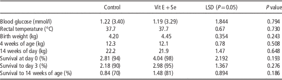
There was no significant difference in the gestation length of control and vitamin E plus selenium ewes (148.5 v. 149.2 days) or of single- or multiple-bearing ewes (150.1 v. 149.0 days). There was no significant effect of maternal supplementation on lamb weight, blood glucose or rectal temperature at birth or liveweight of lambs at 4 weeks of age or 14 weeks of age (Table 1). Survival to 14 weeks of age was also not significantly different, despite there being an 11% difference in survival between control and vitamin E plus selenium lambs. Single born lambs were heavier than twin born lambs at birth (5.0 v. 4.0 kg; P<0.001), at 4 weeks of age (13.4 v. 10.5 kg; P<0.001) and 14 weeks of age (23.9 v. 19.5 kg; P<0.001). Male lambs were heavier than female lambs at birth (4.6 v. 4.3 kg; P<0.01) but not at 4 weeks of age.
Average blood glucose concentration and rectal temperature of the lamb at birth, lamb weights from birth to 14 weeks of age and survival to weaning from ewes in the control group or ewes that were supplemented with vitamin E and selenium (Vit E+Se)

Values in parentheses are back transformed means with the LSD referring to the transformed data.
Vitamin E status in the ewe and lamb
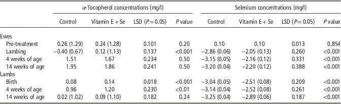
There was no significant difference in the concentration of α-tocopherol between control and vitamin E plus selenium ewes before treatment (Table 2). During late pregnancy the concentration of α-tocopherol was maintained for ewes supplemented with vitamin E plus selenium but decreased by ~50% for control ewes. At lambing the concentration of α-tocopherol was higher for vitamin E plus selenium ewes compared with controls (P<0.001). At 4 weeks of age the concentration of α-tocopherol was still higher in the supplemented ewes (P<0.001) than the control ewes, however there was no difference at 14 weeks of age. The concentration of α-tocopherol in colostrum was higher in the vitamin E plus selenium ewes compared with the controls (10.6 v. 4.1 mg/l; P<0.001), however there was no significant difference in the concentration of α-tocopherol in milk at 4 weeks of age (1.1 v. 1.0 mg/l). There were also no significant differences in the concentration of α-tocopherol in plasma, colostrum or milk when single- and twin-bearing ewes were compared.
Average concentration of α-tocopherol and selenium (mg/l) in plasma of ewes at day 111 (pre-treatment) of gestation and of ewes and lambs from lambing until 14 weeks of age. The ewes were supplemented with vitamin E plus selenium on days 111, and then again with vitamin E on day 124 and 140 of gestation

Values in parentheses are back transformed means with the LSD referring to the transformed data.
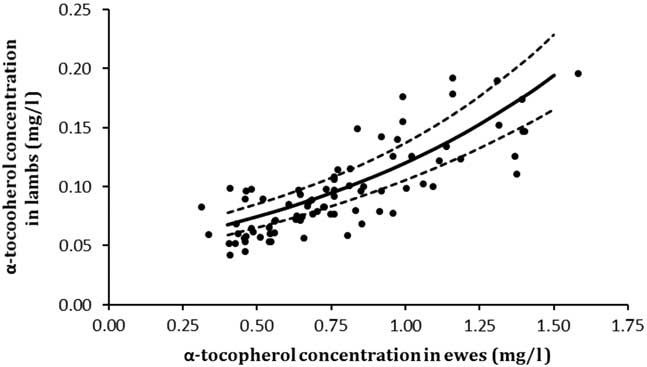
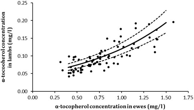
Lambs born to ewes supplemented with vitamin E plus selenium had higher α-tocopherol concentrations at birth compared with the controls (Table 2; P<0.001). The concentration of α-tocopherol increased 10-fold during the 4-week period to marking and remained higher in vitamin E plus selenium lambs compared with the control lambs (P<0.01), however this difference was not significant at 14 weeks of age. There was no effect of sex on the concentration α-tocopherol between birth and 14 weeks of age, but the concentration α-tocopherol was higher for twin born than single born lambs at 4 weeks of age (1.27 v. 1.10; P=0.05). There was a positive correlation between the concentration of α-tocopherol in plasma from ewes and their lambs at birth (Figure 1).
The relationship between the concentration of α-tocopherol in plasma from ewes and their lambs immediately at birth and before suckling using the linear function; lamb α-tocopherol (natural log transformed)=−3.08 (±0.084)+0.964 (±0.0700) ewe α-tocopherol. The fitted line and 95% confidence intervals (dashed line) are presented in the back transformed state. The dots represent the raw data for the plasma α-tocopherol of an individual ewe and her lamb.
Estimated selenium concentration in whole blood in the ewe and lamb
There was no significant difference in estimated selenium concentration in blood of control or vitamin E plus selenium ewes before treatment (Table 2). During late pregnancy the concentration of selenium in blood increased slightly for ewes supplemented with vitamin E and selenium but decreased by ~40% for control ewes (Table 2). At lambing, 4 weeks of age and 14 weeks of age the concentration of selenium in blood was significantly higher for ewes supplemented with vitamin E plus selenium compared with the controls. There were no significant differences in the concentration of selenium in blood when single and twin bearing ewes were compared.
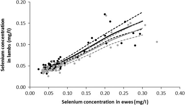
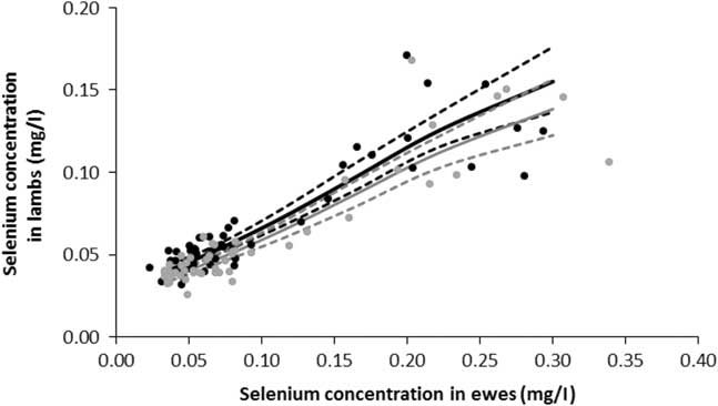
Maternal supplementation with vitamin E plus selenium in late gestation increased the concentration of selenium in the blood of lambs at lambing, 4 weeks of age and 14 weeks of age by 50% to 100% compared with lambs from control ewes (Table 2; P<0.001). The blood selenium concentration was marginally higher in female lambs than male lambs at birth (0.06 v. 0.05 mg/l; P<0.05), but there was no effect of birth type or sex of lamb on the concentration of selenium in blood between birth and 14 weeks of age. There was a positive correlation between selenium concentrations in the ewes and their lambs at birth, and this relationship differed for female and male lambs (P<0.001; Figure 2). A 1-unit change in the selenium concentration in the ewes resulted in a 0.41 and 0.46 unit change in the selenium concentration in their female and male lambs, respectively.
The relationship between the concentration of selenium (Se) in blood from ewes and their female (black lines, Lamb Se=−3.52 (±0.062)+9.31 (±1.028) ewe Se−12.6 (±3.18) ewe Se squared) and male (grey lines, Lamb Se=−3.64 (±0.059)+9.31 (±1.028) ewe Se−12.6 (±3.18) ewe Se squared) lambs at birth and before suckling. The fitted lines and 95% confidence intervals (dashed lines) are presented in the back transformed state. The dots represent the raw data for the plasma selenium of an individual ewe and her female (black) and male (grey) lambs, respectively.
Innate immunity
The total number of white blood cells in whole blood of control ewes did not differ from vitamin E plus selenium supplemented ewes at birth (Table 3). There was also no significant difference in the relative proportion, total number or phagocytic activity of the monocytes or neutrophils when the control and vitamin E plus selenium lambs were compared at birth or 4 weeks of age. As a result, the total number of phagocytic monocytes in whole blood of control lambs did not differ to that of the vitamin E plus selenium lambs at birth or 4 weeks of age, and the total number of phagocytic neutrophils did not differ at birth or 4 weeks of age (Table 3). Across all lambs, on no occasion was there a significant relationship between the actual concentrations of α-tocopherol or selenium in the lambs at birth or 4 weeks of age and their innate immune responses.
The total number (×109/l) of white blood cells, lymphocytes, and phagocytic neutrophils and monocytes in lambs at birth and 4 weeks of age. The ewes were supplemented with vitamin E plus selenium on days 111, and then again with vitamin E on day 124 and 140 of gestation

Values in parentheses are back transformed means with the LSD referring to the transformed data.
a For the transformed variates there was a value added to the data before transformation. For Monocytes at day 0, 1.01 was added whereas for all others 1 was added.
Adaptive immunity
There was no significant difference in the total number of lymphocytes when control lambs and vitamin E plus selenium lambs were compared at birth or 4 weeks of age (Table 3). There was no significant difference between the control and vitamin E plus selenium lambs in the concentration of IgG in colostrum (80.4 v. 72.2 mg/ml). The total concentrations of IgG in the plasma of lambs born to control or supplemented ewes did not differ significantly at 1 week (15.7 v. 16.9 mg/ml), 4 weeks (8.0 v. 8.5 mg/ml), 6 weeks (9.3 v. 9.3 mg/ml) or 14 weeks (15.6 v. 14.7 mg/ml) of age. There was a difference in plasma IgG concentrations between consecutive time points of analyses (P<0.001), but there was no significant interaction with treatment.
There were no significant differences in the absorbances of plasma samples for anti-tetanus toxoid antibodies when control and vitamin E plus selenium lambs were compared at 1 week (0.58 v. 0.58 nm), 4 weeks (0.33 v. 0.33 nm), 6 weeks (0.56 v. 0.49 nm) or 14 weeks (1.40 v. 1.57 nm) of age. Absorbance’s of plasma samples differed between consecutive time points of analysis (P<0.001), however there was no significant interaction with treatment. On no occasion was there a significant relationship between the concentrations of α-tocopherol or selenium in the lambs and their adaptive immune responses.
Discussion
Maternal supplementation with vitamin E plus selenium during late gestation increased the concentrations of α-tocopherol in plasma and selenium in whole blood of the ewe and her lambs at lambing, supporting our first hypothesis. However, supplementation of ewes with vitamin E plus selenium during late gestation did not influence the production of maternal antibodies in the colostrum or the innate phagocytic or adaptive antibody immune responses of the lambs between birth and 14 weeks of age. Therefore, our results suggest that immune function of the lamb was not enhanced by supplementing ewes with vitamin E plus selenium during late gestation. The survival of lambs to 14 weeks of age from ewes supplemented with vitamin E and selenium was 11% higher than the survival of control lambs, but this effect was not statistically significant and there was no evidence that any changes in survival resulted directly from the supplements boosting the lamb’s innate or adaptive immune responses. Our second hypothesis was therefore rejected.
The concentrations of α-tocopherol and selenium decreased by 40% to 50% in ewes during late pregnancy when no vitamin E and selenium supplements were provided, which is consistent with Hatfield et al. (Reference Hatfield, Daniels, Kott, Burgess and Evans2000), Rock et al. (Reference Rock, Kincaid and Carstens2001) and Hall et al. (Reference Hall, Voracheck, Stewart, Gorman, Dosher, Pirelli and Bobe2013). This decline in α-tocopherol and selenium concentrations during late pregnancy was due to low intakes of vitamin E and selenium in the diet, and increased demand for these nutrients from the rapidly growing foetus and the production of colostrum, which has a high concentration of α-tocopherol and selenium (Njeru et al., Reference Njeru, McDowell, Wilkinson, Linda and Williams1994; Rock et al., Reference Rock, Kincaid and Carstens2001; Stewart et al., Reference Stewart, Bobe, Voracheck, Stang, Pirelli, Mosher and Hall2013). Based on the changes in ewe liveweight and the amount of supplement fed, the estimated total intakes of vitamin E and selenium by ewes during late pregnancy were ~8.50 and 0.033 mg/day, respectively. These estimates are significantly below the recommended requirements of vitamin E and selenium for pregnant sheep (Freer et al., Reference Freer, Moore and Donnelly2007). Consequently, at lambing more than 60% of the ewes that were not supplemented had plasma α-tocopherol concentrations lower than 0.7 mg/l, which is considered the critical threshold for deficiency (White and Rewell, Reference White and Rewell2007); however, 50% of ewes had adequate blood selenium concentrations (>0.06 mg/l) with the remaining 50% marginal (>0.02 and <0.06 mg/l; Paynter, Reference Paynter1996). Thus our results demonstrate that low vitamin E status may be a more widespread problem for ewes lambing onto dry pasture before the break of season in late autumn in Western Australia.
Maternal supplementation of ewes with vitamin E plus selenium during late pregnancy increased the concentrations of both α-tocopherol in plasma and selenium in whole blood by about 70% at lambing compared with ewes that were not supplemented. However, 9% of supplemented ewes were still deficient in vitamin E and 18% were still marginal in selenium at lambing. The maximum concentrations of α-tocopherol in plasma of supplemented ewes were significantly lower than that achieved in numerous other studies (Kumagai and White, Reference Kumagai and White1995; Daniels et al., Reference Daniels, Hatfield, Burgess, Kott and Bowman2000; Rooke et al., Reference Rooke, Robinson and Arthur2004; Capper et al., Reference Capper, Wilkinson, Kasapidou, Pattinson, Mackenzie and Sinclair2005; Anugu et al., Reference Anugu, Petersson-Wolfe, Combs and Petersson2013), despite supplementing with higher levels of vitamin E than some studies and using dl-α-tocopherol acetate which is considered to have the highest bioavailability (Hatfield et al., Reference Hatfield, Daniels, Kott, Burgess and Evans2000). The lower concentrations of α-tocopherol in plasma of supplemented ewes in the current study may reflect that ewes actually gained condition score and hence fat during late pregnancy enabling greater storage of vitamin E or alternatively enabling a higher requirement. This is consistent with findings that animals are less susceptible to nutritional myopathy caused by vitamin E deficiency when nutrition is poorer and more susceptible when they are well fed with a diet containing low concentrations of vitamin E (Allen et al., Reference Allen, Steele, Master and D’Antuono1986). The concentrations of selenium in supplemented ewes were also lower than expected in comparison with other studies (Rock et al., Reference Rock, Kincaid and Carstens2001; Stewart et al., Reference Stewart, Bobe, Voracheck, Stang, Pirelli, Mosher and Hall2013). This may be attributed to the lower doses of selenium administered in the current study. Furthermore, ewes were supplemented with an inorganic form of selenium which is less effective at increasing the concentration of selenium in the blood compared with organic selenium (Stewart et al., Reference Stewart, Bobe, Voracheck, Stang, Pirelli, Mosher and Hall2013). Hence, the limited response of ewes to supplementation with vitamin E and selenium in this study may be associated with the ewes being in positive energy balance during late pregnancy, the lower concentration of vitamin E and selenium in the base diet and the dose rate, form and time of supplementation.
Before suckling the concentration of α-tocopherol in plasma and selenium in whole blood was higher in lambs born to ewes supplemented with vitamin E plus selenium compared with control lambs, indicating that both α-tocopherol and selenium were able to cross the placenta. However the concentrations of α-tocopherol and selenium in the lambs was generally lower than other studies (Kumagai and White, Reference Kumagai and White1995; Rock et al., Reference Rock, Kincaid and Carstens2001; Stewart et al., Reference Stewart, Bobe, Voracheck, Pirelli, Mosher, Nichols, Van Saun, Forsberg and Hall2012, Reference Stewart, Bobe, Voracheck, Stang, Pirelli, Mosher and Hall2013) and suggest that all lambs were still deficient in vitamin E and 40% of lambs were marginal in selenium even when ewes were supplemented. The efficiency of maternal transfer varied from 0.08 to 0.15 for α-tocopherol depending on the maternal concentration and from 0.40 to 0.46 for selenium, depending on sex of lamb. The upper level of placental transfer of α-tocopherol closely aligns with Kumagai and White (Reference Kumagai and White1995), who reported plasma concentrations in perinatal foetuses were ~15% of those in their mothers. By contrast the efficiency of placental transfer of selenium was lower in our study due to the combined effects of lower selenium concentrations in the ewes and the use of inorganic selenium. The transfer efficiency of selenium is known to be positively correlated with the selenium concentration in the ewe (Stewart et al., Reference Stewart, Bobe, Voracheck, Pirelli, Mosher, Nichols, Van Saun, Forsberg and Hall2012) and inorganic selenium can result in up to 50% less selenium being transferred across the placenta (Rock et al., Reference Rock, Kincaid and Carstens2001; Stewart et al., Reference Stewart, Bobe, Voracheck, Pirelli, Mosher, Nichols, Van Saun, Forsberg and Hall2012). The lower concentrations of α-tocopherol and selenium in the lambs in the current study are therefore indicative of the lower concentrations achieved in the supplemented ewes for reasons outlined previously plus lower efficiency of placental transfer compared with other studies, especially for selenium.
Maternal supplementation with vitamin E plus selenium in late gestation did not influence the total number or ability of lamb monocytes or PMNL to engulf S. aureus bioparticles between birth or 14 weeks of age. Furthermore, maternal supplementation had no effect on the passive transfer of immunity to the lamb, the total number of lymphocytes or the concentrations of IgG or anti-tetanus toxoid antibodies in the plasma of lambs. It is clear that the innate and adaptive immune responses of the lambs in this study were not influenced by maternal supplementation during late pregnancy, despite evidence across species that improving vitamin E and selenium status can enhance immune function (Finch and Turner, Reference Finch and Turner1996; Meydani and Beharka, Reference Meydani and Beharka1996; Rooke et al., Reference Rooke, Robinson and Arthur2004). Liu et al. (Reference Liu, Lei, Hancock, Scanlan, Broomfield, Kearney, Currie and Thompson2014) reviewed the effects of vitamin E and selenium supplementation on the responses of the immune system and concluded that dietary selenium was more critical than vitamin E, however it is not clear from the literature what concentration of α-tocopherol in the plasma and selenium in the blood are required to elicit the immune responses. In a study investigating the effect of dietary vitamin E in rats, the requirement for optimal lymphocyte proliferation was 3 to 13 times that considered to avoid a deficiency (Meydani and Beharka, Reference Meydani and Beharka1996). As there was no direct relationship between the actual concentrations of α-tocopherol or selenium in the lambs and their innate or adaptive immune response, it appears that the concentration of α-tocopherol and selenium of the lambs and or their level of exposure to environmental or pathogenic stress may have been too low to elicit measurable changes in immune function.
Maternal supplementation had no significant effect on the overall metabolic status of lambs at birth or on the growth of lambs to 14 weeks of age. Lamb survival to 14 weeks of age was 11% higher in the vitamin E plus selenium group compared with the controls, but this was not significant. Intensive sampling limited the number of animals that could be measured in the experiment plus survival outcomes were also probably influenced by the higher levels of intervention at lambing. Literature concerning the effects of vitamin E and selenium supplementation on lambs survival is variable, but using larger numbers of ewes, Kott et al. (Reference Kott, Ruttle and Southward1983) and Thomas et al. (Reference Thomas, Roader, Bohn, Kott and Evans1995) reported increases in survival of 8% to 20%. The potential for vitamin E and selenium supplementation to increase the survival of lambs born in autumn following long periods of dry feed therefore requires further validation at a paddock scale across a range of environmental conditions.
Acknowledgements
The authors acknowledge Meat and Livestock Australia for providing funding for this project, Australian Wool Education Trust for the undergraduate project scholarship and Moojepin Multi-Purpose Merino stud and Merinotech (WA) Ltd for supplying the semen used to artificially inseminate the ewes. Murdoch University Clinical Pathology laboratory staff is also thanked for their assistance with the blood sample analysis.