Left ventricular dysfunction can occur following permanent pacing in children with isolated congenital complete atrioventricular block, Reference Karpawich, Rabah and Haas1–Reference Silvetti, Drago and Ravà4 especially from right ventricular apex and right ventricular free wall/outflow tract. Reference Gebauer, Tomek and Kubus5,Reference Janoušek, van Geldorp and Krupičková6 Alternative pacing sites have been proposed to prevent or reduce it, through different approaches: left ventricular epicardial Reference Janoušek, van Geldorp and Krupičková6–Reference Silvetti, Muzi and Unolt10 or selective right ventricular endocardial pacing. Reference Karpavich, Singh and Zelin11–Reference Silvetti, Pazzano and Battipaglia13 This study aimed to compare the functional outcome (left ventricular systolic function and synchrony) in paediatric patients with congenital complete atrioventricular block and left ventricular apical epicardial or right ventricular endocardial pacing from alternative sites.
Methods
This single-centre, retrospective study was conducted on paediatric patients without CHDs requiring permanent pacing for congenital complete atrioventricular block. Inclusion criteria were congenital complete atrioventricular block, absence of CHD, selective right ventricular endocardial septal pacing guided by 3D electroanatomic mapping system or left ventricular apex epicardial pacing, and substantial anticipated ventricular pacing requirements.
Exclusion criteria were other arrhythmias (sinus node dysfunction and sporadic/intermittent atrioventricular block), presence of CHD, traditional pacing sites or biventricular pacing, and non-substantial anticipated ventricular pacing requirements.
Patients underwent pacemaker implantation between 2010 and 2020 at the Cardiac Arrhythmias Unit of Bambino Gesù Children’s Hospital. According to the policy of the centre, neonates and infants underwent left ventricular epicardial pacing; children (>15 kg) underwent selective right ventricular endocardial septal pacing. Autoantibodies (anti-SSa and anti-SSb) were searched in newborns and in some children with congenital complete atrioventricular block.
The pacing system implanted was the first one in all patients, and some of them were previously included in other studies. Reference Silvetti, Muzi and Unolt10,Reference Silvetti, Pazzano and Battipaglia13
Demographics data, procedure data, electrocardiographic and echocardiographic findings, complications, and clinical status at follow-up were recorded.
This study complies with the Declaration of Helsinki and was approved by the local Ethics Committee. Informed consent was obtained from the guardians of all patients.
Implant procedure, pacemakers, and leads
All patients underwent electrocardiogram (ECG) and echocardiogram evaluation before implantation. Indications for pacing were in accordance with the guidelines of the European Society of Cardiology. Reference Brignole, Auricchio and Baron-Esquivias14 The surgical technique and implant procedures have been already reported in detail in previous studies Reference Silvetti, Muzi and Unolt10,Reference Silvetti, Pazzano and Battipaglia13 and are described below briefly.
Epicardial pacing system
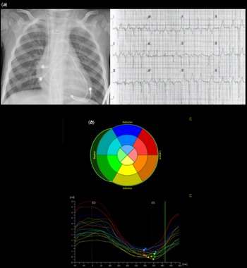
Through a midline sternotomy, the steroid-eluting pacing leads were directly affixed and sutured to the epicardial surface of the heart, and tunneled to the abdomen, in a pocket beneath the posterior fascia of the rectus abdominis. Leads were implanted into the left ventricular apex beyond left anterior descending coronary artery (minimum distance of 5 mm) (Figure 1). The location of the stimulating electrode (cathode) was considered the site of implantation of bipolar leads. Dual-chamber pacemakers were implanted in patients with a body weight ≥3 kg, single chamber in those <3 kg.
( a ) Left panel, chest X-ray showing dual-chamber pacing (DDD) epicardial pacing with bipolar leads positioned into right atrium free wall and left ventricular apex; right panel, ECG showing DDD pacing with the ventricular lead in left ventricular apex. ( b ) Echocardiographic analysis of speckle-tracking left ventricular global longitudinal strain. The figure shows the systolic left ventricle longitudinal strain curves of the 16-segment model (Bull’s eye plot, top panel) calculated as the average of the regional end-systolic strains in a four-chamber apical section (bottom panel). All segmental longitudinal strain peak curves are lined up without significant delay between representative strain peaks, with a negative concordant pattern indicating a synchronous and effective systolic shortening: global longitudinal strain −31, ejection fraction, 65%. (ES = end systolic). (Epiq 7G, QLab 10.4, aCMQ module; Philips Healthcare North America, Andover, MA, USA). All images are from the same epicardial pacing patient.

Endocardial pacing systems
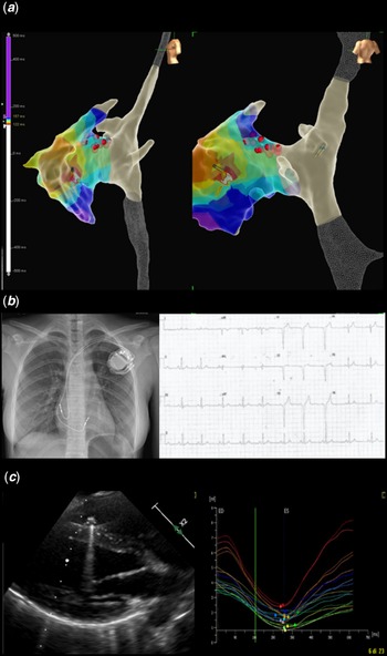
Implantation procedure was performed with 3D electroanatomic mapping systems, EnSite Velocity™ Cardiac Mapping System 4.0.2 or Precision™ 5.0.1 (Abbott MedicalTM). Via the femoral vein, a quadripolar steerable 6-Fr catheter acquired geometric reconstruction of the right atrium and right ventricular. A 3D right ventricular local pacing map was created by measuring the QRS duration obtained by pacing with the roving catheter. Pacing output was as low as possible (up to 5 V/1 ms) to ensure stable ventricular capture. The 3D right ventricular map was coded white to purple: the white colour shows the pacing site corresponding to the narrowest paced QRS and the purple colour shows the broadest QRS (Figures 2, 3). Screw-in bipolar leads were connected to the navigation system through alligator cables and introduced through axillary vein puncture. Hand-fashioned metallic stylets were used to implant the lead on the desired ventricular site (narrowest paced QRS). Single-chamber pacemakers were implanted in children and dual-chamber were implanted in post-puberty patients.
( a ) 3D electroanatomic map of right chambers showing the position of the ventricular/atrial leads implanted into the mid-septum/right atrial appendage and the colour-coded pacing map of the right ventricle. The white area shows the narrowest paced QRS area, and the violet area shows the broadest paced QRS area. The His bundle recording area is tagged with red dots. Left lateral (left panel) and posterior (right panel) view. ( b ) Left panel, chest X-ray; right panel, ECG showing DDD pacing. ( c ) Left panel, echo long axis view of the left ventricle, showing the lead tip on the interventricular septum. Right panel, longitudinal strain curves. GLS −24, EF 61%. All images are from the same endocardial pacing patient. See text and caption of Figure 1 for further details. GLS, global longitudinal strain.

Follow-up evaluation
Clinical evaluation, ECG, and telemetric pacemaker interrogation have been performed 1 month after implantation, and then every 6 months. QRS duration has been measured manually in standard 12-lead ECG (paper speed of 25 mm/s). Reference Silvetti, Ammirati, Placidi, Di Mambro, Ravà and Drago9
Echocardiography has been performed every year. Interventricular dissynchrony and left ventricular dissynchrony have been determined by calculating the interventricular mechanical delay and the septal to posterior wall motion delay, respectively. Reference Van der Hulst, Delgado and Blom15 Speckle tracking echocardiography examined global and regional left ventricular function by evaluating left ventricular strain. Global longitudinal strain was assessed (Figs 1 and 2). Normal values of strain in children are age-related. Reference Marcus, Mavinkurve-Groothuis and Barends16 3D echocardiography measured ejection fraction and the systolic dissynchrony index. Reference Van der Hulst, Delgado and Blom15
Statistical analysis
Categorical variables are reported as count and percentage. Continuous variables are described as median (25th–75th percentile). The difference between continuous variables was tested with the non-parametric Wilcoxon test (Mann–Whitney and signed-rank test). Categorical variables were compared with χ2 test or Fisher’s exact test, as appropriate. QRS duration, global longitudinal strain, and ejection fraction were compared between epicardial and endocardial pacing groups, and before and after pacemaker implantation, with patients serving as their own controls, in the same group. A p value of <0.05 was considered statistically significant. All statistical analyses were performed using StataSE 13.0 (StataCorp, College Station, Texas, USA).
Results
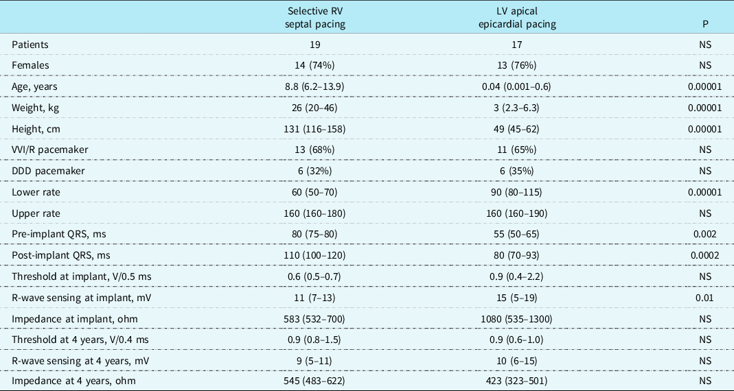
The study included 36 consecutive patients, 19 with endocardial systems and 17 with epicardial systems, all those who performed selective site pacing. During the study period, the total number of patients without CHD that underwent first pacemaker implantation was 65. Demographic and procedure data are reported in Table 1.
Demographic and procedure data

DDD = dual-chamber pacing, LV = left ventricular, mV = millivolt, NS = not significant, RV = right ventricular, V = volt, VVI = single-chamber ventricular pacing. See text for further explanations.
Autoantibodies (anti-SSa and anti-SSb) were present in 7 of 15 epicardial patients (47%) and in 0 of 5 endocardial patients.
The leads were implanted at left ventricular apex for epicardial systems (Figure 1) and right ventricular (RV) mid-septum for endocardial systems (Figure 2). Procedure time for endocardial implantation was 170 (135–184) min. There were no failures to implant the epicardial and endocardial lead at the required selective pacing sites. Pacing mode and pacing rate data, as well as pacing thresholds, R-wave sensing, and impedance data are reported in Table 1. All patients showed a percentage of ventricular pacing >95%.
All patients were in good clinical conditions at last follow-up visit without signs or symptoms of left ventricular dysfunction and without drug therapy.
Electrocardiographic data
Pre-implantation, all endocardial system patients showed narrow QRS junctional escape rhythm. Three epicardial system patients showed broad QRS escape rhythm: 90, 110, and 110 ms. One of these patients had presence of autoantibodies. Despite this finding, pre-operative QRS complexes of epicardial system patients were significantly narrower than those of endocardial system patients (Table 1).
Post-implantation, QRS significantly widened either in endocardial (p = 0.0002) or in epicardial patients (p = 0.0015), and QRS complexes of epicardial patients were significantly narrower than those of endocardial patients (Table 1). However, the increase of paced QRS duration (expressed as percentage of pre-operative values) was not significantly different between the two groups: endocardial systems 40 (25–50)% and epicardial systems 40 (21–50)%.
Echocardiographic data
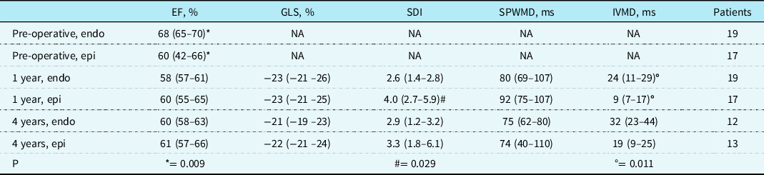
Pre-implantation, all endocardial patients showed normal pre-operative ejection fraction. Whereas, in the newborn groups, pre-operative ejection fraction (EF) was within normal limits, although seven patients showed impaired (<55%) ejection fraction (Table 2). Two of these seven patients had also broad QRS.
Echocardiographic data

EF = ejection fraction; endo = endocardial; epi = epicardial; GLS = global longitudinal strain; IVMD = interventricular mechanical delay; NA = not available; SDI = systolic dyssynchrony index; SPWMD = septal to posterior wall motion delay.
Post-implantation (1-year) ejection fraction increased in all epicardial system patients with low pre-operative values. However, overall ejection fraction did not significantly change in these patients (pre-post, p = 0.088). Conversely, 1-year ejection fraction significantly decreased in endocardial pacing patients (pre-post, p = 0.0003), although most patients remained within normal limits. Only one patient with transvenous system showed ejection fraction 51% at 1 year and 55% at 4 years. However, 1-year and 4-year ejection fraction did not show significant differences between the two groups and among patients of the same group.
The other echo parameters, such as global longitudinal strain, systolic dyssynchrony index, septal to posterior wall motion delay, and interventricular mechanical delay, were generally within normal limits (Figs 1b, 2c), showed only few significant differences between the two groups (systolic dyssynchrony index and interventricular mechanical delay, Table 2), and did not show significant changes between 1 and 4 years of follow-up.
Complications
Complications occurred in three epicardial systems. There were as follows:
-
Two lead fractures: one repaired (fracture site proximal to generator) and the other substituted with a new epicardial implantation. This latter patient was censored.
-
One abdominal pocket infection treated with antibiotics and solved.
Discussion
To our knowledge, this is the first study so far comparing two groups of congenital complete atrioventricular block patients without other CHDs and without prior pacing, who received epicardial or transvenous pacing systems implanted in alternative pacing sites, the left ventricular apex and the right ventricular mid-septum.
The right ventricular septal implantation was guided by 3D electroanatomic mapping systems and 3D right ventricular local pacing map. Due to the pacing policy of the centre, age and body size at implantation were significantly different. Some neonates showed impaired ejection fraction before pacing, and left ventricular function increased with left ventricular apical pacing reaching normal limits. Consequently, a positive functional effect of left ventricular pacing can be predicted in infants or small children with congenital complete atrioventricular block and impaired left ventricular function. Reference Silvetti, Muzi and Unolt10 On the other hand, older patients with congenital complete atrioventricular block before pacemaker implantation showed enhanced systolic function, Reference Kertesz, Friedman and Colan17 and left ventricular function decreased after transvenous selective right ventricular pacing although remaining within normal limits. These findings have been already reported. Reference Silvetti, Muzi and Unolt10,Reference Silvetti, Pazzano and Battipaglia13 Therefore, post-operative left ventricular ejection fraction of the two pacing groups did not show significant differences.
Global longitudinal strain as well was within normal limits and not significantly different between the two groups. Moreover, ejection fraction and global longitudinal strain did not significantly differ within the same endocardial or epicardial pacing group between 1-year and 4-year follow-up.
Synchrony indexes were mostly within normal limits, although showing some significant differences between the two groups, favouring transvenous pacing systems. These results showed that both pacing system seemed effective in preserving left ventricular systolic function and synchrony in children with congenital complete atrioventricular block at short-/mid-term follow-up. Paced QRS complexes widened significantly in comparison with pre-operative QRS complexes, and the increase of paced QRS duration was not significantly different between the two groups. The finding that epicardial pacing showed narrower both intrinsic and paced QRS than endocardial pacing could be related to the lower cardiac mass of the newborns rather than to the pacing itself.
Other paediatric studies focused on left ventricular function and pacing, either comparing the results of various epicardial lead positions Reference Gebauer, Tomek and Kubus5 or comparing multiple endocardial and epicardial lead positions. Reference Janoušek, van Geldorp and Krupičková6,Reference Van Geldorp, Vanagt, Prinzen and Delhaas8 It was found a better outcome for left ventricular apex and left ventricular free wall epicardial pacing rather than for right ventricular sites, either endocardial or epicardial. Reference Gebauer, Tomek and Kubus5,Reference Janoušek, van Geldorp and Krupičková6,Reference Van Geldorp, Vanagt, Prinzen and Delhaas8 Moreover, prospective studies showed that left ventricular apical epicardial pacing in neonates and infants preserved left ventricular contractility and synchrony. Reference Silvetti, Ammirati, Placidi, Di Mambro, Ravà and Drago9,Reference Silvetti, Muzi and Unolt10 Among right ventricular pacing from endocardial and epicardial sites, right ventricular apex showed a less negative effect on left ventricular performance, and non-targeted right ventricular septal pacing did not show any advantage over right ventricular apical pacing. Reference Janoušek, van Geldorp and Krupičková6,Reference Silvetti, Ammirati and Palmieri18
In patients with congenital complete atrioventricular block, autoantibodies have been identified as risk factors for the development of dilated cardiomyopathy. Reference Villain, Coastedoat-Chalumeau, Marijon, Boudjemline, Piette and Bonnet19 Transient myocarditis or immune-mediated myocardial damage have been proposed, although endomyocardial biopsies did not reveal signs of myocarditis. Reference Moak, Barron and Hougen3,Reference Udink ten Cate, Breur and Cohen20 In the current study, the presence of anti-SSa/SSb antibodies was not associated with impaired left ventricular function as other studies reported. Reference Kim, Friedman and Eidem2–Reference Silvetti, Drago and Ravà4,Reference Janoušek, van Geldorp and Krupičková6,Reference Silvetti, Muzi and Unolt10 Moreover, a study showed that patients with congenital complete atrioventricular block who were not paced did not develop dilated cardiomyopathy. Reference Udink ten Cate, Breur and Cohen20 Therefore, the pacing site seems to be the most important determinant of left ventricular function in children with congenital complete atrioventricular block, regardless of the serological pattern.
Left ventricular apical epicardial pacing in paediatric patients should be now considered a consolidated procedure to preserve left ventricular function, probably due to the homogeneous spread of activation from the apex towards the left ventricular base. Reference Silvetti, Muzi and Unolt10
Data from this study showed that right ventricular septal implantation guided by 3D electroanatomic mapping system and right ventricular pacing map is comparable to left ventricular apical pacing in terms of preserved left ventricular contractility and synchrony. A possible explanation of such result might be due to the propagation of the right ventricular impulse through the septum that depolarises both the myocardial fibres and the conduction system. The fusion of the propagation obtained this way would start left ventricular activation across the base of the mitral septal papillary muscle, as in normal activation. Reference Silvetti, Pazzano and Battipaglia13
Therefore, both procedures could be satisfactory approaches in order to implant a permanent pacing system in paediatric patients with congenital complete atrioventricular block, and the selection of endocardial or epicardial system would rely on body characteristics and centre’s policy.
Limitations
This is a retrospective, single-centre study, and the number of patients included is small, as most paediatric studies. The follow-up may be too short, as left ventricular dysfunction may become manifest after longer time. Reference Janoušek, van Geldorp and Krupičková6 However, the finding of preserved synchrony may be a dependable predictor of preserved contractility after longer follow-up. The measurement of ejection fraction to evaluate ventricular function can be biased and subjected to individual interpretation. For this reason, 2D speckle-tracking echo and the derived myocardial strain were used. The automated quantification of myocardial strain reduces the measurement errors, the inter- and intra-observer variability, and improves the accuracy and reproducibility of this method.
Although patients of both groups had the same disease (congenital complete atrioventricular block) and underwent the same therapy (pacemaker implantation), age at implantation significantly differed, including neonates and infants in the epicardial pacing group, and children and adolescents in endocardial pacing group. This seems the main limitation of this study. Really, the risk of pacing-induced left ventricular dysfunction of the two groups may differ, being probably higher in the smaller subjects. Reference Silvetti, Drago and Ravà4 However, these data do not show significant differences between the two groups, after chronic pacing of similar duration. Hence, the comparison of the results of a new approach to alternative endocardial pacing sites with those of an established pacing therapy (left ventricular epicardial pacing in infants) seems to be encouraging in terms of left ventricular systolic function and synchrony preservation.
Conclusions
Left ventricular apical epicardial pacing and right ventricular mid-septal pacing guided by 3D electroanatomic mapping systems and pacing map preserved left ventricular contractility and synchrony in children and adolescents with congenital complete atrioventricular block at short-/mid-term follow-up and did not show significant differences between the two pacing groups. Therefore, right ventricular septal pacing guided by 3D electroanatomic systems seems a good alternative to left ventricular apical epicardial pacing in older paediatric patients.
Acknowledgements
The authors thank Dr. Elisa Del Vecchio and Dr. Ilaria Cazzoli, MD, for their valuable collaboration in the editorial revision.
Financial support
This research received no specific grant from any funding agency, commercial or not-for-profit sectors.
Conflict of interest
None.