Introduction
Significant economic losses in sheep production are associated with parasitism by gastrointestinal helminths, mainly Haemonchus contortus. The annual world economic losses due to infections caused by gastrointestinal nematodes are estimated in millions of dollars (Amarante et al., Reference Amarante, Susin, Rocha, Silva, Mendes and Pires2009; Silva et al., Reference Silva, Araújo, Braga, Frassy, Tavela, Carvalho and Castejon2009). In addition, the frequent use of anthelmintics for the prophylaxis of gastrointestinal nematode (GIN) infections has led to the dissemination of populations of resistant parasites (Getachew et al., Reference Getachew, Dorchies and Jacquiet2007). Research worldwide looks for alternative measures for control of domestic animal helminthiasis, aiming to reduce the usage of chemotherapeutics (Silva et al., Reference Silva, Araújo, Braga, Frassy, Tavela, Carvalho and Castejon2009). The application of biological control using nematophagous fungi has become a viable alternative and has presented itself as promising. Some work done with the species Duddingtonia flagrans and Monacrosporium thaumasium demonstrate their effectiveness in the control of nematodes in the laboratory (Waller et al., 1994; Larsen, Reference Larsen1999; Terril et al., Reference Terril, Larsen, Samples, Husted, Miller, Kaplan and Gelaye2004). The objective of this work was to evaluate in vitro activity of fungal conidia of D. flagrans and M. thaumasium on H. contortus infective larvae.
Materials and methods
Fungal cultures and experimental assays
The isolates of the predator fungal species D. flagrans (AC001) and M. thaumasium (NF34a) were kept in test tubes containing 2% corn–meal–agar (CMA), in the dark, at 4°C for 10 days. The isolates were previously stored at the Laboratory of Parasitology in the Department of Veterinary Medicine, Federal University of Viçosa, Minas Gerais, Brazil. Petri dishes containing 2% water-agar (2% WA) were inoculated with one of the isolates and incubated at 26°C for 10 days. After that, the conidia were collected according to Araújo et al. (Reference Araújo, Santos, Ferraz and Maia1993).
The present work consisted of two experimental assays (A and B). In assay A, the predatory activity of the predator fungi D. flagrans and M. thaumasium on H. contortus L3 was evaluated in an in vitro assay on Petri dishes containing 2% WA. In assay B, the predatory capacity of the fungal conidia of the isolates D. flagrans and M. thaumasium on sheep faeces naturally infected by H. contortus was evaluated.
Collection of L3 of Haemonchus contortus
The H. contortus L3 were obtained by the Baermann method after coprocultures were carried out for 10 days, according to Gordon & Whitlock (Reference Gordon and Whitlock1939), using naturally infected sheep faeces that were positive for the superfamily Strongyloidea. Using light microscopy ( × 10 objective lens) the samples with 100% H. contortus were used in the experiment, having been washed with distilled water and centrifuged five times to remove the supernatant. The coprocultures were carried out, for each sheep, according to the methodology described by Roberts & O'Sullivan (Reference Roberts and O'Sullivan1950). The identification of the infective larvae in the coprocultures was performed according to Keith (Reference Keith1953).
Collection of conidia
Culture discs (4 mm in diameter) were removed from the fungal isolates kept in test tubes containing 2% CMA and transferred to 9.0-cm Petri dishes containing 20 ml of 2% potato dextrose agar and kept at 25°C in the dark for 10 days. After growth, new culture discs (4 mm in diameter) were transferred to 9.0-cm diameter Petri dishes containing 20 ml of 2% water-agar (2% WA) and 1 ml of distilled water containing 1000 larvae of Panagrellus sp. was added daily for 21 days to induce conidia formation. When fungal development was complete, 5 ml of distilled water were added to each Petri dish, and the conidial and mycelial fragments were removed as described by Araújo et al. (Reference Araújo, Santos, Ferraz and Maia1993).
Assay A
Two treatments and one control were made and kept at 26°C for 7 days. Each treatment consisted of 1000 conidia of a fungal isolate and 1000 H. contortus L3, with 1000 L3 without fungus as the control, and ten repetitions per group. Ten random fields of each plate were counted daily, checking the number of predated larvae until the end of the experiment (seventh day). After the last reading of the plates, the Baermann method was used to count the non-predated larvae of all treated groups and control plates.
Assay B
In coprocultures made from 20 g of faeces from sheep naturally infected by H. contortus, 1000 conidia of the fungal isolates D. flagrans (AC001) and M. thaumasium (NF34a) were homogenized and plated. In the control group, only coprocultures made from 20 g of faeces from sheep naturally infected by H. contortus were used. There were six replicates per group. At the end of 7 days, the H. contortus larvae were recovered from the treated and control groups using the Baermann method, according to the technique described by Araújo et al. (Reference Araújo, Santos, Ferraz and Maia1993).
Data analysis
Means of recovered H. contortus L3 were calculated. Data were examined by analysis of variance at significance levels of 1 and 5% probability (Ayres et al., Reference Ayres, Ayres, Ayres and Santos2003). Predation efficiency of L3 relative to the control group was evaluated by the Tukey's test at 5% probability. The reduction percentage of L3 means was calculated according to the following equation: Reduction % = ((Mean of L3 recovered from control − Mean of L3 recovered from treatment)/Mean of L3 recovered from control) × 100.
Results and discussion
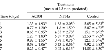
The mean of non-predated H. contortus L3 per 4 mm diameter field during the experiment is shown in table 1. The D. flagrans (AC001) and M. thaumasium (NF34a) fungal isolates predated the larvae throughout the experimental assay. The predation was observed on the first reading of the two treated groups, 24 h after the interaction between the larvae and the fungal isolates. However, the presence of fungus and formed traps predating the L3 were not observed in the control group Petri dishes.
Daily means and (±) standard error of non-predated infective larvae (L3) of Haemonchus contortus per 4 mm diameter field in 2% water-agar during 7 days of treatment with the fungal isolates Duddingtonia flagrans (AC001), Monacrosporium thaumasium (NF34a) and control without fungus.
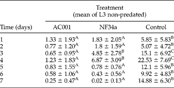
Means followed by the same letter in the same column are not significantly different (P>0.05) by Tukey's test.
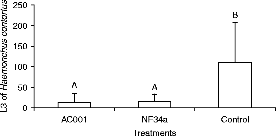
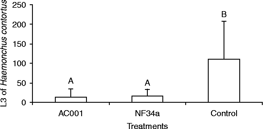
At the end of the experimental assay, no difference was found (P>0.05) between the groups treated with the fungi D. flagrans (AC001) and M. thaumasium (NF34a); nonetheless, differences were observed between the treated and the control groups (fig. 1). The reduction percentages of the L3, obtained using the Baermann method at the end of the experiment, with the fungi D. flagrans (AC001) and M. thaumasium (NF34a), were 87.75 and 85.57%, respectively, compared to the control group.
Mean number of non-predated Haemonchus contortus infective larvae (L3) recovered in 2% water-agar by the Baermann method on the seventh day after interaction with the fungal isolates Duddingtonia flagrans (AC001), Monacrosporium thaumasium (NF34a) and control (without fungus). Lines on bars represent standard deviations. Means accompanied by at least one common capital letter (A) are not significantly different by Tukey's test at a 5% probability level.
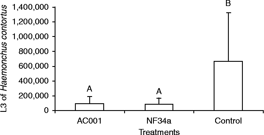
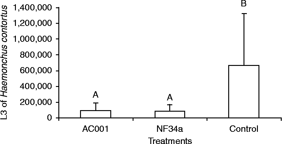
At the end of 7 days, the conidia of D. flagrans (AC001) and M. thaumasium (NF34a) isolates reduced the number of recovered L3 from Assay B. However, no difference (P < 0.05) was found between the treatments. Moreover, a difference (P < 0.05) was observed between the treatments and the control group (fig. 2). The reduction percentages due to the D. flagrans (AC001) and M. thaumasium (NF34a) isolates were 85.82 and 87.32%, respectively.
Mean number of non-predated Haemonchus contortus infective larvae (L3) recovered in coprocultures by the Baermann method on the seventh day after interaction with the fungal isolates Duddingtonia flagrans (AC001), Monacrosporium thaumasium (NF34a) and control (without fungus). Lines on bars represent standard deviation. Means accompanied by at least one common capital letter (A) are not significantly different by Tukey's test at a 5% probability level.
In the presence of nematodes, the fungi are able to produce traps in a short period of time. This predation was already seen in the first reading of the Petri dishes of the treated groups 24 h after the interaction between the larvae and the fungal isolates. However, the presence of fungus and of formed traps predating the L3 present in the control group Petri dishes was not observed.
During the present work, the temperature was kept constant at 26°C, and it was observed that there was an effective reduction of the number of H. contortus L3 due to the presence of D. flagrans and M. thaumasium fungal isolates throughout and after 7 days of interaction (table 1 and fig. 1). According to Gronvold et al. (Reference Gronvold, Henriksen, Larsen, Nansen and Wolstrup1996), after the capture of the larvae by the trapping structures of the predatory fungi, a process of hyphal penetration into the cuticle occurs, followed by the digestion of the nematodes' interior. The most commonly observed differentiated structures of these fungi along the mycelium are non-adhesive constricting rings, non-constricting rings, buttons, and three-dimensional adhesive networks. These results are in accordance with the present work, since the production of traps on H. contortus L3 was observed in the experimental assay.
Furthermore, M. thaumasium (NF34a) destroyed the H. contortus L3, with a reduction of 85.57% of the larvae at the end of the experimental assay, demonstrating its effectiveness. Fungi of the genus Monacrosporium (M. sinense and M. thaumasium), have been evaluated by several authors, demonstrating effectiveness in the control of gastrointestinal nematodiosis of different animal species (Araújo et al., Reference Araújo, Santos, Ferraz, Maia and Magalhães1992; Braga et al., Reference Braga, Carvalho, Araujo, Silva, Araújo, Lima, Tavela and Ferreira2009).
In assay B, a reduction of the number of H. contortus L3 recovered from the faeces was observed, caused by conidia of the D. flagrans (AC001) and M. thaumasium (NF34a) isolates. These results are in accordance with Silva et al. (Reference Silva, Araújo, Braga, Frassy, Tavela, Carvalho and Castejon2009) who also observed that these isolates could be used in the control of sheep haemonchosis in natural conditions. In the same work, H. contortus was the most prevalent gastrointestinal parasitic nematode, demonstrating its importance in sheep production.
The results obtained demonstrated that fungal conidia of D. flagrans (AC001) and M. thaumasium (NF34a) were effective in the in vitro control of sheep H. contortus infective larvae (L3) and could be used in the biological control of this nematode.