Introduction
Horses are economically important animals with varying roles in different parts of the globe. Even today they are still used as working animals in agriculture and transportation in some parts of the world, for the provision of meat in others and especially in the racing and leisure industries in more affluent countries (British Horseracing Authority, 2012). Maintaining their health is therefore crucial. However, as with other domestic livestock, gastrointestinal (GI) helminths are a major cause of morbidity in horses and enormous sums are spent annually in treating horses for worm infections (Matthews et al., Reference Matthews, Hodgkinson, Dowdall and Proudman2004; Matthews, Reference Matthews2014). Parasitic nematodes such as Parascaris, the strongyloids and the cyathostomins are important equine pathogens, with effects that can be severe enough to cause marked distress to the animals. Infection with the equine cestode Anoplocephala perfoliata has also been recognized as a significant cause of morbidity in horses (Proudman & Trees, Reference Proudman and Trees1999). Intestinal problems may range from mild colic ((Proudman et al., Reference Proudman, French and Trees1998) to necrosis (Rodriguez-Bertos et al., Reference Rodriguez-Bertos, Corchero, Castano, Pena, Luzon, Gomez-Bautista and Meana1999), and there has even been a report of a fatal case of caecal rupture attributable this cestode (Ryu et al., Reference Ryu, Bak, Kim, Yoon, Seo, Kim, Park and Lee2001).
Control of the GI helminths of horses is principally by chemotherapy (Behnke et al., Reference Behnke, Buttle, Stepek, Lowe and Duce2008) and, not surprisingly with the wide-scale usage and overuse of anthelmintics, resistance has arisen among equine nematodes, just as it has in those of cattle and sheep. Anthelmintic resistance among horse nematodes is now a serious problem of considerable economic significance (Kaplan, Reference Kaplan2002, Reference Kaplan2004), although we are unaware of any current reports of resistance among horse tapeworms. Nevertheless, the development of resistance to praziquantel in other platyhelminths suggests that this may simply be a matter of time, and it is pertinent therefore to consider alternatives to the currently available anthelmintics for use in the equine industry (Ismail et al., Reference Ismail, Botros, Metwally, William, Farghally, Tao, Day and Bennett1999).
Conventional chemotherapy is largely based on synthetic products, but an alternative source of anthelmithic agents may reside in the promising developments of plant-based anthelmintics, such as the cysteine proteinases (CPs) found naturally in plant latices and the juices of fruits, as for example in pineapples, figs and papaya (Behnke et al., Reference Behnke, Buttle, Stepek, Lowe and Duce2008). Plant-derived CPs have been shown to have powerful anthelmintic effects on various parasitic nematodes ranging from murine (Stepek et al., Reference Stepek, Lowe, Buttle, Duce and Behnke2007b, Reference Stepek, Lowe, Buttle, Duce and Behnkec) to ovine (Buttle et al., Reference Buttle, Behnke, Bartley, Elsheikha, Bartley, Garnett, Donnan, Jackson, Lowe and Duce2011) and porcine (Levecke et al., Reference Levecke, Buttle, Behnke, Duce and Vercruysse2014), and recent work shows that they are also effective against tapeworms (Stepek et al., Reference Stepek, Lowe, Buttle, Duce and Behnke2007d; Mansur et al., Reference Mansur, Luoga, Buttle, Duce, Lowe and Behnke2014, Reference Mansur, Luoga, Buttle, Duce, Lowe and Behnke2015). In this paper we present data assessing in vitro the efficacy of papaya latex and pineapple extracts against the equine cestode A. perfoliata, a species that is not easy to treat with conventional anthelmintics (Matthews et al., Reference Matthews, Hodgkinson, Dowdall and Proudman2004).
Materials and methods
Enzyme preparation
The enzymes used in this study were cysteine proteinases (CPs) found in papaya latex: chymopapain, glycyl endopeptidase, caricain and papain (in order of relative abundance (Buttle et al., Reference Buttle, Dando, Coe, Sharp, Shepherd and Barrett1990)), pineapple fruit bromelain (FB), pineapple stem bromelain (SB) and papain. Papaya latex supernatant (PLS) was prepared as described previously by Buttle et al. (Reference Buttle, Behnke, Bartley, Elsheikha, Bartley, Garnett, Donnan, Jackson, Lowe and Duce2011) and aliquoted into individual vials and stored at − 80°C. The total molar concentration of the active cysteine proteinases present in PLS was measured by active-site titration adapted from Barrett et al. (Reference Barrett, Kembhavi, Brown, Kirschke, Knight, Tamai and Hanada1982) and Zucker et al. (Reference Zucker, Buttle, Nicklin and Barrett1985) with increasing concentrations of the cysteine proteinase specific inhibitor, l-trans-epoxysuccinyl-l-leucylamido-(4-guanidino) butane (E-64) (Apollo Scientific Ltd, Stockport, Cheshire, UK) with 4 mm l-cysteine as a reducing agent and benzoyl-arginyl-p-nitroanilide (BAPNA) (Bachem Ltd, St. Helens, Merseyside, UK) as the substrate.
For preparation of FB we adapted the method of Rowan et al. (Reference Rowan, Buttle and Barrett1990). Ripe fruits were purchased from Sainsbury's supermarket, UK, peeled, cut into small pieces and then crushed in a laboratory juicer (Philips juicer HR 1861; Royal Philips, Amsterdam, The Netherlands) and filtered. The filtrate was then subjected to vacuum filtration (Whatman®quantitative filter paper, hardened ashless, Grade 540 diameter 110) to remove the remaining fine debris. The filtered juice was concentrated by dialysis over polyethylene glycol and the concentrated juice was aliquoted into vials and stored at − 80°C for future use. The active enzyme concentrations from each stage of preparation were determined by active-site titration with E-64 using benzyloxycarbonyl-Phe-Arg-pNA (Z-Phe-Arg-pNA) (Bachem Ltd) as the substrate.
Pineapple SB was obtained from Hong Mao Biochemical Co. Ltd, Thailand, kindly provided as a gift. Two hundred grams of SB powder were dissolved in 1 litre of distilled water and centrifuged at 17,700 × g at 4°C and the pellets were discarded while the supernatant was aliquoted into vials and stored at − 80°C. The molar concentration of functionally active SB was assessed by active site titration with E-64 using benzyloxycarbonyl-Arg-Arg-pNA (Z-Arg-Arg-pNA) (Bachem Ltd) as its substrate. On the day of each experiment, individual vials were thawed and assessed for active enzyme concentration before use, and diluted according to the experimental requirements.
The same purified papain (catalogue no. P3125; Sigma-Aldrich, Gillingham, Dorset, UK) used in earlier publications (Stepek et al., Reference Stepek, Buttle, Duce, Lowe and Behnke2005, Reference Stepek, Lowe, Buttle, Duce and Behnke2006, Reference Stepek, Curtis, Kerry, Shewry, Clark, Lowe, Duce, Buttle and Behnke2007a) was used in the experiments described in this paper. It was diluted in Hanks' saline to the exact molar concentration of active CPs required for individual in vitro experiments.
Parasites
The horse tapeworm A. perfoliata was collected from Turner's abattoir, Cheshire and from the Lawrence J. Potter abattoir, Somerset. Upon receipt of the whole horse GI tract, which was removed by slaughterhouse personnel, a small incision ~5 cm was made at the ileo-caecal junction so as to create an opening. About 500 ml of caecal fluid were collected and stored in a flask, the fluid acting as a maintenance medium during transportation of the worms to the laboratory. Worms attached to the mucosal surface of the intestine were removed and transferred into the flask containing the caecal fluid. The flask was then stored inside a polystyrene box pre-warmed with water bottles containing water that had been boiled earlier. Worms were then conveyed rapidly back to the University of Nottingham and transferred immediately into Hanks' saline at 37°C before commencing in vitro motility assays or imaging experiments.
Experiment 1. Effect of PLS on worm motility
Worms were transferred individually and incubated in 12-well plates (1 worm/well) containing Hanks' saline (without phenol red), pH 7.2 and 1 mm l-cysteine (Sigma-Aldrich) and one of the following enzyme preparations: 0, 10, 30, 100, 300, 1000 or 3000 μm active CP in PLS. In all cases molarity here refers to that of the functional proteinase active site as determined by active-site titration with the selective CP inactivator E-64 (Apollo Scientific Ltd). The control wells were incubated in parallel and contained: one of the concentrations of enzyme without cysteine (as specified in the Results section); no enzyme but with cysteine; enzyme pre-treated with a molar excess of E-64 (3500 μm, negative control) that was subsequently mixed with cysteine (1 mm) prior to incubation with worms; or Hanks' saline alone. The worms were incubated at 37°C for 2 h and their motility was recorded every 15 min using a standard motility scale from 0 to 5 adapted from Stepek et al. (Reference Stepek, Buttle, Duce, Lowe and Behnke2005), where zero was assigned to completely motionless worms not responding to manual stimulation; 1, movement only when prodded; 2, spontaneously active only at the ends, i.e. scolex and end of strobila; 3, slowly spontaneously active throughout; 4, more active; and 5, highly active.
Experiment 2. Scanning electron microscopy of worms exposed to PLS
Adult worms were incubated in well plates containing either 1000 μm active CP in PLS +1 mm cysteine or Hanks' saline. At 0, 10, 20 and 60 min worms were removed from the wells and placed into bijou bottles containing 2.5% glutaraldehyde in 0.1 m phosphate buffer (pH 7.2). After 1 h the glutaraldehyde was removed and replaced with 0.1 m phosphate buffer (pH 7.2) and left for another 1 h. The worms were then prepared for scanning electron microscopy (SEM) by further fixation in 1% osmium tetrachloride for 1 h, followed by dehydration in increasing ethanol concentrations from 30 to 100%. They were next subjected to critical-point drying with CO2. The dried worms were mounted on disc stubs with quick-drying silver paint and sputter coated with gold before examination in a Jeol JSM 840 SEM (JEOL USA, Inc., Peabody, Massachusetts, USA).
Experiment 3. Effect on A. perfoliata of CPs from four different plant sources
Worms were transferred individually and incubated in 12-well plates (1 worm/well) containing Hanks' saline (without phenol red), pH 7.2 and 1 mm l-cysteine (Sigma-Aldrich), and one of the following enzyme preparations: 0, 10, 30, 100 or 300 μm fruit bromelain (FB); 0, 10, 30, 100, 300 or 1000 μm active stem bromelain (SB); and 0, 10, 30, 100, 300 or 1000 m active papain. For the assays of each of these preparations all the relevant control treatments, as specified in Experiment 1 above, were also included and the concentrations of E-64 were 350 μm in exposure to FB and 1200 μm for both papain and SB, to ensure in each case an excess of the inhibitor at the highest enzyme concentration used. The worms were assessed for motility as described above for exposure to PLS.
Data analysis
All enzyme concentrations mentioned in the Results section refer to the molar concentration of active CP, as determined by active-site titration with E-64, the selective inactivator of cysteine proteinases. Where relevant, data are shown as mean values with standard errors of the mean (SEM). In vitro motility data were analysed using repeated measures general linear models (rmGLM) in SPSS (version 19.0; SPSS Inc., Chicago, Illinois, USA) with time after introduction of experimental treatment being fitted as a within-subject factor and concentration of PLS, FB, papain or SB as a covariate. The Huynh–Feldt adjustment to the degrees of freedom was used to interpret significance on the side of caution when the data did not meet the requirements of Mauchley's Test of Sphericity. P ≤ 0.05 was accepted as indicating a significant difference. All statistical models were checked for approximately normal distribution of residuals. The enzyme concentrations causing 50% and 95% reduction in motility (50% and 95% inhibitory concentration, IC50 and IC95) were determined by fitting the data for inhibition of motility, with increasing concentration of CPs after 30 min of incubation, in GraphPad Prism 6 (GraphPad Software, La Jolla, California, USA) using a non-linear regression with variable slope and the lower plateau set at 0 and the upper plateau set at the maximum motility for that experiment. Comparison of IC50s from the concentration–response curves was by an extra sum of squares F-test in GraphPad Prism 6.
Results
Efficacy of PLS on worm motility
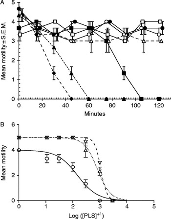
Worm motility decreased with increasing concentration of PLS in the incubation medium. There was no loss of activity in worms incubated in Hanks' saline with or without 1 mm cysteine or in PLS at 3000 μm+E-64. The lowest concentration of PLS tested (10 μm PLS) also caused no detectable loss of motility; however, higher concentrations of PLS caused a dose-related loss of motility, as shown in fig. 1A. Analysis by rmGLM indicated that the motility of A. perfoliata declined significantly with time (main effect of time, F 3.2, 60.5= 14.6, P < 0.001), but loss of motility with time varied between the different concentrations of PLS (time × concentration, F 3.2, 60.5= 3.16, P = 0.028). There was also a highly significant overall effect of concentration of PLS (main effect of concentration, F 1,19= 13.9, P = 0.001). Exclusion of exogenous cysteine from the incubation media containing active CPs (3000 μm PLS) made no difference to their efficacy: whether with or without cysteine (mean motility at time zero = 3.7 ± 0.33 and 4.3 ± 0.3, respectively, P= non-significant (NS)), all worms were immotile within 15 min of incubation.
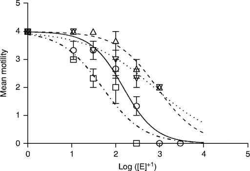
Motility of adult A. perfoliata, Hymenolepis diminuta and H. microstoma in varying concentrations of papaya latex supernatant (PLS) in vitro. (A) Motility of adult A. perfoliata incubated in varying concentrations of active PLS in vitro. Only values for concentrations of active enzyme up to 300 μm PLS are shown and values are slightly offset to avoid obscuring error bars. At 1000 μm PLS, the mean motility at 15 min was just 1.0 ± 0.6, and zero at 30 min, and in 3000 μm PLS the worms were not active at 15 min. Key to symbols used: open circles = Hanks alone; open squares = Hanks + cysteine; filled circles = 10 μm PLS; filled squares = 30 μm PLS; filled triangles = 100 μm PLS; filled diamonds = 300 μm PLS; open triangles = 300 μm PLS +E-64. (B) Concentration–inhibition curves of PLS activity against A. perfoliata, H. diminuta and H. microstoma at 30 min post-incubation. Note the greater efficacy of PLS against A. perfoliata, with IC50 values that are lower than those of the murine cestodes. Error bars represent the standard error of the mean, n= 3, for all treatments. Data for H. diminuta and H. microstoma are taken from Mansur et al. (Reference Mansur, Luoga, Buttle, Duce, Lowe and Behnke2014). Key to symbols used: open circles = A. perfoliata; inverted open triangles = H. microstoma; upright open triangles = H. diminuta.

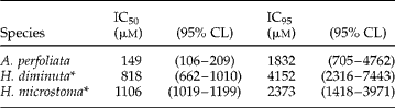
Figure 1B shows the dose response at 30 min and compares this to dose responses reported earlier for Hymenolepis diminuta and Hymenolepis microstoma (Mansur et al., Reference Mansur, Luoga, Buttle, Duce, Lowe and Behnke2014). It can be seen from these data, and from IC50 and IC95 values in table 1, that A. perfoliata was considerably more susceptible to PLS, losing motility at lower concentrations of the enzymes compared with the rodent species (sum of squares F-test for IC50s, A. perfoliata vs. H. diminuta, P < 0.0001; A. perfoliata vs. H. microstoma, P < 0.0001).
IC50 and IC95 values for the efficacy of PLS against three species of cestodes.
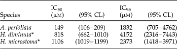
CL, confidence limits.
* Data derived from Mansur et al. (Reference Mansur, Luoga, Buttle, Duce, Lowe and Behnke2014).
SEM of cestodes incubated in PLS
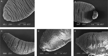
Worms incubated in PLS showed marked generalized erosions as early as 10 min post-exposure (fig. 2C). After 20 min of incubation in PLS the progressive erosion was more apparent and the tegument appeared to undergo ‘exfoliation’ (fig. 2D). After 60 min of incubation in PLS the tegument was severely damaged and appeared chapped and flaky with detachment of sections of the tegument from the underlying muscles (fig. 2E).
SEM images of A. perfoliata in papaya latex supernatant (PLS). Anoplocephala perfoliata incubated in vitro in Hanks' saline, (A) at 0 min, and (B) at 60 min; and in 1 mm PLS +1 mm cysteine, (C) at 10 min, (D) at 20 min, and (E) at 60 min. Note the progressive damage to the tegument in worms incubated in PLS and contrast with intact worms incubated in Hanks' saline.

Effects on A. perfoliata of CPs from four plant sources
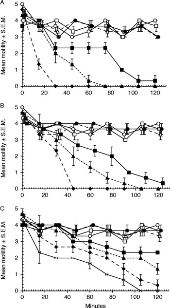
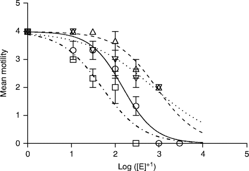
All the CPs tested (FB, SB and papain, respectively) reduced the motility of adult A. perfoliata rapidly as the concentration of active CPs in the incubation medium increased (fig. 3A–C), and this was reflected in the highly significant main effects of concentration (F 1,13 = 29.6, P < 0.001; F 1,16 = 45.6, P < 0.001; F 1,16 = 7.32, P = 0.016, respectively) and significant two-way time × concentration interactions in the case of FB (F 3.0, 38.7 = 6.3, P = 0.001) and SB (F 3.6, 57.6 = 7.4, P < 0.001) but not papain (F 2.6, 41.6 = 1.82, P = 0.17), indicating that the rate of loss of motility over time varied with CP concentration in the case of FB and SB but not papain. However, as can be seen from table 2 and figs 3 and 4, there was significant variation between the CPs from different plant sources. Although all CPs tested were efficacious, FB was the most efficacious and SB the least (table 2). However, the dose responses for FB, SB and papain all differed significantly from that of PLS (fig. 4; comparison of IC50s by sum of squares F-test gave PLS vs. FB, P= 0.0009; PLS vs. papain, P < 0.0001; and PLS vs. SB, P < 0.0001). In all cases motility was unaffected when worms were incubated in the presence of low concentrations of active CPs (10 μm), in the control solutions of Hanks' saline with or without cysteine, and also in the presence of each CP pre-treated with E-64 (fig. 3A–C). Exclusion of exogenous cysteine from the incubation media containing active CP made no difference to their efficacy (confining analysis by rmGLM to treatments with the highest concentration of enzyme in each case, with and without cysteine, gave for main effect of treatment: F 1,4= 0.14, P= 0.7 for FB at 300 μm; F 1,4= 0.03, P= 0.87 for SB at 1 mm and F 1,4= 0.25, P= 0.64 for papain at 1 mm). There were no significant interactions between time and treatment.
Motility of adult A. perfoliata incubated in various CPs in vitro: (A) pineapple fruit bromelain (FB); (B) papain; (C) pineapple stem bromelain (SB). Error bars represent the standard error of the mean, n= 3. Key to symbols used: open circles = Hanks' saline alone; open squares = Hanks' saline + cysteine; filled circles = 10 μm CP; filled squares = 30 μm CP; filled triangles = 100 μm CP; filled diamonds = 300 μm CP; line without symbols = 1000 μm stem bromelain (C only); open triangles = fruit extract +E-64 (in A, FB at 300 μm; in B, papain at 1000 μm; and in C, SB at 1000 μm).

IC50 and IC95 values for the efficacy of cysteine proteinases from four different plant-derived sources against A. perfoliata.

CL, confidence limits.
Concentration–inhibition curves of various CPs against A. perfoliata in vitro at 30 min post-incubation. The concentration of active CP expressed in μm is the molar equivalent of the inhibitor E-64 used to block active CP in a 10 μl volume in a titration assay. Error bars represent the standard error of the mean, n= 3. Key to symbols used: open squares = fruit bromelain (FB); open upright triangles = stem bromelain (SB); open inverted triangles = papain; open circles = papaya latex supernatant (PLS).
Discussion
An important consideration in the development of novel classes of anthelmintic is broad-spectrum activity against target nematodes and even parasites of other taxonomic groups, notably the platyhelminths, because the broader the spectrum of activity, the greater the likelihood of acceptance by end-users and hence commercial success (Behnke et al., Reference Behnke, Buttle, Stepek, Lowe and Duce2008). Papaya latex has been demonstrated to be efficacious against lumen-dwelling stages of murine (Stepek et al., Reference Stepek, Lowe, Buttle, Duce and Behnke2007c), porcine (Satrija et al., Reference Satrija, Nansen, Bjorn, Murtini and He1994; Levecke et al., Reference Levecke, Buttle, Behnke, Duce and Vercruysse2014), ovine (Buttle et al., Reference Buttle, Behnke, Bartley, Elsheikha, Bartley, Garnett, Donnan, Jackson, Lowe and Duce2011) and canine (Stepek et al., Reference Stepek, Lowe, Buttle, Duce and Behnke2007d) nematodes, and even nematodes of poultry (Mursof & He, Reference Mursof and He1991). It has also been shown to affect the juvenile stages of plant parasitic nematodes (Stepek et al., Reference Stepek, Curtis, Kerry, Shewry, Clark, Lowe, Duce, Buttle and Behnke2007a) and some efficacy has been observed against rodent-parasitic tapeworms at both in vitro and in vivo levels (Mansur et al., Reference Mansur, Luoga, Buttle, Duce, Lowe and Behnke2014, Reference Mansur, Luoga, Buttle, Duce, Lowe and Behnke2015). Efficacy against the free-living Caenorhabditis elegans has also been reported (Phiri et al., Reference Phiri, de Pomerai, Buttle and Behnke2014). These findings suggest a broad spectrum of activity and hence a promising prospect for CPs in conventional therapy.
The experiments reported in this paper extend the range of efficacy to the equine cestode A. perfoliata, the motility of which was severely curtailed in vitro with increasing concentrations of the enzymes in the incubation medium. We have demonstrated previously that the major anthelmintic principles in some fruit and latices of plants are the enzymatically active forms of cysteine proteinases (Stepek et al., Reference Stepek, Buttle, Duce, Lowe and Behnke2005). These enzymes require a reducing agent to maintain activity, but there is always a proportion of the enzyme that has been irreversibly oxidized and cannot be reactivated by the presence of a reducing agent (Drenth et al., Reference Drenth, Jansonius, Koekoek, Swen and Wolthers1968), and therefore has lost anthelmintic efficacy. This is one reason why we have chosen to standardize our enzyme preparations by carefully determining the concentration of the active principles in our enzyme preparations. Incubation of A. perfoliata in CPs with accurately quantified active cysteine proteinase activity resulted in severe disruption of the structural integrity of the worms and death at the highest concentrations that were tested. The reduction in motility was both time and dose dependent but, interestingly, independent of the presence of cysteine, as has already been reported for both rodent cestodes (Mansur et al., Reference Mansur, Luoga, Buttle, Duce, Lowe and Behnke2014) but differing from the effects on nematodes, which are dependent upon the presence of cysteine in the incubation medium (Stepek et al., Reference Stepek, Buttle, Duce, Lowe and Behnke2005). As was discussed in Mansur et al. (Reference Mansur, Luoga, Buttle, Duce, Lowe and Behnke2014), the most likely explanation of this lack of dependence on exogenous cysteine is that the excretory/secretory products of cestodes include a reducing agent that, in turn, activates CPs in the surrounding medium.
Equally interesting was the finding that CPs appeared to be more efficacious against A. perfoliata than against the other cestodes, with IC50 values of 3–4 times lower than those reported earlier for rodent cestodes (see table 1). This greater sensitivity of A. perfoliata to CPs may be explained by the relatively diminutive length of A. perfoliata, which ranged between 2 and 5 cm, compared to 14-day-old adult H. diminuta which were almost 15 cm long. Since CPs are known to exert their anthelmintic effects through damage to the surface of their target organisms, the exposed surface area may have been a factor, but it is also possible that the tegument itself is different from that of rodent cestodes in terms of its constituent proteins, as a result of the different diets, and consequently physiological conditions, in the intestines of horses compared to rodents. Efficacy also differed between different types of CPs, with naturally occurring CPs such as FB and PLS being more efficacious than other CPs such as papain and SB. These findings generally agree with those reported earlier for rodent cestodes (Mansur et al., Reference Mansur, Luoga, Buttle, Duce, Lowe and Behnke2014).
It has been reported that CPs damage the tegument of murine cestodes, possibly by hydrolysing particular proteins that make up the tegument, resulting in generalized tegumental digestion (Behnke et al., Reference Behnke, Buttle, Stepek, Lowe and Duce2008). Consequently, at weakened points, CPs eventually cause breaches of tegumental integrity and the subsequent release of internal contents. The SEM observations presented in this paper show damage to the tegument of A. perfoliata which is very similar in nature, beginning with superficial generalized erosion of the surface of the tegument and, with time, a much deeper and widespread generalized destruction. Visually, the images documenting damage to the surface of rodent cestodes and those in the current paper to A. perfoliata were indistinguishable in terms of the sequence and extent of the changes that were observed. This is quite different from conventional cestocidal drugs, such as praziquantel which causes contraction in platyhelminths (Becker et al., Reference Becker, Mehlhorn, Andrews and Thomas1980; Mehlhorn et al., Reference Mehlhorn, Becker, Andrews, Thomas and Frenkel1981), or benzimidazoles which interfere with β-tubulin synthesis, resulting in blebbing of the tegument (Schmidt, Reference Schmidt1998). It seems likely that the CPs are attacking more than one tegumental protein, suggesting that resistance to these new potential drugs would take some time to develop.
The current results constitute the first evidence of CPs affecting an equine helminth, although our results so far are based only on exposure of A. perfoliata to CPs in vitro. Nevertheless, they provide an interesting possibility for the development of CP-based anthelmintics for cestodes of horses, the latter being recognized as a significant risk factor in equine health (Proudman et al., Reference Proudman, French and Trees1998), but the ultimate test of the usefulness of CPs in the treatment of equine cestodes will depend on the outcome of in vivo trials. This prospect of such trials is particularly compelling for a number of reasons. First, the greater efficacy of PLS against A. perfoliata, reflected in more potent effects on the worms at lower concentrations of the enzymes compared to rodent cestodes, must be a positive feature. Second, CPs have been shown to accumulate in the hindgut region, where A. perfoliata resides, for longer periods of time than in more proximal regions, and this is another positive feature since it should ensure prolonged and more intense exposure to CPs for tapeworms dwelling in this region of the gut. Last, it has also been demonstrated that large animals, such as small ruminants, including sheep (Buttle et al., Reference Buttle, Behnke, Bartley, Elsheikha, Bartley, Garnett, Donnan, Jackson, Lowe and Duce2011) and pigs (Levecke et al., Reference Levecke, Buttle, Behnke, Duce and Vercruysse2014), required less drug per kilo body weight than small animals such as mice (Stepek et al., Reference Stepek, Lowe, Buttle, Duce and Behnke2007c), thus minimizing, or perhaps even eliminating, any problem of toxicity. More importantly, our results provide further evidence that the spectrum of activity of CPs is not restricted to nematodes, supporting the idea that they can be developed into useful broad-spectrum anthelmintics, which may be refractory to the development of resistance.
Acknowledgements
We thank Tim Smith for technical assistance with the scanning electron microscopy reported in this paper. We are grateful to Dr Ian Mellor for fitting our dose–response data in GraphPad Prism 6 and for the associated statistical analysis.
Dedication
We dedicate this paper to Dr Wenceslaus Luoga who sadly was killed in a road accident in July 2013 shortly after returning to Tanzania on the successful completion of his postgraduate studies at the University of Nottingham.
Financial support
F.M. was supported by a postgraduate studentship from the government of Malaysia, and W.L. from the Government of Tanzania
Conflict of interest
None of the authors of this paper has a financial or personal relationship with other people or organisations that could inappropriately influence or bias the content of the paper.