Highlights
-
The difference in biological characteristics of X and Y sperm.
-
The difference in transcriptomics and proteomics of X and Y sperm.
-
Recent progress in research on sex control technology.
Introduction
Sperm are produced in the seminiferous tubules of the testes of male animals and are differentiated from spermatogonial stem cells. Haploid round spermatids differentiate into mature sperm containing only an X- or Y-bearing chromosome, which is referred to as an X or Y sperm, respectively (Roldan and Teves, Reference Roldan and Teves2020). During division from spermatogonia to sperm cells, sperm cells at the same stage form a syncytium and are connected by intercellular bridges until the end of spermatogenesis when sperm cells differentiate into mature spermatozoa. During sperm development, small molecules (including genes and proteins) can shuttle freely between the cells of different genotypes via the intercellular bridges. The maturation of X and Y sperm is synchronized. Therefore, sperm carrying X or Y chromosome have essentially the same origin, developmental stage, maturity level and function (Li et al., Reference Li, Mao, Wu, Lian, Ge, Silvestrini and Cheng2018). Spermatogonia differentiate into primary and secondary spermatocytes, and the cytoplasm does not divide completely. Therefore, it is reasonable to speculate that there may be a difference between X and Y sperm.
Mature sperm are expelled during ejaculation and fuse with oocytes during fertilization to deliver half of the genetic material for the resulting offspring (Hazzouri et al., Reference Hazzouri, Rousseaux, Mongelard, Usson and Sèle2015). To date, there have been many studies on the differences between X and Y sperm, however their results are discordant, which may have been caused by the different research methods and platforms used (Hossain et al., Reference Hossain, Barik and Kulkarni2001). The selection of sperm types using the sex control technology could be used to accelerate livestock breeding and maximize livestock potential for lactation, reproduction, and production. Therefore, this technology is of great significance in animal husbandry. The difference between the two types of sperm provides a basal theory and spurs research on the separation of X and Y sperm. This review summarizes several differences between X and Y sperm, including their morphology, physiological, and molecular characteristics; genomics; and proteomics to provide references for studying the biological characteristics of sperm and development of new technologies for sex control.
Search scheme and article selection
The PubMed search engine was used to thoroughly search the MEDLINE database for literature on X and/or Y spermatozoa using the following search terms: ratio, shape, size, gender selection, motility, swimming pattern, velocity, CASA, FISH, flow cytometric analysis, Percoll gradient, swim-up method, viability, electrophobicity, electronegativity, pH tolerance, surface properties, Y-specific antigen, HY antigen, stress response, oxidative stress, DNA damage, chromosomal abnormality, proteomics, disease, gender-specific disease, and genomics. Full-text articles and abstracts in the English language on X and Y spermatozoa published before October 2021, were included in the review after screening their content. All article types, such as original articles, reviews, letter to the editor, editorials, opinions, and debates, were included in this review.
Differences between X and Y sperm
Shape and size
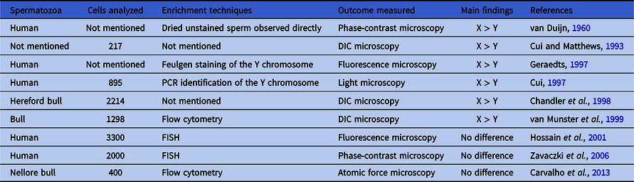
X and Y sperm have the same autosomes but distinct sex chromosomes (X or Y) (Rahman and Pang, Reference Rahman and Pang2019). Shettles (Reference Shettles1960) first observed the two separate sperm types using electron microscopy. Y sperm have small and round heads, whereas X sperm have relatively large and long heads. These differences are caused by the different sizes of the X and Y chromosomes; however, as the study included no reliable method to verify X and Y sperm, the results needed to be validated (Shettles, Reference Shettles1960). Since then, researchers have used Feulgen staining, microscopic measurements, flow cytometry analysis, and fluorescence in situ hybridization (FISH) to explore the difference in the size of X and Y sperm. Human X sperm was found to have 2.8% more DNA than Y sperm, whereas livestock X sperm contains 3–4.2% more DNA than Y sperm (van Duijn, Reference van Duijn1960; Ortega-Ferrusola et al., Reference Ortega-Ferrusola, Gil, Rodríguez-Martínez, Anel, Peña and Martín-Muñoz2017). Hossain and colleagues (2001) used FISH technology to compare the two germ cell types, X and Y. No significant change in the sex ratio from 1:1 was seen in either the mature or immature germ cell population. Similarly, the haploid, X-specific round cells did not differ from Y-specific cells when defined by their size (diameter) and shape. Carvalho and co-workers (2013) used atomic force microscopy to generate detailed three-dimensional and cell surface images of sperm cells and identified 23 structural differences between bovine X and Y sperm; however, no significant differences in shape and size were observed. The different research methods and results are summarized in Table 1.
Sperm size and shape research statistics

Movement under different incubation conditions in vitro
Gadêlha et al. (Reference Gadêlha, Hernández-Herrera, Montoya, Darszon and Corkidi2020) examined sperm movement using high-speed three-dimensional microscopy and described the movement of human sperm similar to that of a sea otter swimming in a rotating manner, with the head rotating counterclockwise and the flagella rotating synchronously around the swimming direction axis. When incubated in vitro, sperm swam to the upper layer of the incubated diluent. Under different incubation conditions, the motility of X and Y sperm differed. According to Mendel’s law of separation, the ratio of X and Y sperm produced by meiosis in mammalian spermatogenesis is 1:1 and their natural sex ratio is 1:1. Researchers successively used Leishman staining (Benet et al., Reference Benet, Genescà, Navarro, Egozcue and Templado1992), FISH (Williams et al., Reference Williams, Ballenger, Malter, Bishop, Tucker, Zwingman and Hassold1993), quantitative polymerase chain reaction (Lobel et al., Reference Lobel, Pomponio and Mutter1993), and other methods to determine the ratio of X and Y sperm in semen after routine incubation and found that the number of the two sperm types was nearly the same, with a ratio close to 1:1. However, changing the in vitro incubation conditions had different effects on the movement of X and Y sperm, with incubation altering their ratio in the upper layer.
Umehara and co-workers (2020) found that activating the X chromosome-specific TLR7/8 receptor reduced the ATP production and motility of X sperm without affecting Y sperm. It was also found that Y sperm accounted for up to 90% of the population in the upper semen portion of the incubation fluid. In addition, You and colleagues (Reference You, Kwon, Saidur Rahman, Park, Kim and Pang2017) examined the effect of different incubation temperatures (4°C, 22°C, or 37°C), pH (6.5, 7.5, or 8.5), and incubation times (3 or 5 days) on the ratio and motility of the two sperm types and found that the number of live X sperm was greater at low temperatures (4°C and 22°C) and acidic pH (6.5). Oyeyipo and co-workers (2017) also examined the effects of different semen incubation temperatures, pH, and hydrogen peroxide concentrations (50, 750, and 1000 μM) on the ratio of X and Y sperm and their motility in the upper semen portion; the results indicated that X sperm were enriched upon incubation in acidic medium at higher temperatures with elevated H2O2 concentration, but they did not count the difference in the sex ratio as significant. When sperm were exposed to chemicals (such as endocrine disruptors) or pesticides [such as 2,3,7,8-tetrachlorodibenzo-p-dioxin (TCDD) and atrazine (ATZ)], the viability and motility of Y sperm were significantly reduced compared with those of X sperm (You et al., Reference You, Kwon, Saidur Rahman, Park, Kim and Pang2017; Song et al., Reference Song, Mohamed, Pang, Kang, Ryu, Rahman and Pang2018).
Activity in a dynamic female reproductive tract environment
Sperms are driven through the female reproductive tract, which is essential for reproduction, by their flagella (Wallmeier et al., Reference Wallmeier, Nielsen, Kuehni, Lucas, Leigh, Zariwala and Omran2020). Sperm viability is closely related to the environment of the female reproductive tract (Hook and Fisher, Reference Hook and Fisher2020). Tung and colleagues (2014) established an in vitro mammalian reproductive tract-fluid model and found that the characteristics of the female reproductive tract environment (such as liquid composition, pH, and ion concentration) could affect the motility as well as the plasma membrane, acrosome membrane integrity, and fertilization ability of X and Y sperm; the identification of these characteristics may aid in selecting the most dominant sperm for fertilization. In addition to the DNA content, X sperm are slightly larger than Y sperm; therefore X sperm showed a stronger buffering capacity and adaptability during stress conditions (Carvalho et al., Reference Carvalho, Silva, Sartori and Dode2013). In addition, Y sperm exhibited a higher survival rate than X sperm in simulated in vitro female reproductive tracts and were able to strongly bind to the zona pellucida (Van Dyk et al., Reference Van Dyk, Mahony and Hodgen2001). In addition, Y sperm may be able to produce more energy due to the high expression of proteins primarily involved in energy metabolism, including ATP synthase units (Chen et al., Reference Chen, Zhu, Wu, Han, Hao, Zhao, Du, Qin, Liu and Wang2012).
’Omics research
The identification and quantification of gene and protein in cells provide a novel technological platform for identifying cellular functions and are convenient for studying gene expression, protein content, protein type, and function of sperm. There are proteomic differences between X and Y sperm, which result from their different sex chromosomes. Although most genes expressed in the early stages of sperm development are shared between the two sperm types via intercellular bridges, certain gene products are sperm-type specific. X and Y sperm can therefore be distinguished based on mRNA data and protein content (Rahman and Pang, Reference Rahman and Pang2019).
Microamplification of RNA from sperm is required to study sperm cell gene expression due to the small amount of total RNA present in a single sperm (Chen et al., Reference Chen, Zhu, Wu, Han, Hao, Zhao, Du, Qin, Liu and Wang2012). Using whole genome sequencing, 31 differentially expressed genes were identified in bovine X and Y sperm, with 27 and 4 genes upregulated in X and Y sperm, respectively (Chen et al., Reference Chen, Yue, He, Zhu, Hao, Zhao, Qin and Wang2014). In mice, RNA sequencing-based analysis revealed that <700 genes are encoded by the Y chromosome and >3000 genes by the X chromosome (Soh et al., Reference Soh, Alföldi, Pyntikova, Brown, Graves, Minx, Fulton, Kremitzki, Koutseva, Mueller, Rozen, Hughes, Owens, Womack, Murphy, Cao, de Jong, Warren, Wilson, Skaletsky and Page2014). These X chromosome genes include Taz (encoding tafazzin, a transcriptional regulator activated by physical stimulation), Xiap (encoding X-apoptotic protein that inhibits apoptosis signal activation), and G6PDX (encoding X-glucose-6-phosphate dehydrogenase that catalyzes the oxidation of the pentose phosphate pathway). By introducing X sperm or Y sperm populations into the two separate oviducts of single female pigs using bilateral laparoscopic insemination, Almiñana and co-workers (2014) found that the spermatozoa did indeed elicit sex-specific transcriptomic responses. In addition, either type of spermatozoa elicits sex-specific signal transduction signalling by oviductal cells. There are significant differences in the mRNA levels of GSTM3, DNMT3A, and PGRMC1 in bovine blastocysts produced via in vitro fertilization with the bovine X or Y sperm, thereby indicating that oocytes also have sperm fertilization-regulating mechanisms (Bermejo-Alvarez et al., Reference Bermejo-Alvarez, Rizos, Rath, Lonergan and Gutierrez-Adan2010). Cocquet and colleagues (2012) found that knocking out Slx/Slx1 or Sly elicited antagonistic effects on this pair of linked genes in X and Y sperm; this evidence was the first regarding the existence of this phenomenon in mammalian sperm. Using gene editing, Vernet and co-workers (2012) found that Zfy2 on the short arm (Yp) of the mouse Y sperm chromosome had many functions, including inducing apoptosis in sperm that contained unpaired or mismatched chromosomes during the first differentiation phase, promoting the second meiotic phase, and acting during sperm head and tail development.
Proteomic analysis is more suitable for mature sperm because of the small amount of transcription and protein synthesis. Protein levels in X and Y sperm could be used to identify additional differences (Kwon et al., Reference Kwon, Rahman, Lee, Kim, Yoon, Park, You, Hwang and Pang2014). To date, reports on the proteomics of X and Y sperm have been very limited. Chen and colleagues (2012) used two-dimensional electrophoresis and tandem mass spectrometry (2DE–MS/MS) and identified 42 differentially expressed proteins in bovine X and Y sperm, with 11 proteins upregulated and four proteins downregulated in X sperm. Scott and co-workers (2018) identified six differentially expressed proteins in bovine X and Y sperm. In Y sperm, one protein related to embryonic development contained an EF-hand domain protein 1. Moreover, highly expressed proteins in Y sperm were predominantly related to guanylate cyclase, cell fate determination (Notch), actin cytoskeleton assembly, and glutathione metabolism signalling pathways. In X sperm, the following proteins were highly expressed: FUN14, domain protein 2, mitochondrial NADH dehydrogenase iron–sulfur protein, acetyl-coenzyme A carboxylase, and cytochrome c oxidase. These highly expressed proteins are closely related to glucose metabolism, glycolysis, mTOR signals, tricarboxylic acid (TCA) cycle, and oxidative phosphorylation signalling pathways (Chen et al., Reference Chen, Zhu, Wu, Han, Hao, Zhao, Du, Qin, Liu and Wang2012; Scott et al., Reference Scott, de Souza, Aristizabal, Hethrington, Krisp, Molloy, Baker and Dell’Aqua2018). Umehara and co-workers (2019) analyzed the mouse sperm RNA database and chose Tlr7, Tlr8, Ar, Gpr174, Gpr34, and Edr2a from the 492 expressed genes on the X chromosome for further study. The activation of Tlr7 and Tlr8 receptors expressed during the late stage of X sperm formation could inhibit ATP production and enhance glycogen synthase kinase 3α/β (GSK3α/β) and NF-κB phosphorylation, thereby affecting sperm movement.
Studies on X and Y sperm separation methods
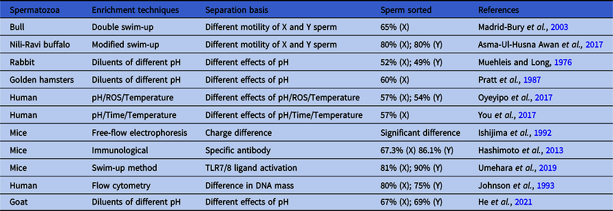
Sex control technology, in which female animals produce offspring of the desired sex, is a vital technology in animal husbandry. This technology has always been an area of intense research in animal husbandry research. Through sex control technology, the selection intensity of livestock breeding can be greatly improved and the breeding process can be accelerated, enabling the sole use of female animals for reproduction and lactation and male animals for meat or labour performance to improve efficiency. Separating X and Y sperm using their unique characteristics and then fertilizing oocytes is the most direct way to intervene and select the sex of offspring (Xie et al., Reference Xie, Xu, Wu and Hong2020). To achieve sex control, researchers have applied different methods for sperm separation. The principles and results of the different separation methods are summarized in Table 2.
Different separation methods and result statistics

Sperm swim-up method
The most commonly used method in assisted reproduction to separate highly viable sperm is the sperm swim-up method, which includes the regular upstream process and swim-up process (Heidari et al., Reference Heidari, Lakpour, Darbandi, Darbani, Shani, Goharbakhsh, Cheshmi, Akhondi and Sadeghi2018). Sperm in their natural state will swim forward (upward) and can be separated by modifying the incubation conditions (Yang and Yang, Reference Yang and Yang2020). Madrid-Bury and colleagues (2003) separated X and Y sperm using the double swim-up method and additional heparin. In this case, Y sperm accounted for 35% of the total sperm in the upper semen after incubation for 150 min, demonstrating ideal separation. Asma-Ul-Husna Awan and co-workers (Reference Asma-Ul-Husna Awan, Mehmood, Sultana, Shahzad, Ansari, Rakha, Saqlan Naqvi and Akhter2017) used a modified swim-up method to separate buffalo X and Y sperm (change temperature, TALP solution, the isolated tube dimensions, and the number of spermatozoa layered per tube). The final result showed that the proportion of Y sperm in the upper incubation liquid was four-fold that of X sperm, validating this method’s efficiency to separate X and Y sperm.
The pH of the incubation liquid affects the movement of mammalian sperm, therefore X and Y sperm can be easily and effectively separated by altering the pH of the incubation liquid. When rabbit sperm were incubated in buffers at pH 5.4, 6.9, and 9.6, after insemination, 48%, 63%, and 49% of offspring, respectively, were male. However, the difference in the sex ratio in this result was not statistically significant and needs to be validated (Muehleis and Long, Reference Muehleis and Long1976). When the pH value of a female hamster reproductive tract was increased, the proportion of male offspring decreased, suggesting that the proportion of male offspring is significantly negatively correlated with pH (Pratt et al., Reference Pratt, Huck and Lisk1987). When human sperms were incubated in an acidic environment, the proportion of X sperm in the upper semen layer increased by 12.7%, and the total sperm motility decreased. When incubation was performed in an alkaline environment, the proportion of Y sperm in the upper semen layer increased by 8.9%, and the total sperm motility increased (Oyeyipo et al., Reference Oyeyipo, van der Linde and du Plessis2017; You et al., Reference You, Kwon, Saidur Rahman, Park, Kim and Pang2017). Our research results showed that the percentages of the X sperm cells in the upper sperm layer were 67.24 ± 2.61% at sperm dilution pH of 6.2 and 30.45 ± 1.03% at sperm dilution pH of 7.4, which was significantly different from 52.35 ± 1.72% of the control group (pH 6.8), and no significant effect was observed on sperm cell performance and quality. The percentages of female embryos after in vitro fertilization of the isolated upper sperm with mature oocytes at pH 6.2 and 7.4 were 66.67 ± 0.05% and 29.73 ± 0.04%, respectively (He et al., Reference He, Wu, Huang, Wang, Zhang, Kang, Zhang and Quan2021). These results indicated that the pH of the diluent had different effects on X and Y sperm, but the underlying mechanism requires further investigation.
Separation of X and Y sperm by electrophoresis
Many studies have separated X and Y sperm as per their charge differences. The epididymal epithelium secretes sialic acid in the form of glycoproteins, which alters the surface charge on sperm (Lepine et al., Reference Lepine, McDowell, Searle, Kroon, Glujovsky and Yazdani2019). Human X and Y sperm separate as they move toward positively charged electrodes during electrophoresis; the mobility of X sperm is significantly higher than that of Y sperm towards the positively charged electrode, indicating that the negative charge of X sperm is higher than that of Y sperm. However, test results consistently show that electrophoretic separation significantly weakens the motility and fertilization ability of sperm, making the application of separated sperm difficult (Yetunde and Vasiliki, Reference Yetunde and Vasiliki2013).
Separation of X and Y sperm via immunological methods
The basic concept of immunological methods for sperm sexing is based on the different proteins present on the surface of X and Y sperm. The observed genomic DNA differences among X and Y sperm across different species have led to the possibility that these DNA differences might result in protein differences as well. X and Y sperm can be separated by binding specific ligands to their corresponding receptors. SRY encodes for a protein in the high mobility group (HMG) that binds to DNA in the nucleus. Hashimoto and colleagues (2013) labelled mouse spermatozoa with the Cy3-SRY antibody and used the selected sperm for intracytoplasmic sperm injection (ICSI). In addition, spermatozoa with the SRY antibody were conjugated to magnetic beads (Mag) and pulled to the bottom of the medium. The supernatant of the medium was used for in vitro fertilization (IVF). The rate of males reproduced following ICSI and using the spermatozoa conjugated with the Cy3-SRY antibody was 86.1%. The female proportion reproduced by IVF using spermatozoa separated in the supernatant after Mag-SRY antibody conjugation was 67.3%. Soleymani and co-workers (2021) produced mouse anti-rbSRY mAb for separating Y sperm from X sperm. They used whole bull semen to show specific binding of mouse SRY2mAb. SRY2mAb is capable of specifically binding Y sperm whereas it does not bind X-bearing sperm. Conversely, their study surprisingly demonstrated surface expression of SRY (or at least some of its fragments or epitopes) on Y sperm.
There have been several conflicting reports about a possible difference between X and Y sperm in expressing H-Y antigen. Several researchers have reported the preferential binding of anti-H-Y antibody to Y sperm (Ali et al., Reference Ali, Eldridge, Koo and Schanbacher1990; Yadav et al., Reference Yadav, Gangwar, Singh, Tikadar, Khanna, Saini, Dholpuria, Palta, Manik, Singh and Singla2017). Ali and colleagues (1990) identified and separated X sperm from Y sperm using monoclonal H-Y antibodies. They observed that specific H-Y antibody could bind to around half the sperm population. However, the sex ratio of the offspring was not altered after treatment of sperm with the same antibody (Ali et al., Reference Ali, Eldridge, Koo and Schanbacher1990). Moreover, others researchers did not find evidence for the H-Y antigen to be preferentially present on Y sperm (Ohno and Wachtel, Reference Ohno and Wachtel1978). Furthermore, Srinivasa Prasad and colleagues (2010) concluded that H-Y antigen was present on the cell membranes of both X sperm and Y sperm under normal circumstances. Therefore, the probability of sexing mammalian semen using H-Y antibody was not appropriate. Umehara and co-workers (2019) activated the TLR7/8 receptor expressed on the X chromosome using specific antibodies and found that the production of ATP in X sperm was inhibited and that the proportion of Y sperm in the upper dilution was as high as 90%. In summary, immunological methods can effectively separate the two sperm types, however, due to the cumbersome and expensive steps required, such approaches have not been performed at the commercial level.
Separation of X and Y sperm via flow cytometry
The first successful separation of X and Y sperm performed via flow cytometry remains the most effective and commonly used method for sperm separation (Johnson et al., Reference Johnson, Flook and Hawk1989). The basic principle is to incubate sperm with Hoechst 33342 stain, which stains living cells. This dye penetrates the sperm and binds to DNA at the sperm head. Due to the >3% difference in the DNA content of X and Y sperm, the resulting levels of bound fluorescent dyes vary and X and Y sperm can be separated using a flow cytometer (Ortega-Ferrusola et al., Reference Ortega-Ferrusola, Gil, Rodríguez-Martínez, Anel, Peña and Martín-Muñoz2017). However, sperm vitality decreased after staining and sorting. In addition, flow cytometry equipment is expensive and difficult to operate. Therefore, its application in actual production is limited (Soares et al., Reference Soares, Martins, Macedo, Cardoso, Martins, Caetano, Fonseca, Silvério, Cardoso and Freitas2019) and this technology needs further improvement.
Research on animal sex control using gene-editing technology
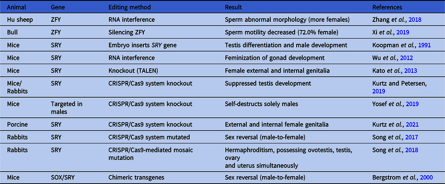
In recent years, more intensive studies on the mechanism of mammalian sex determination have been conducted and a series of genes and signalling pathways related to sex determination and differentiation have been identified. The transcription levels of these genes affect the expression of specific proteins at the meiotic stage, which plays critical role in animal sex determination, gonadal differentiation, reproduction, and development. Gene editing can therefore be used to control the sex of animal offspring (Schmid and Steinlein, Reference Schmid and Steinlein2001). Studies on sex control via gene editing are listed in Table 3.
Studies on sex control via gene editing

Zfy, located on the short arm (Yp) of the mammalian Y chromosome, is an important sex-determining gene as changes in its mRNA levels alter the sex ratio of offspring (Mitchell et al., Reference Mitchell, Simon, Affara, Ferguson-Smith, Avner and Bishop1989). RNA interference (RNAi) is a powerful research tool for gene silencing (Boettcher and McManus, Reference Boettcher and McManus2015). Xi and co-workers (2019) injected the Zfy interference vector into bull testis, using three injections. By modifying the expression of the Zfy gene during spermatogenesis, semen was collected and used for the artificial insemination of dairy cows. Results showed that, after exposure to interference vector, sperm motility decreased (P < 0.01), but the sperm density was similar (P > 0.05) to that of the non-treated control semen. Injection of the Zfy gene interference vector resulted in 72.0% female offspring, and was greater than the 49.4% female calves in the control (P < 0.01). Zhang and colleagues (Reference Zhang, Du, Sun, Wang, Liu, Xi, Li, Ying, Jiang, Wang, Shen and Jia2018) used RNA interference (RNAi) to generate ZFY-interrupted Hu sheep by injecting short hairpin RNA (shRNA) into round spermatids. ZFY mRNA expression was decreased significantly in Hu sheep with interrupted ZFY compared with wild-type Hu sheep. The sex ratio of lambs also exhibited a bias toward females. The resulting spermatozoa exhibited abnormal sperm morphology, including spermatozoa without tails and others with head and tail abnormalities. Therefore, further research is needed if this approach is to be used in actual livestock production.
SRY is a specific sex-determining gene located on the short arm (Yp) of the Y chromosome, which is currently believed to be a testicular determining factor serving as a master switch for mammalian sex determination (Mäkelä et al., Reference Mäkelä, Koskenniemi, Virtanen and Toppari2019). Structurally, the SRY gene consists of a single exon, which encodes for testis-determining transcription factor (TDF). The SRY gene contains a centrally located HMG domain, which is presumed to be the main functional domain of the SRY protein. This protein is expressed in the male genital ridge at the time of sex determination and serves as the main factor triggering the sex determination cascade. Its expression causes the formation of primary precursor cells of tubuli seminiferi, leading to the development of testes from undifferentiated gonads (Hogarth et al., Reference Hogarth, Itman, Jans and Loveland2005). Koopman and colleagues (1991) successfully introduced a part of SRY sequence into mouse embryos and, in the resulting offspring, female mice showed male phenotypes with normal size and weight with no reproductive ability. It was shown that the 14-kb mouse genomic DNA fragment containing SRY is sufficient to guide the formation of testis in XX transgenic embryos, and subsequently produce complete phenotypic sex reversal in XX transgenic adults. Wu and co-workers (2012) injected a recombinant RNAi vector into the tail vein of pregnant female mice to affect SRY expression. The study revealed that knocking down SRY resulted in feminization of gonad development in mouse embryos and that the Sox9 and Wt1 genes were directly regulated by the SRY gene. Kato and colleagues (2013) generated a knockout mouse for Sry, a sex-determining gene on the Y chromosome, using microinjection of transcription activator-like effector nuclease (TALEN) RNA into pronuclear-stage oocytes. The knockout mouse had female external and internal genitalia, a female level of blood testosterone and a female sexually dimorphic nucleus in the brain. The knockout mouse exhibited an estrous cycle and performed copulatory behaviour as females, although it was infertile or had reduced fertility. A male-to-female sex reversal was induced by a mutation of the Sp1-binding site in the 5′ flanking region of the rabbit SRY gene by microinjecting CRISPR/Cas9 plasmids into the cytoplasm of pronuclear-stage zygotes. A dramatically reduced number of follicles (only one follicle was found) and a smaller size of ovaries, accompanied by reduced expression of the SRY gene, was observed in SRY knockout rabbits compared with the male wild-type (Kato et al., Reference Kato, Miyata, Sonobe, Yamashita, Tamano, Miura, Kanai, Miyamoto, Sakuma, Yamamoto, Inui, Kikusui, Asahara and Takada2013). The above results indicated that SRY plays an important role in sex determination and that sex transformations can be achieved by editing SRY. However, this process often affects reproductive development and causes infertility.
Yosef and co-workers (2019) used the clustered regularly interspaced short palindromic repeats/CRISPR-associated protein 9 (CRISPR/Cas9) system to encodes a functional Cas9 protein on an autosomal chromosome, whereas the paternal line encoded guide RNAs on the Y chromosome targeting vital mouse genes. After fertilization, the presence of both the Y-encoded guide RNAs from the paternal sperm and the Cas9 protein from the maternal egg targeted the vital genes in males. They showed that these genes were specifically targeted in males and that this breeding consequently self-destructed solely males (Yosef et al., Reference Yosef, Edry-Botzer, Globus, Shlomovitz, Munitz, Gerlic and Qimron2019). Furthermore, explorations on the sex-determining genes in X and Y sperm have revealed additional genes, including Sox9, Dmrt1, WT1, SF1, Gata4, DAX1, and AMH (Sekido and Lovell-Badge, Reference Sekido and Lovell-Badge2009). Sex determination is a cooperative process governed by multiple genes. A deletion or mutation in any sex-determining gene will greatly effect the sex of individual animals. Although the technology of animal sex control via gene editing has existed for many years, there is still significant progress to be made for satisfactory results that can be applied to animal production.
Conclusion
In animal husbandry, the use of livestock sex control technology to produce livestock of the expected sex could enable maximization of milk production by female animals and meat production by male animals. Current research has revealed differences in the DNA content between X and Y sperm, but little information is known about other potential distinguishing characteristics of the two sperm types, including the basic idea of the spermatozoal structure, physical characteristics, chemical characteristics, and functional differences. Currently, there is only one method used for commercial sex sorting, which consists of the individual separation of X- and Y-chromosome-bearing sperm using flow cytometry, however sperm vitality decreases after staining and sorting. This development requires a focus on improving hardware, optics, electronics, and handling protocols before flow cytometry such as reducing the sheath pressure of the sorter, implementation of pulsed lasers, or transportation of red deep sperm at high sperm concentration without staining before sorting (Xie et al., Reference Xie, Xu, Wu and Hong2020). A report using TLR7/8 antibody to sort X/Y sperm has also attracted the attention of researchers. As mentioned by Navarro-Costa and colleagues (2020), TLR7/8 biology in vivo must differ significantly from this in vitro situation to allow these genes to persist in the genome. The most parsimonious explanation for the TLR7/8 conundrum is that both genes escape sharing and this asymmetric distribution is not selected against, because both receptors have no functional significance for in vivo fertilization (Navarro-Costa et al., Reference Navarro-Costa, Molaro, Misra, Meiklejohn and Ellis2020). When taking changing the pH of the dilution solution for enrichment of X/Y sperm as an example, the change in pH will affect the movement of X and Y sperm, and then cause the ratio of X/Y sperm to be enriched in the upper layer to change. It can be hypothesized that exposure of spermatozoa to external stress results in their differential survival, however it is unclear whether stress provides a selective survival advantage to X or Y sperm (You et al., Reference You, Kwon, Saidur Rahman, Park, Kim and Pang2017).
Researchers are attempting to use gene-editing technology to produce male animals that produce only normal X or Y sperm, which could enable the control of offspring sex to improve the efficiency of animal husbandry. The CRISPR/Cas system can identify and induce mutations on target loci with high specificity. This system is superior to the use of zinc-finger nucleases (ZFNs) and TALENs due to its ease of use and cost–effectiveness. Although these gene editors cannot be used in livestock production presently, among them the CRISPR/Cas system is the most suitable technique for sex selection. Gene-editing technology requires highly skilled personnel, and the lack of a clear role of the sex-determining gene in livestock sex determination hinders its application in this field. However, optimization of the existing gene editors, the development of new gene editors, and continued research on the sex-determining gene will aid in breaking the barrier (Xie et al., Reference Xie, Xu, Wu and Hong2020). In addition, RNAi is a simple method that has had a significant effect on the expression of the sex-determining gene. Future studies should therefore explore the mechanisms of sex-determining genes in spermatogenesis and sex determination, improve transfection efficiency, and reduce electropuncture damage (Douglas and Turner, Reference Douglas and Turner2020). In addition, researchers have focussed on the development of sex-controlled DNA vaccines. The theory behind this concept is that isolation/identification of such a marker(s) could lead to antibodies then being developed against X- and/or Y-specific surface protein(s). Subsequently, the use of magnetic beads, affinity chromatography, and sperm identification FACS techniques would provide a batch separation process for the same result (Yadav et al., Reference Yadav, Gangwar, Singh, Tikadar, Khanna, Saini, Dholpuria, Palta, Manik, Singh and Singla2017). However, the latter three methods are still at the research stage and their application in animal husbandry still faces some challenges.
There is reason to believe that with continuous progress in life sciences, particularly with the integration and development of interdisciplinary new technologies and methods, resolution of related issues (including the biological characterization of X and Y sperm), clarification and tracing of the in vitro development of mammalian sperm, determination of metabolic differences between X and Y sperm and their sex determination mechanisms and the establishment of efficient, fast, accurate, and economical sex control methods are not far away. Methodological research and technological innovation in this field will have important impacts on animal husbandry and human reproductive health control in the future.
Acknowledgements
This research was supported by grants from Shaanxi Provincial Agricultural Science and Technology Research Project [2020NY-019].
Author contributions
QFH wrote the original manuscript. MH, XYC, and KZ discussed the main ideas and structure of the manuscript. JL and FSQ edited the manuscript.
Declaration of competing interest
The authors report no declarations of interest.