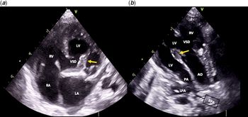
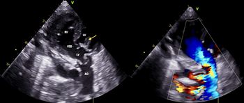
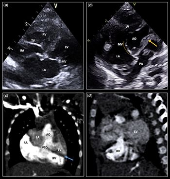
A three-month-old infant, born at 33 weeks’ gestation, and weighting 2070 g (10–50th percentile) presented to our hospital with central cyanosis (peripheral oxygen saturation of 80%) and tachypnoea. Cardiac auscultation revealed a grade 3/6 systolic ejection murmur at the left upper sternal edge. The two-dimensional echocardiogram showed normal situs and atrio-ventricular connections. He had a large subaortic ventricular septal defect. Both great arteries had their origin in a side-by-side fashion from the morphological left ventricle with the aorta dextroposed and anterior to the pulmonary artery (D-malposition). There was a subpulmonary conus separating both great arteries (Fig 1). The pulmonary valve was dysplastic and stenotic, anterior to the muscular conus, and distant from the ventricular septal defect. The continuous-wave Doppler study showed a 20 mmHg gradient across the subpulmonary conus, a 50 mmHg gradient across the pulmonary valve, and streaming of blood from the right ventricle to the aorta causing pulmonary hypoperfusion (Fig 2). An angio thoracic CT confirmed the superior–inferior cardiac ventricular arrangement. The superiorly positioned ventricle was identified as the left ventricle due to its smooth endocardial surface with aortic-mitral continuity (Fig 3b). The inferiorly positioned ventricle was more trabeculated (Fig 3d) and its valve inserted more apically (Fig 3a), hence, the right ventricle. Another rare finding is the evidence of a double-outlet left ventricle, as in most cases of superior–inferior cardiac ventricles, a double-outlet right ventricle is a more common anomaly.
(a) 2D echocardiogram, 4-chamber view, failed to show arterial-ventricular concordance. A large ventricular septal defect (VSD) was the only outlet of the right ventricle (RV). A subpulmonary conus (yellow arrow) divided the left ventricle (LV) in two chambers. The right (RA) and left atria (LA) are separated by an intact atrial septum. (b) 2D echocardiogram, modified parasternal long axis view, shows both great arteries arising from the left ventricle in a parallel fashion. The aorta (AO) is anterior and to the right of the pulmonary artery (PA). Semilunar valves are separated by a fibrous ridge namely subpulmonary conus (yellow arrow). LV = left ventricle; RV = right ventricle; VSD = ventricle septal defect; LPA = left pulmonary artery; RPA = right pulmonary artery.

2D echocardiogram with color Doppler study, subcostal view: aorta (AO) arising from the left ventricle (LV) in close relationship to the ventricular septal defect (VSD) causing streaming of deoxygenated blood from the right ventricle (RV) to the aorta. Subpulmonary conus (yellow arrow) is also seen.

(a) 2D echocardiogram, subcostal 4-chamber view: tricuspid valve (TV) related to the right ventricle (RV) with the annulus being inserted more inferiorly toward the cardiac apex than the mitral valve (MV). (b) 2D echocardiogram, subcostal 5-chamber view shows the MV in relationship with the left ventricle (LV) with the mitral-aortic continuity being evident (yellow line). The pulmonary artery (PA) is left-sided and posterior to the aorta (AO). The semilunar valves are separated by a fibrous ridge, namely subpulmonary conus (yellow arrow) partially dividing the left ventricle. (c and d) Angio thoracic CT: superior-inferior ventricular arrangement. The LV is characterized by smooth endocardial surface and is posterosuperior to the RV, with is distinctive trabeculated endocardial surface. The interventricular septum (blue arrow) is horizontal. The figure c shows also streaming of blood from the RV through the subaortic ventricular septal defect (VSD) to the AO.

Acknowledgements
None.
Financial Support
This research received no specific grant from any funding agency, commercial, or not-for-profit sectors.
Conflicts of Interest
None.