INTRODUCTION
Trypanosoma evansi is a digenetic flagellate implicated in the infection of a large number of domestic and wild animals, such as equines, canines, felines, rabbits, capybaras, ring-tailed coatis, bovines and buffaloes (Dávila and Silva, Reference Dávila and Silva2000; Herrera et al. Reference Herrera, Davila, Norek, Abreu, Souza, Andrea and Jansen2004; Tarello, Reference Tarello2005; Da Silva et al. Reference Da Silva, Costa, Lopes and Monteiro2008) and humans (Joshi et al. Reference Joshi, Shegokar, Power, Herder, Katti, Salkar, Dani and Bhargava2005). This protozoan is the agent of trypanosomosis, a disease with broad distribution in Africa, Asia, and Latin America (Lun and Desser, Reference Lun and Desser1995). The trypomastigotes present in blood vessels of vertebrate hosts are transmitted by blood-sucking insects during feeding. The insect vectors are most commonly tabanide species (Tabanus sp., Chrysops sp., and Hematopota sp.) and vampire bats (Hoare, Reference Hoare1972).
Two features of the disease were reported in Brazil: the acute syndrome, responsible for the death of equines and non-treated canines, and the chronic syndrome, which affects many wild animals such as Hydrochaeris hydrochaeris and Nasua nasua (Herrera et al. Reference Herrera, Davila, Norek, Abreu, Souza, Andrea and Jansen2004). The acute form is characterized by intermittent fever, subcutaneous widespread oedema, progressive anaemia and blindness. Clinical signs disappear during the subacute phase and the trypanosomosis may often go undiagnosed during clinical examination. Accurate diagnosis is only possible during the chronic stage of the disease, where clinical signs are more evident and the animal's condition is more severely affected (Silva et al. Reference Silva, Seidl, Ramirez and Dávila2002).
Adenosine deaminase (ADA: EC 3.5.4.4) is considered to be a key enzyme in purine metabolism, catalysing the irreversible deamination of adenosine and deoxyadenosine to inosine and deoxyinosine, respectively and closely regulating extracellular adenosine concentrations (Franco et al. Reference Franco, Casado, Ciruela, Saura, Mallo, Canela and Llouis1997). Adenosine deaminase activity has been detected on the surface of haematopoietic cells (Aran et al. Reference Aran, Colomer, Matutes, Vives-Corrons and Franco1991). Researchers have described a family in which there is a dominantly inherited form of haemolytic anaemia associated with a notable increase of ADA activity in erythrocytes but with normal ADA levels in other blood cells, including lymphocytes (Valentine et al. Reference Valentine, Paglia, Tartaglia and Gilsanz1977).
ADA is present in all cell types, but high activity is present in the thymus, lymphoid tissues and peripheric lymphocytes. It has been demonstrated that this enzyme plays an important role in lymphocyte function and is essential for the normal growth, differentiation and proliferation of T lymphocytes (Franco et al. Reference Franco, Casado, Ciruela, Saura, Mallo, Canela and Llouis1997; Codero et al. Reference Codero, Salgado, Fernández-Alonso, Herrera, Lluis, Franco and Nogueira2001). The observation that ADA deficiency leads to severe combined immunodeficiency syndrome points to the physiological importance of controlling extracellular adenosine levels in the immune system (Aldrich et al. Reference Aldrich, Blackburn and Kellems2000).
Anaemia caused by T. evansi is often described and is characterized by decreased levels of erythrocytes, haemoglobin and haematocrit. However, its causes are not completely understood (Silva et al. Reference Silva, Herrera, Domingos, Ximenes and Dávila1995; Aquino et al. Reference Aquino, Machado, Alessi, Marques, Castro and Malheiros2002). In infections by this protozoan, leukocyte changes are described as neutropenia, neutrophilia, monocytosis, lymphopenia, lymphocytosis (Silva et al. Reference Silva, Herrera, Domingos, Ximenes and Dávila1995; Marques et al. Reference Marques, Machado, Alessi, Aquino and Pereira2000; Wolkmer et al. Reference Wolkmer, Da Silva, Carnelutti, Paim, Traesel, Lopes and Monteiro2009).
Considering the functions of ADA in leukocytes and the haematopoietic system, this study aimed to evaluate the activity of this enzyme in serum, erythrocytes and lymphocytes of rats infected with T. evansi.
MATERIALS AND METHODS
A total of 30 adult rats, males, with a mean age of 90 days and weighing on average 300 (±29) grams were used in this study. The animals were kept in cages with 10 animals each, in a room experiment with controlled temperature and humidity (25°C; 70%). They were fed with commercial ration and water ad libitum. All animals received a formulation containing pyrantel pamoate, praziquantel and fenbendazole and were submitted to a period of 15 days of adaptation. All animals were apparently healthy when the experimental period began (day 0).
These rats were divided into 3 groups of 10 animals each. The rats in groups A and B were inoculated intraperitoneally (Day 1) with a strain of T. evansi that had been obtained from a naturally infected dog (Colpo et al. Reference Colpo, Monteiro and Stainki2005) and had been kept in liquid nitrogen. The number of inoculated flagellates was estimated by using a Neubauer chamber (Wolkmer et al. Reference Wolkmer, Da Silva, Carnelutti, Costa, Traesel, Lopes and Monteiro2007). This study aimed to evaluate the acute and chronic disease in the rat, so the infectious dose used for groups A and B was 2×10⁶ trypomastigotes/animal in fresh blood and cryopreserved blood, respectively (Da Silva et al. Reference Da Silva, Wolkmer, Gressler, Otto, Bess, Tavares, Zanette and Monteiro2009a). The collection of samples from animals in group A was performed on day 4 post-infection (p.i.) and group B was on day 20 p.i. Group C (negative control) consisted of healthy rats, not infected by T. evansi, but received a physiological solution administered in the same way. Group C was divided into 2 groups (C1 and C2) and the material was collected on day 4 and day 20 p.i. in order to compare with the infected groups (A and B). Parasitaemia was estimated daily by microscopic examination of smears. Each slide was mounted with blood collected from the tail vein, stained by the panoptic method, and visualized at a magnification of 1000×.
The animals were anaesthetized in a chamber with isoflurane for collection of blood by cardiac puncture (8 ml). The storage of the samples was considered according to the analysis. Thus, part of the material collected was allocated in tubes containing anticoagulant for separation of lymphocytes (4 ml), separation of erythrocytes (2 ml) and analysis of haemogram (1 ml). The volume of 1 ml was stored in a tube without anticoagulant to obtain serum.
Erythrocyte count, haematocrit (Ht), haemoglobin concentration (Hb), mean corpuscular volume (MCV), mean corpuscular haemoglobin concentration (MCHC) and total leukocytes were evaluated. Smears were mounted and stained by the panoptic method. The determination of microhematocrit was performed according to the technique described by Feldman et al. (Reference Feldman, Zinkl and Jain2000), and blood smears were stained with the panoptic method to perform differential leukocyte counts. Erythrocyte counts and haemoglobin concentrations were determined using an electronic counter.
Erythrocytes were obtained from whole blood with EDTA, according to the technique described by Hostetter and Johnson (Reference Hostetter and Johnson1989). Erythrocytes were resuspended to haematocrit in Hepes-buffer at 10%. Lymphocytes were also obtained from whole blood with EDTA by gradient separation using Ficoll-Histopaque™ plus, according to the technique described by Böyum (Reference Böyum1968). The samples stored in tubes without anticoagulant were centrifuged for 10 min, and the serum was obtained. The ADA activity was measured immediately after obtaining the erythrocytes, lymphocytes and serum.
ADA activity was measured spectrophotometrically in serum, lymphocytes and erythrocytes by the method of Giusti and Gakis (Reference Giusti and Gakis1971). The reaction was started by addition of the substrate (adenosine) to a final concentration of 21 mmol/l and incubations were carried out for 1 h at 37°C. The reaction was stopped by adding 106 mmol/l/0·16 mmol/l phenol-nitroprusside/ml solution. The reaction mixtures were immediately mixed to 125 mmol/l/11 mmol/l alkalinehypochlorite (sodium hypochlorite) and vortexed. Ammonium sulphate at a concentration of 75 μmol/l was used as ammonium standard. The ammonia concentration is directly proportional to the absorption of indophenol at 650 nm. The specific activity is reported as U/L in serum and lymphocytes and U/g of Hb in erythrocytes.
The data were submitted to one-way analysis of variance (ANOVA) followed by the Tukey's test (P<0·05). The effect of ADA in erythrocytes and lymphocytes on haematocrit and lymphocytes was analysed by linear regression, respectively. The analyses were performed using SAS statistical package (SAS Institute, Cary, NC, USA) with a significance level of 5% (P<0·05).
The procedure was approved by the Animal Welfare Committee of Federal University de Santa Maria (UFSM), number 23081.012513/2009-52, in accordance to Brazilian laws and ethical principles published by the Colégio Brasileiro de Experimentação Animal (COBEA).
RESULTS
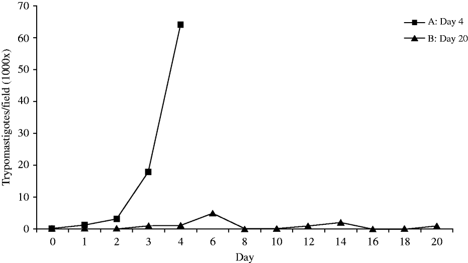
Examination of peripheral smear blood showed that the pre-patent period in experimentally infected rats varied between 24 and 72 h. The peak of parasitaemia at day 4 p.i. in group A (63 trypanosomes per microscopic field at 1000×magnification) and irregular waves of parasitaemia were observed in group B, ranging from 0 to 3 trypomastigotes per microscopic field (Fig. 1). Seven (7/10) rats in group A demonstrated apathetic slow movements, disorientation and gasping on day 4 p.i. In group B, the animals did not show any clinical change, neither did the non-infected rats (group C).
Parasitaemia of Trypanosoma evansi in infected rats at day 20 post-inoculation. The collection of material was performed in groups A and B on day 4 and day 20 post-infection at high and low parasitaemia, respectively.

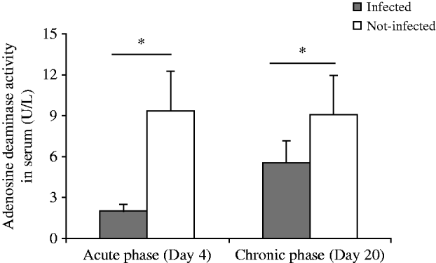
Means and standard deviation of the adenosine deaminase activity in serum of rats infected with Trypanosoma evansi (day 4 and day 20 p.i.) compared to non-infected rats (n=10). (*P<0·05).

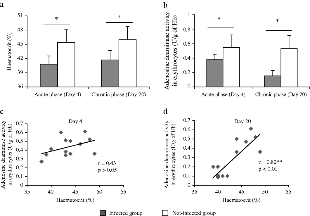
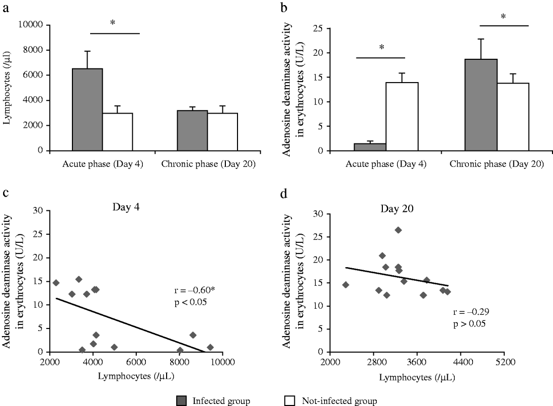
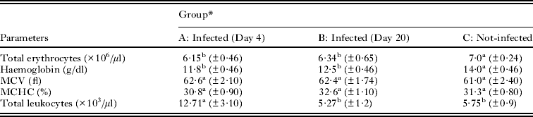
Haematological changes were observed in the infected rats as a decrease in haematocrit (Fig. 3a), erythrocyte count and haemoglobin content (Table 1 – P<0·05). The mean MCV and the mean MCHC did not differ among groups, characterizing a normocytic–normochromic anemia (Table 1). Simultaneously with haematological changes, the rats of group A showed leukocytosis (Table 1) and lymphocytosis (Fig. 4a), as compared to groups B and C.
Means and standard deviation of the haematocrit (a) and adenosine deaminase activity in erythrocytes (b) of rats infected with Trypanosoma evansi (day 4 and day 20 p.i.) compared to non-infected (n=10). Linear regression analysis of individual infected rat haematocrit with adenosine deaminase activity in erythrocytes during the acute phase (c) and chronic phase (d). (*P<0·05.)

Means and standard deviation of the lymphocytes (a) and adenosine deaminase activity in lymphocytes (b) of rats infected with Trypanosoma evansi (day 4 and day 20 p.i.) compared to non-infected (n=10). Linear regression analysis of individual infected rat number of lymphocytes with adenosine deaminase activity in lymphocytes of the acute phase (c) and chronic phase day 20 (d). (*P<0·05.)

Means and standard deviation of the haematological parameters of rats experimentally infected with Trypanosoma evansi

* Means in the same line followed by different letters are statistically different among them by Tukey's test at 5% probability.
The ADA activity in serum was reduced in the groups infected with T. evansi compared to the non-infected rats (P<0·001 – Fig. 2). In erythrocytes, the ADA activity was reduced on day 4 and day 20 (Fig. 3b), but was more pronounced after 20 days when the parasitaemia was low (Fig. 1). In the chronic phase a positive correlation (r 2: 0·82) was observed between the ADA activity in erythrocytes and the haematocrit values (P<0·001 – Fig. 3d).
In lymphocytes, the activity of ADA was reduced in the acute phase (day 4), when the parasitaemia was high, but showed an increase of ADA activity after 20 days (Fig. 4b), when the parasitaemia was low (Fig. 1) and the number of lymphocytes was normal (Fig. 4-a). In the acute phase a negative correlation (r 2: −0·60) was observed between the ADA activity in lymphocytes and total number of lymphocytes (P<0·05).
DISCUSSION
Many studies have reported changes in adenosine transport and ADA activity associated with infections by Trypanosoma brucei, T. evansi, T. vivax, Leishmania donovani and L. infantum (Okochi et al. Reference Okochi, Abaelu and Akinrimisi1983; De Koning and Jarvis, Reference De Koning and Jarvis1999; Suswam et al. Reference Suswam, Ross and Martin2003; Tripathi et al. Reference Tripathi, Kumar, Bharti, Kumar, Shrivastav, Sundar and Pai2008). However, a study that correlates the acute and chronic effect of experimental infection with T. evansi in the ADA activity in the serum, erythrocytes and lymphocytes of rats has not been found in the literature.
An increased ADA activity has been observed in serum samples, erythrocytes, leukocytes and in plasma haemoglobin concentrations with vivax malaria as compare to the control group (Ozcan et al. Reference Ozcan, Abdurrahim, Adnan, Senel and Necmeddin1997). Authors described significantly increased ADA activity in visceral leishmaniasis patients compared to healthy controls (Khambu et al. Reference Khambu, Mehta, Rijal, Lamsal, Majhi and Baral2007). Researchers showed that intraperitoneal injection of the adenosine analogue cordycepin (3´-deoxyadenosine) for treatment of the encephalitic stage of human African trypanosomosis, together with an adenosine deaminase inhibitor (coformycin or deoxycoformycin), cures T. brucei infection in mice (Rottenberg et al. Reference Rottenberg, Masocha, Ferella, Petitto-Assis, Goto, Kristensson, McCaffrey and Wigzell2005). In this study we demonstrate that ADA activity may be associated with trypanosome infection, since the T. evansi-infected rat had reduced enzyme activity both in blood cells and in serum.
In the acute phase of this study we observed a reduction of ADA activity in serum, erythrocytes and lymphocytes of rats infected with T. evansi compared to healthy rats. At this stage, rats had 4 days of infection and high parasitaemia. The reduction in ADA activity would have caused an increase in the extracellular concentrations of adenosine, which would then be converted to inosine. Adenosine acts as a sensor and provides information to the immune system regarding the tissue damage or acute inflammatory changes occurring in the vicinity of the immune system (Kumar and Sharma, Reference Kumar and Sharma2009). The reduction in ADA activity in lymphocytes would lead to interaction of adenosine with adenosine receptors that exist in many cell types, with possible anti-inflammatory effects, among them the inhibition of the Th1 immune response. In acute infection caused by T. cruzi there is a predominance of Th1 and cellular response with production of interferon-γ (Kumar and Tarleton, Reference Kumar and Tarleton2001). Therefore, inhibition of this response by the action of extracellular adenosine in purinergic receptors could be a compensatory effect, attenuating inflammation and tissue damage. The treatment of macrophages with interferon-γ upregulates the expression of the adenosine receptor, A2B, and the activation of A2B receptors is involved with the deactivation of macrophages, possibly through an increase of cAMP (Xaus et al. Reference Xaus, Valledor, Card, Marques, Beleta, Palacois and Celada1999). This reinforces the concept of the anti-inflammatory action of adenosine as a way to preserve cells and tissues.
The ADA may be expressed as an ectoenzyme on the surface of lymphocytes. In the serum there is another isoenzyme, ADA2, which has a low affinity for the substrate (Muraoka et al. Reference Muraoka, Katsuramaki, Shiraishi and Yokoyama1990). Thus, it will only increase its activity in higher concentrations of substrate, unlike the lymphocyte isoform. The concentration of extracellular adenosine in this study seems to be sufficient for activation of the enzyme in serum, assuming that it could be binding on the adenosine receptors in blood cells. Regarding the ADA activity in erythrocytes, reduction of enzyme activity seems to follow the decrease in total erythrocytes, which could be verified in future studies by observing the expression of ectoenzymes on the surface of red blood cells.
Researchers mentioned that red blood cells are relatively well supplied with ADA and circulating damaged erythrocytes release significant amounts of ADA, a process that may predispose to vaso-occlusive events (Muraoka et al. Reference Muraoka, Katsuramaki, Shiraishi and Yokoyama1990). Based on this information, we hypothesized that ADA activity was increased in serum and/or plasma, as a consequence of the decrease in red blood cells due to a haemolytic process, causing anaemia in trypanosomosis (Jackson et al. Reference Jackson, Mi, Koehler, Carcillo and Herzer1996). However, this was not observed in our study. Therefore, despite the positive correlation between haematocrit and activity levels in erythrocytes, further studies are necessary to confirm the role of ADA in anaemia caused by T. evansi in rats.
In addition, our experiment showed that in the chronic phase the parasitaemia had stabilized and fluctuated between 0 and 3 parasites per field (1000×). At the same time we found that the ADA activity in serum and erythrocytes was reduced in rats with trypanosomosis, and in lymphocytes ADA activity was increased when compared to non-infected rats. The low parasitaemia suggests that the compensatory effect of an anti-inflammatory like adenosine is no longer necessary. Thus, increased ADA activity in lymphocytes, reduces the extracellular concentrations of adenosine, promoting an inflammatory response that would be sufficient to contain the spread of the parasite without major tissue damage. The low concentration of extracellular adenosine also prevents activation of the isoforms present in erythrocytes and serum, probably due to a low affinity for the substrate.
Other workers recently observed that ADA activity in the serum of patients with idiopathic Parkinson's disease was significantly higher than in normal controls, suggesting that high serum ADA activity may be involved in the pathogenesis of Parkinson's disease via peripheral T-lymphocyte activation (Chiba et al. Reference Chiba, Matsumoto, Saitoh, Kasahara, Matsuya and Kashiwagi1995). An increase in serum ADA activity in patients with cutaneous leishmaniasis (Ozcan et al. Reference Ozcan, Abdurrahim, Salih, Vedat, Adnan and Yuksel1998), and a change in activity of this enzyme may also be related to pathogenesis of the parasite, also in Parkinson's disease. In contrast, this study showed reduced ADA activity in the serum of rats infected with T. evansi, compared with infection by Leishmania sp. (Ozcan et al. Reference Ozcan, Abdurrahim, Salih, Vedat, Adnan and Yuksel1998; Khambu et al. Reference Khambu, Mehta, Rijal, Lamsal, Majhi and Baral2007).
An important aspect to be discussed is that T. evansi caused an acute infection in rats, in contrast to a chronic infection in some rodents. These protozoa are Salivarian trypanosomes which are usually more virulent and pathogenic than Stercorarian trypanosomes (Menezes et al. Reference Menezes, Queiroz, Gomes, Marques and Jansen2004). The detection of parasitaemia in rats (24 h) occurred earlier than previously reported in experimentally infected rats (Queiroz et al. Reference Queiroz, Cabello and Jansen2000; Al-Mohammed, Reference Al-Mohammed2006; Omer et al. Reference Omer, Mousa and Al-Wabel2007), probably due to the high pathogenicity of this strain. Although authors reported that R. norvegicus is a suitable model for the study of the parasitaemic wave of T. evansi (Queiroz et al. Reference Queiroz, Cabello and Jansen2000), the typical undulating course of parasitaemia was observed in group B of this experiment. According to Da Silva et al. (Reference Da Silva, Wolkmer, Gressler, Otto, Bess, Tavares, Zanette and Monteiro2009a) the longevity of rats may be related to the type of inoculum used. When they are made by successive infections in rats, there is an acute phase with high parasitaemia and death of animals within 5 days p.i. Now, when the inoculum used was cryopreserved in liquid nitrogen the longevity of rodents could increase considerably, resulting in a chronic phase. In rats the disease was characterized by high levels of parasitaemia along with clinical signals of apathy, weakness, ataxia and severe anaemia (Wolkmer et al. Reference Wolkmer, Da Silva, Carnelutti, Paim, Traesel, Lopes and Monteiro2009), similar to what occurred in the acute phase of this experiment.
Previous studies from our laboratory showed that in the infection with T. evansi haematological changes are commonly related to other factors, but major changes depend on the degree of parasitaemia and period of infection (Wolkmer et al. Reference Wolkmer, Da Silva, Carnelutti, Costa, Traesel, Lopes and Monteiro2007; Da Silva et al. Reference Da Silva, Zanette, Wolkmer, Costa, Gressler, Faccio, Santurio, Lopes and Monteiro2009b). Our research group has already found decreased activity of acetylcholinesterase in erythrocytes in cats (Da Silva et al. Reference Da Silva, Spanevello, Stefanello, Wolkmer, Costa, Zanette, Lopes, Santurio, Schetinger and Monteiro2010) and an increased lipid peroxidation in rats (Wolkmer et al. Reference Wolkmer, Da Silva, Carnelutti, Paim, Traesel, Lopes and Monteiro2009) infected with T. evansi. It is important to state that these alterations might be related to pathogenesis of the disease or may just be a consequence of anaemia, because adenosine is related to maturation of erythrocytes (Franco et al. Reference Franco, Aran, Colomer, Matutes and Vivis-Corrons1990; Jackson et al. Reference Jackson, Mi, Koehler, Carcillo and Herzer1996). This question also persists for the decreased ADA activity in erythrocytes found in this study.
Based on these results, we conclude that parasitism by T. evansi alters the activity of ADA in serum, erythrocytes and lymphocytes of experimentally infected rats, suggesting that trypanosomiasis can interfere with purinergic signalling. Other studies need to be performed to verify the expression of ectoenzymes on the surface of red blood cells and lymphocytes in trypanosomiasis, in order to understand the relationship of the ADA with anaemia and lymphocytosis in this disease.