Only a few years ago, the differential diagnosis of mitochondrial disorders with cardiomyopathy based on clinical and biochemical approaches led to a merely limited number of possible diagnoses.Reference Honzik, Tesarova and Magner 1 The arrival of next-generation DNA sequencing methods challenged this concept – for instance, infantile- or childhood-onset mitochondrial cardiomyopathy alone has been associated with mutations in >50 genes.Reference Finsterer and Kothari 2 Meanwhile, various studies have documented the presence of cardiomyopathy in many mitochondrial diagnoses, and the spectrum is growing rapidly. In 2015, nine new mitochondrial disorders with cardiomyopathy or known mitochondrial disorders with new phenotypes including heart involvement were recognised.Reference Powell, Kopajtich and D’Souza 3 – Reference Brisca, Fiorillo and Nesti 11 Cardiomyopathy is very frequent in mitochondrial disorders, with a minimum occurrence of 20–30%.Reference Holmgren, Wahlander, Eriksson, Oldfors, Holme and Tulinius 12 , Reference Yaplito-Lee, Weintraub, Jamsen, Chow, Thorburn and Boneh 13 This frequency varies with age, being more frequent in neonates and children – for example, 40% of 120 neonates in the study by Honzik et alReference Honzik, Tesarova and Magner 1 and 58% of 113 children in the study by Scaglia et alReference Scaglia, Towbin and Craigen 14 – compared with adults with various mitochondrial DNA or nuclear DNA mutations – for example, cardiac involvement was present in 30% of 109 patients in the study by Wahbi et alReference Wahbi, Bougouin and Behin 15 . The prognosis of neonates with mitochondrial disorders and cardiomyopathy is poor with usually fatal outcomes within the 1st year of life. In the largest study of Scaglia et alReference Scaglia, Towbin and Craigen 14 , there was an 18% survival rate in children with cardiac involvement versus 95% survival rate in children without cardiac manifestation at the 16-year horizon; however, some exceptions exist – for example, transmembrane protein 70 deficiency or Barth syndrome.Reference Honzik, Tesarova and Mayr 16 – Reference Magner, Dvorakova and Tesarova 18 This fact places extreme importance on rapid clinical and molecular diagnosis.
The mitochondrial thymidine kinase 2 is involved in the first step of deoxypyrimidine nucleoside phosphorylation required for mitochondrial DNA replication, and its disturbance results in overall mitochondrial DNA depletion. The phenotypic spectrum of thymidine kinase 2 deficiency is broad, but its principal clinical feature is isolated progressive muscle disease.Reference Chanprasert, Wang and Weng 19 Heart hypertrophy is extremely rare: only two patients have been reported thus far.Reference Chanprasert, Wang and Weng 19 Incorrect tRNA aminoacylation caused by the deficiency of mitochondrial alanyl-tRNA synthetase 2 is another rare cause of perinatal or infantile fatal cardiomyopathy, with only eight patients reported thus far.Reference Gotz, Tyynismaa and Euro 20 , Reference Taylor, Pyle and Griffin 21 In this study, we characterise in detail the third and ninth known patients with thymidine kinase 2 and alanyl-tRNA synthetase 2 deficiencies, respectively, with lethal cardiomyopathy and provide a review of the literature. Exome sequencing was used for rapid diagnosis, enabling prenatal diagnosis within the affected families.
Methods
Enzymatic analyses
Heart and muscle mitochondria were isolated from bioptic or autoptic tissues by differential centrifugation.Reference Darley-Usmar, Rickwood and Wilson 22 The activities of the respiratory chain complexes and the activity of the control enzyme citrate synthase were determined spectrophotometrically.Reference Rustin, Chretien and Bourgeron 23 , Reference Srere and Brooks 24 Blue-Native electrophoresis was used to separate mitochondrial membrane protein complexes on polyacrylamide 6–15% (w/v) gradient gels.Reference Schagger and von Jagow 25
Histochemistry
Muscle tissues were snap-frozen in isopentane (2-methylbutane; Sigma–Aldrich Co., Munich, Germany) and cooled in liquid nitrogen. To evaluate the histopathological changes in the muscle tissue, at least five 3-µm cryosections from each tissue block were examined by routine haematoxylin–eosin staining and a conventional spectrum of histochemical reactions was evaluated, including adenosine triphosphatase, at pre-incubation pH values of 10.4, 4.6, and 4.3, nicotinamide adenine dinucleotide dehydrogenase-tetrazolium reductase, succinate dehydrogenase, and periodic acid-Schiff with diastase digestion, detailed elsewhere.Reference Sheehan and Hrapchak 26
Molecular-genetic analyses
Quantification of mitochondrial DNA in muscle was performed by the method previously detailed by Pejznochova et alReference Pejznochova, Tesarova, Honzik, Hansikova, Magner and Zeman 27 . Exome sequencing of the trio – proband-case 1 and unaffected parents – was performed at Axeq (Rockville, Maryland, United States of America) using an Illumina HiSeq 2000 system (Illumina, San Diego, California, United States of America) and SeqCap EZ Exome Enrichment kit v3.0 (Roche NimbleGen, Indianapolis, Indiana, United States of America). Raw sequence reads were aligned to the reference genome (hg19) using Novoalign version 2.08.03 (Novocraft, Selangor, Malaysia) with the default parameters. The conversion of sequence alignment map format to binary form of sequence alignment map and duplicate removal were performed using Picard Tools (1.99). Sequence variants in the analysed samples were identified using GATK package version 3.3. The high-confidence variants list was annotated using ANNOVAR annotation tool (hg19).
Targeted sequencing of the characterised mitochondria-related genes was performed for case 2 on a SOLiDTM 4 System (Life Technologies, Foster City, California, United States of America) using a custom SeqCap Enrichment kit according to the manufacturer’s protocol. Sequence reads were aligned to the reference genome (hg19) using NovoalignCS version 1.08 (Novocraft, Selangor, Malaysia) with the default parameters. The conversion of sequence alignment map format to the binary form of sequence alignment map, duplicate removal, and sequence variants were identified using the sequence alignment map tools package version 0.1.12. The high-confidence variants list was annotated using ANNOVAR annotation tool (hg19).
For further analyses, we prioritised sequence variants present in affected individuals that were not found in unaffected parents and that had a frequency <0.05 in the dbSNP, 1000 Genomes, Exome Variant Server, ExAC, and internal exome databases. Candidate variants were visualised using the Integrative Genomics Viewer version 2.3.32. TK2 and AARS2 mutations were confirmed by Sanger sequencing in both probands and their parents.
Case reports
Case 1 – a girl with thymidine kinase 2 deficiency
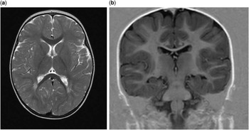
A child was born at term after an uncomplicated pregnancy by spontaneous delivery as the second child of healthy, unrelated parents. Her birth weight (3800 g) and length (50 cm) were normal. Her development was uneventful until the age of 13 months, when she presented with regression of gross motor development and hypotonia. Neurological symptoms progressed into severe myopathy with serious peripheral hypotonia and markedly decreased deep tendon reflexes. Failure to thrive required feeding via gastrostomy. She suffered from respiratory failure during an acute respiratory infection at the age of 18 months and remained dependent on ventilatory support. Psychomotor development was arrested. Brain MRI documented diffuse leucoencephalopathy (Fig 1a and b). Echocardiographic studies performed at the age of 18 and 20 months revealed progressive hypertrophic cardiomyopathy with an interventricular septum (148–178%) and left ventricular posterior wall thickening (148–182%), with preserved contractility and ejection fraction. Laboratory findings included mild lactic acidosis (lactate 4.2 mmol/L, controls <2.1), elevated creatine kinase (10.7 μkat/L, controls <3.1), and hepatopathy (alanine aminotransferase 2.04 μkat/L, controls <0.6; aspartyl aminotransferase 1.73 μkat/l, controls <0.6). She succumbed at the age of 22 months because of cardiac arrest.
Extensive, diffuse signal abnormalities in cerebral white matter with subcortical preponderance. ( a ) T2-weighted and ( b ) T1-weighted images in a 14-month-old girl with thymidine kinase 2 deficiency.

Case 2 – a girl with alanyl-tRNA synthetase 2 deficiency
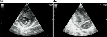
The first child of healthy, non-consanguineous parents was born at term after an uneventful pregnancy with a birth weight of 2720 g and length of 47 cm. The foetal ultrasound revealed no sign of heart involvement. There were no complications immediately after birth. Her condition worsened at the 12th hour of life, when severe respiratory distress requiring intubation occurred. She presented with severe hypotonia, lethargy, and poor feeding. Echocardiography showed hypertrophic cardiomyopathy with progressive thickening of the interventricular septum (115–155%) and left ventricular posterior wall (135–155%), which was documented together with Wolff–Parkinson–White syndrome by cardiological examination (Fig 2a and b). Laboratory findings showed severe lactic acidosis (lactate level up to 29 mmol/L, controls <2.1), elevated creatine kinase (41.43 μkat/L, controls <3.1), and poor glycaemic control (levels between 1.5–22.4 mmol/L). Cardiorespiratory failure led subsequently to death at the age of 9 weeks.
Echocardiography findings in a 8-week-old girl with alanyl-tRNA synthetase 2 deficiency (from Case 2) ( a ) Left ventricle in the short-axis view. ( b ) Left ventricle in the long-axis view. Both images show left ventricular hypertrophy to ~155% of standard values.

Results
Case 1 – thymidine kinase 2 deficiency
The respiratory chain complex activities in isolated muscle mitochondria showed profound reduction in the activities of complex I (122 nmol/min/mg of protein, controls 194–354) and complex IV (190 nmol/min/mg of protein, controls 287–1077) with decreased ratio to citrate synthase (0.33, controls 1.1–2.22). Blue-Native electrophoresis confirmed the decrease in the amount of complex IV. Severe destructive myopathy with cytochrome c oxidase-negative fibres in the muscle and hypertrophy of the heart tissue were found at autopsy (heart weight 68 g, controls 56 g). At autopsy, histological analysis of brain tissue revealed demyelination and neuronal degeneration, mainly in the cerebellum (brain weight 820 g, controls 1060 g). Liver histology showed steatosis. Targeted sequencing of the mitochondrial exome revealed heterozygosity for missense mutations c.416 C>T (p.Ala139Val) and c.209T>C (p.Phe70Ser) in the TK2 gene. The mutation c.209T>C is novel, and the variant was not present in the ExAC database or the 1000 genome project and was predicted to be pathogenic by in silico analysis using MutationTasterReference Schwarz, Cooper, Schuelke and Seelow 28 , PolyPhen 2Reference Adzhubei, Schmidt and Peshkin 29 , and SIFT.Reference Kumar, Henikoff and Ng 30 A significantly diminished mitochondrial DNA copy number in the muscle, 12% of age-matched controls (n=8), was found.
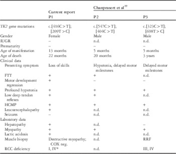
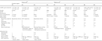
The clinical and laboratory data of our patient are summarised and compared with previously described patients with thymidine kinase 2 deficiency manifesting with hypertrophic cardiomyopathy in Table 1.
Clinical and laboratory data in the Czech patient with thymidine kinase 2 (TK2) deficiency compared with other patients with this diagnosis manifesting hypertrophic cardiomyopathy.

COX neg.=cytochrome c oxidase-negative fibres; FTT=failure to thrive; HCMP=hypertrophic cardiomyopathy; IUGR=intrauterine growth retardation; n.d.=not determined; P=patient; RCC=respiratory chain complexes; RRF=ragged red fibres
* Reduced activities of complexes I and IV in muscle, 63 and 66% of lower reference range, respectively. Reduced amount of complex IV
Case 2 – alanyl-tRNA synthetase 2 deficiency
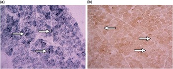
Biochemical analysis of a muscle biopsy and heart autopsy showed combined respiratory chain complex deficiency. Decreased activity of complex I (152 nmol/min/mg of protein, controls 194–354) and reduction in the amount of complex IV were found by Blue-Native electrophoresis and were the predominant findings in the muscle. In isolated mitochondria from the heart autopsy, a severe reduction in the activities of complex I+III (7 nmol/min/mg of protein, controls 50–140) and complex IV (94 nmol/min/mg of protein, controls 1000–1500) and decreased amounts of complex I, complex IV, and adenosine triphosphate synthase were observed (Table 2). Histological findings revealed general myocardial hypertrophy. Although the presence of typical, ragged red fibres was not observed in enzyme skeletal muscle histochemistry, the severity of the subsarcolemmal accumulation of succinate dehydrogenase product in a proportion of muscle fibres was profound for this age (Fig 3a). In cytochrome c oxidase histochemistry in the skeletal muscle (Fig 3b) and in the heart tissue, the reaction was weak in a portion of fibres, but no completely cytochrome c oxidase-negative fibres were found. Mild steatosis and pericellular centrilobular fibrosis were found in liver histology.
In muscle biopsy, ( a ) massive subsarcolemmal accumulation of succinate dehydrogenase product (exemplified by arrows) was observed in a portion of muscle fibres (succinate dehydrogenase histochemistry, original magnification 400×). ( b ) Focal subsarcolemmal accumulation of mitochondria was also observed in cytochrome c oxidase histochemistry. Cytochrome c oxidase-negative fibres were not observed; however, the reaction was weakened focally (cytochrome c oxidase histochemistry, original magnification 200×, indicated by arrows).

Clinical and laboratory data in the Czech patient with alanyl-tRNA synthetase 2 (AARS2) deficiency compared with the patients described in the literature.

COX neg.=cytochrome c oxidase-negative fibres; FTT=failure to thrive; HCMP=hypertrophic cardiomyopathy; IUGR=intrauterine growth retardation; n.a.=not applicable; n.d.=not determined; P=patient; RCC=respiratory chain complexes; RRF=ragged red fibres; SDH poz.=succinate dehydrogenase-positive blood vessels; W-P-W=Wolf–Parkinson–White syndrome
* Reduced activities of complex I in muscle, 78% of lower reference range. Reduced amount of complex IV; reduced activities of complexes I+III and IV in the heart, 14 and 9% of lower reference range, respectively. Reduced amount of complexes I, IV, and V
Whole-exome sequencing revealed the homozygous mutation c.1774C>T (p.Arg592Trp) in the AARS2 gene. The clinical and laboratory data on our patient are summarised and compared with previously described patients with fatal courses in Table 2.
Discussion
We present two different examples of early-onset mitochondrial cardiomyopathy due to deficiency in thymidine kinase 2 and alanyl-tRNA synthetase 2. Our findings expand the phenotypic and genetic spectrum of these mitochondrial disorders, but they also illustrate the demanding process of diagnosis due to their rarity and non-specificity of particular symptoms. Although gene targeting may be used in some mitochondrial disorders with heart involvement such as transmembrane protein 70 deficiency or Barth syndrome, the majority require direct indication by next-generation sequencing methods.
Thymidine kinase 2 is a nuclear-encoded enzyme with an intramitochondrial localisation. It catalyses the phosphorylation of deoxypyrimidine nucleotides in their salvage pathway. Therefore, mutations in the TK2 gene lead to the depletion of deoxynucleotides and cause a reduction in amounts of mitochondrial DNA in the affected tissues.Reference Copeland 31 The phenotypic spectrum of thymidine kinase 2 deficiency is broad, with isolated progressive muscle disease as a main clinical feature.Reference Chanprasert, Wang and Weng 19 Apart from the pure myopathic form of thymidine kinase 2 deficiency, other less-frequent symptoms have been described in the literature, including neurological features such as epilepsy, spinal muscular atrophy-like manifestation, proximal muscle weaknessReference Oskoui, Davidzon and Pascual 32 – Reference Galbiati, Bordoni and Papadimitriou 34 , chronic progressive external ophthalmoplegiaReference Tyynismaa, Sun and Ahola-Erkkila 35 or sensorineural hearing lossReference Marti, Nascimento and Colomer 36 , and hepatic involvement.Reference Chanprasert, Wang and Weng 19 , Reference El-Hattab, Scaglia and Mitochondrial 37 Progressive myopathy with gross motor development regression was also the main symptom in our patient. Further findings of hypertrophic cardiomyopathy are extremely rare in thymidine kinase 2 deficiency and have been reported in only two cases so farReference Chanprasert, Wang and Weng 19 and, as in our patient, led to early death (Table 1). From the laboratory perspective, increased activity of creatine kinase may be considered a constant finding in thymidine kinase 2 deficiency; on the other hand, lactic acidosis has been found in <50% of cases. This finding further illustrates the fact that the absence of lactic acidosis does not exclude the possibility of mitochondrial disorder.Reference Magner, Szentivanyi and Svandova 38 Hepatopathy, detected in our patient, is rare.Reference Chanprasert, Wang and Weng 19
Examination of the skeletal muscle, the most affected tissue, detected severe destructive myopathy with combined complex I and complex IV deficiencies (Table 1). Moreover, muscle histochemistry revealed numerous cytochrome c oxidase-negative fibres. No ragged red fibres were detected in the muscle of our patient. This finding contrasts with the current literature, in which cytochrome c oxidase-negative and/or ragged red fibres are frequently observedReference Mancuso, Salviati and Sacconi 33 , Reference Galbiati, Bordoni and Papadimitriou 34 , Reference Marti, Nascimento and Colomer 36 , Reference Spinazzola, Invernizzi and Carrara 39 – Reference Blakely, He and Gardner 43 , accompanied by severe reduction in the amounts of complexes I and IV.Reference Chanprasert, Wang and Weng 19 , Reference Mancuso, Salviati and Sacconi 33 , Reference Galbiati, Bordoni and Papadimitriou 34 , Reference Marti, Nascimento and Colomer 36 , Reference Gotz, Isohanni and Pihko 40 , Reference Blakely, He and Gardner 43
The predominant involvement of cerebral white matter (Fig 1) and autoptic findings of cerebellar neuronal degeneration and hepatopathy with hepatic steatosis in our patient are unique. Rapid loss of supratentorial white and grey matter with severe cerebral atrophy and cerebellar neuronal degeneration were noted in only one patient each.Reference Galbiati, Bordoni and Papadimitriou 34 , Reference Knierim, Seelow, Gill, von Moers and Schuelke 44 Hepatic steatosis was not previously mentioned in the literature, although fatty infiltration in muscle is quite common in patients with thymidine kinase 2 deficiency.Reference Galbiati, Bordoni and Papadimitriou 34 , Reference Marti, Nascimento and Colomer 36 , Reference Blakely, He and Gardner 43 , Reference Alston, Schaefer and Raman 45 More than 40 mutations in the TK2 gene have been found to date (HGMD professional);Reference Stenson 46 two missense mutations, c.416C>T and c.209T>C, were confirmed in our patient. Mutation c.416C>T has been previously described in a family with two affected siblings, both without documented cardiac involvement.Reference Galbiati, Bordoni and Papadimitriou 34 , Reference Knierim, Seelow, Gill, von Moers and Schuelke 44 The second mutation, c.209T>C, changing the highly conserved Phe70 into Ser, is novel.
The AARS2 gene encodes the enzyme alanyl-tRNA synthetase 2, which catalyses the alanine aminoacylation of a mitochondrial tRNA for alanine. The reaction involves a two-step mechanism that occurs in the catalytic domain of the enzyme. Alanine is first activated by adenosine triphosphate and subsequently transferred to the acceptor end of tRNA (Alanine). In addition, as aminoacyl-tRNA synthetases are multi-domain proteins, alanyl-tRNA synthetase 2 also edits incorrectly charged amino acids via its editing domain.Reference Konovalova and Tyynismaa 47 , Reference Sokabe, Ose and Nakamura 48 The phenotype of alanyl-tRNA synthetase 2 deficiency shows isolated myopathy, Charcott–Marie–Tooth syndromeReference McLaughlin, Sakaguchi and Giblin 49 , Reference Zhao, Hashiguchi and Hu 50 , or ovario-leucoencephalopathy with cerebellar atrophy.Reference Dallabona, Diodato and Kevelam 51 Cardiac involvement has been associated with severe forms of this disorder, reported in only eight children thus far. Hypertrophic cardiomyopathy and death within the 1st year of life were common characteristics in all the cases: four of them manifested shortly postnatally with no report of cardiac involvement on foetal ultrasound, and one documented case developed hypertrophic cardiomyopathy prenatally, which led to intrauterine death (see Table 2).Reference Gotz, Tyynismaa and Euro 20 , Reference Taylor, Pyle and Griffin 21 The Wolff–Parkinson–White syndrome observed in our case is a frequent finding in mitochondrial disorders – for instance, it was present in six out of 48 children with transmembrane protein 70 deficiencyReference Magner, Dvorakova and Tesarova 18 and four out of 30 patients with Mitochondrial myopathy, encephalopathy, lactic acidosis, and stroke-like episodes syndrome.Reference Sproule and Kaufmann 52 Skeletal muscle involvement was documented clinically, biochemically, and histologically. Surprisingly, creatine kinase was elevated >13 times above the upper normal level in the neonatal period (Table 2). Biochemical and histological findings overall revealed combined deficiencies of complexes I and IV and adenosine triphosphate synthase in our patient. These observations are in accordance with the findings of Götz et alReference Gotz, Tyynismaa and Euro 20 and Taylor et alReference Taylor, Pyle and Griffin 21 , showing combined respiratory chain complex deficiency with markedly decreased activity and, particularly, amounts of complexes I and IV in various tissues. The prevalent histological image is cardiac muscle hypertrophy.Reference Gotz, Tyynismaa and Euro 20 The previously documented finding of cytochrome c oxidase-negative fibres in the cardiomyocytes was absent in our patient; however, massive mitochondrial accumulation creating the impression of a ragged red fibre-like image in skeletal muscle has been documented for the first time in our case (Fig 2). The mutation c.1774C>A has been described previously in patients with a similar phenotypeReference Gotz, Tyynismaa and Euro 20 , Reference Taylor, Pyle and Griffin 21 and is considered to be a founder mutation carried by every patient with the cardiomyopathy phenotype.Reference Euro, Konovalova and Asin-Cayuela 53 The mutation changes Arg592 to Trp and is predicted to result in a dramatic reduction of the aminoacylation rate due to its effects on substrate binding.Reference Euro, Konovalova and Asin-Cayuela 53
Conclusion
Cardiomyopathy is a common symptom in neonates and infants with various mitochondrial disorders. We report on the third and ninth known patients with fatal hypertrophic cardiomyopathy caused by thymidine kinase 2 deficiency and mitochondrial alanyl-tRNA synthetase 2 deficiency. Severe leucoencephalopathy and hepatic steatosis in thymidine kinase 2 deficiency and the finding of ragged red fibre-like images in the muscle biopsy in alanyl-tRNA synthetase 2 deficiency are reported for the first time. Mitochondrial disorders must always be considered in the case of early-onset cardiomyopathy; however, diagnosis is demanding, often requiring methods of next-generation DNA sequencing.
Acknowledgement
None.
Financial Support
This study was supported by grants and by institutional research support from Charles University and General University Hospital in Prague PRVOUK-P24/LF1/3 (TH, MM), RVO-VFN 64165 (VKV, HH, JZ), SVV 260256/2016 (SM, AP), UNCE 204011 (AV), by grants GAUK 1402213 (AP), 14-36804G (MT, VS) from the Grant Agency of the Czech Republic, by the project LQ1604 NPU II and LM2015091 (VS) from the Ministry of Education of the Czech Republic and by 15-28208A (VS) from the Ministry of Health of the Czech Republic.
Conflicts of Interest
None.
Ethical Standards
This study was performed in accordance with the Declaration of Helsinki of the World Medical Association. Owing to the nature of the study, no ethics approval was requested from the local ethics committee. This study did not involve human and/or animal experimentation. Informed consent was obtained from the parents of our patients.