Introduction
Aspergillomas are rare, benign fungal balls which are most commonly associated with lung disease. They are caused by aspergillus species, fungi found in organic debris such as soil, rotting plants and food. Aspergillus fumigatus is the subtype most commonly found in aspergillomas.Reference Soubani and Chandrasekar1 In otolaryngology practice, aspergillomas are usually encountered in the paranasal sinuses, and treatment is surgical. Aspergillomas of the middle ear are extremely rare, with only two previously reported cases.Reference Supiyaphun, Sampatanukul and Sukumalpaiboon2, Reference Supiyaphun, Sampatanukul, Vongfu and Kerekhanjanarong3
In this article, we describe the first reported case of aspergilloma of the middle ear presenting with facial nerve palsy. The investigation and management of this condition are presented, and fungal infections in ENT practice are discussed.
Case report
A 72-year-old, diabetic man presented to the ENT department with a five-week history of worsening right-sided otalgia, aural discharge and hearing loss, associated with a five-day history of right-sided facial weakness and vertigo. His past medical history included a right tympanoplasty 30 years previously.
Examination revealed a red, inflamed external auditory meatus with a small collection of pus. The tympanic membrane was thickened but featureless. The patient had a right facial nerve palsy (House–Brackmann grade IV).
A diagnosis of malignant otitis externa was made, and the patient was commenced on broad spectrum antibiotics, antibacterial ear drops and hyperbaric oxygen therapy.
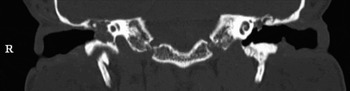
A computed tomography scan (Figure 1) showed extensive soft tissue opacification within the right middle ear and mastoid. There was no evidence of bony destruction.
Coronal computed tomography scan showing opacification of the middle ear. R = right

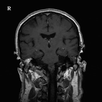
A magnetic resonance imaging scan (Figure 2) showed tissue enhancement in the temporal bone and middle ear, raising the possibility of residual cholesteatoma or acute suppurative otitis media.
Coronal magnetic resonance imaging scan showing enhancement of the right middle-ear cleft. R = right

A decision was made to insert a grommet into the right tympanic membrane in case suppuration was present. No pus was found during the procedure; however, a sheet of Silastic was found in the middle ear surrounded by grey, necrotic material. The Silastic was removed and a biopsy taken. The middle ear was also found to be obliterated by soft tissue.
Histological examination of biopsy material showed fungal hyphae, and A. fumigatus was cultured from an ear swab.
The patient's symptoms failed to resolve on antibiotic therapy, and his facial palsy deteriorated to House–Brackmann grade VI.
A formal exploratory tympanotomy was subsequently performed, which revealed inflammatory tissue in the middle ear and a large fungal pellet. The tympanic portion of the facial nerve was found to be dehiscent, with surrounding mucosal disease. There was no evidence of any invasive disease. The middle ear was cleared, and the mucosal disease dissected off the facial nerve.
Antibiotic treatment and hyperbaric therapy were stopped, and intravenous antifungal treatment commenced.
The patient's facial nerve function improved significantly on the day of surgery, to House–Brackmann grade III, and his pain improved dramatically over the next few days. He was discharged home on antifungal therapy.
On review in the clinic, the patient's otalgia had fully resolved and his facial weakness had improved further (to grade I to II).
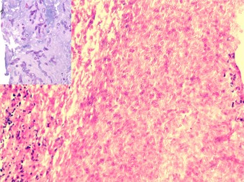
Histopathological examination of tissue taken at the time of surgery confirmed the presence of fungal hyphae and spores, consistent with a diagnosis of aspergilloma (Figure 3).
Photomicrograph of surgical specimen material showing abundant fungal hyphae (H&E; ×20). Inset shows branching hyphae, consistent with aspergillus (periodic acid Schiff; ×20).

Discussion
Aspergillus is one of the commonest airborne fungi, with A. fumigatus being the commonest species encountered. It has been estimated that humans inhale at least several hundred spores per day.Reference Hospenthal, Kwon-Chung and Bennett4 Individuals with normal immune function normally eliminate the fungus before it becomes established. If disease does occur in immunocompetent individuals, it is usually in the form of a non-invasive aspergilloma of the lung or allergic bronchopulmonary aspergillosis. An aspergilloma is defined as a spherical mass of fungal hyphae embedded in a matrix of proteinaceous material with sporing structures at the periphery.Reference Latgé5
In ENT practice, fungal infections are encountered most commonly in the paranasal sinuses. Mycotic disease can affect both the immunocompetent and the immunocompromised; however, the clinical presentation in these two groups varies greatly.
Non-invasive infection usually occurs in the immunocompetent, and can cause either allergic fungal sinusitis or a fungal ball. Fungal balls are usually unilateral and most commonly affect the maxillary sinus.Reference Grosjean and Weber6 Treatment with functional endoscopic sinus surgery is usually effective, and often no antifungal treatment is required.Reference Dufour, Kauffmann-Lacroix, Ferrie, Goujon, Rodier and Karkas7
Invasive disease occurs almost exclusively in immunocompromised patients, and causes an acute, necrotising fungal sinusitis. In this form of the disease, the fungus spreads along vascular channels destroying bone as it progresses, and invades surrounding structures such as the brain and orbit. Invasive fungal disease carries a poor prognosis. Treatment is with antifungal therapy, together with surgical debridement in selected patients.
Fungal colonisation of the ear is common in cases of non-invasive otitis externa, otitis media and post-operative infection. Fungus can be cultured in approximately 30 per cent of these infections. Aspergillus is found in 38 per cent of these positive cultures.Reference Kurnatowski and Filipiak8 However, localised, non-invasive aspergilloma of the middle ear is rare.
A Medline search was conducted to identify other reported cases, using the PubMed electronic database and Medical Subject Headings as the search string ‘[mycoses OR fungus AND ear]’. The retrieved abstracts were reviewed and relevant articles examined. This revealed only two other published cases reporting aspergilloma of the middle ear, both from the same unit in Thailand.Reference Supiyaphun, Sampatanukul and Sukumalpaiboon2, Reference Supiyaphun, Sampatanukul, Vongfu and Kerekhanjanarong3 One patient presented with gradual onset hearing loss and the other with chronic otorrhoea. Both cases occurred after previous tympanoplasty. There was no description of facial nerve weakness in either of these cases.
Thus, to the best of our knowledge, the current case represents the first report of aspergilloma of the middle ear presenting with facial nerve palsy.
• Aspergillomas are rare; in ENT practice, they are usually encountered in the paranasal sinuses
• There have been two previously reported cases of aspergilloma of the middle ear occurring as a complication of surgery, and presenting with chronic otorrhoea and hearing loss
• This paper describes the first reported case of aspergilloma of the middle ear mimicking necrotising otitis externa and causing facial nerve palsy
Facial nerve palsy caused by non-fungal middle-ear disease is well described. Around 8 per cent of the population have a dehiscent fallopian canal, such that acute or chronic suppurative otitis media, with or without cholesteatoma, and malignant otitis externa can all result in facial neuropathy. A dehiscent facial nerve can be congenital, or iatrogenic secondary to previous surgery.
In our patient, we believe that the sheet of Silastic discovered in the middle ear had been present for over 30 years and may have acted as a nidus for fungal infection. Silastic is still routinely used in ear surgery to restore the mucosal lining of the middle ear, and to prevent adhesions between the tympanic membrane and the promontory. Readers should be aware of this potential complication. It is possible that other implants, including those found in ossiculoplasty prostheses, and materials such as permacol, could behave in a similar fashion.
In this article, we present a rare case of an aspergilloma of the middle ear mimicking necrotising otitis externa. It is important to consider alternative diagnoses in patients with presumed necrotising otitis externa who fail to respond to conventional therapy. A low threshold for tympanotomy is suggested for patients in whom the diagnosis is in doubt.
Acknowledgements
We would like to thank Dr Manish Powari, Consultant Histopathologist at Derriford Hospital, for his help in producing the illustrations for this paper.