Introduction
Hypoglossal–facial nerve anastomosis, first described by Körte in 1903,Reference Körte1 is a well-known technique for treating total facial nerve paralysis. There are three options: end-to-end, end-to-side and split anastomoses.Reference Mohamed, Omi, Honda, Suzuki and Ishikawa2 Variations on the original end-to-end procedure were developed to better preserve tongue function, whilst still restoring facial movement. No consensus exists in the literature regarding which technique is the most effective.Reference Mohamed, Omi, Honda, Suzuki and Ishikawa2, Reference Han, Suh, Kim, Cho and Moon3 Indeed, some evidence suggests a trade-off, such that better facial function is at the cost of tongue movement.Reference Han, Suh, Kim, Cho and Moon3
We report on an individual with a complex otological history who presented with bilateral lower motor neuron House–Brackmann grade VI facial nerve paralysis that was refractory to conservative measures, including steroids and intensive facial physiotherapy. He was treated with sequential bilateral end-to-side hypoglossal–facial nerve anastomoses with an interpositional jump graft for facial reanimation. This staged technique required preservation of hypoglossal nerve function, and is therefore more appropriate for an end-to-side or split anastomotic technique. As bilateral lower motor neuron facial nerve palsy is rare, the use of bilateral hypoglossal–facial nerve anastomoses for facial reanimation is described infrequently in the global literature.
Case report
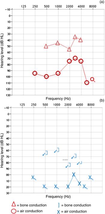
A 46-year-old gentleman was referred to our tertiary care multidisciplinary facial palsy unit with a history of bilateral chronic ear disease for several years. This first started in childhood and required multiple operations in both ears. He had a significant smoking history, which had led to peripheral vascular disease and emphysema, but he was not diabetic or immunocompromised. His bilateral open mastoid cavities had been stable for many years, and although his hearing had deteriorated (Figure 1), he had not sought ENT input. Both ears had recently started discharging again over a period of 18 months, alongside recurrent tinnitus and vertiginous episodes. Symptoms were more troublesome in his right ear on presentation to a district general hospital ENT unit.
Pre-operative audiology findings in the patient's (a) right ear and (b) left ear.

Computed tomography (CT) scanning revealed a possible right lateral semi-circular canal fistula. Radical mastoidectomy, blind sac closure and a bone-anchored hearing aid were suggested. The patient declined such a radical intervention and instead underwent a right-sided revision modified radical mastoidectomy.
Surgery revealed no clear labyrinthine fistula; however, poor quality, possibly osteomyelitic bone was seen over the lateral semi-circular canal, with exposed middle fossa dura. The cavity was obliterated with bone pâté placed over the lateral semi-circular canal. An examination of the left ear was also performed, which showed exposed bone in the floor of the outer ear canal, with a scarred and retracted ear drum. The patient was started on a long course of oral ciprofloxacin, 500 mg twice daily, for presumed osteomyelitis.
Six days post-operatively, the patient developed a contralateral left-sided House–Brackmann grade IV facial palsy, which was managed with steroids and the palsy improved to grade III. Unfortunately, after a month, there was regression to grade VI palsy.
The patient then underwent left revision mastoidectomy and facial nerve decompression as an emergency. This revealed a lateral semi-circular canal fistula on the left side, which was not visible on imaging. This was repaired with fascia and bone pâté. Post-operatively, facial nerve function improved to grade IV palsy with steroids and continued oral antibiotics. There was no clear documentation of cholesteatomas during either ear surgery conducted at the district general hospital.
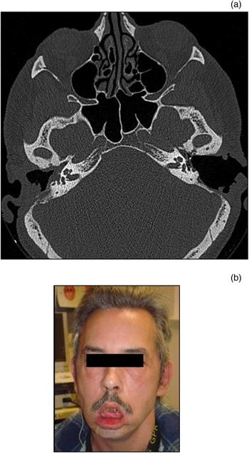
Two months after right-sided ear surgery, the patient developed a right-sided House–Brackmann grade IV lower motor neuron facial palsy, which a course of steroids and ongoing antibiotics did not improve. This left him with bilateral grade IV facial nerve palsies, associated with poorly healing mastoid cavities (Figure 2), with a reasonable but gradually dropping cochlear reserve. This prompted referral to our tertiary unit for further management.
(a) Pre-operative axial computed tomography scan showing bilateral mastoid cavities and a possible right-sided lateral semi-circular canal fistula, and (b) a clinical pre-operative photograph of the patient.

By the time of the review, the patient had deteriorated to a bilateral House–Brackmann grade VI complete facial nerve palsy, with progressive hearing loss, balance disturbance and tinnitus. Audiology revealed an essentially dead left ear. There was severe mixed hearing loss in the right ear, with bone conduction averages of 45 dB HL and air conduction averages of 80 dB HL. Although resurgence of chronic ear disease with osteomyelitis was suspected, such rapid deterioration of facial motor function associated with inner-ear impairment prompted medical input. Immunological, connective tissue and systemic disease were all investigated and ruled out.
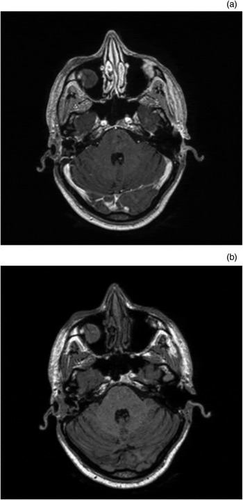
By this stage, he had suffered from paralysis for seven months on the left side and five months on the right side. Imaging of the skull base revealed that the facial nerve was no longer in continuity on the left side, with erosion of the labyrinth extending into the horizontal and proximal vertical portions, but still in continuity on the right (Figure 3). No underlying cause for the progressive facial nerve injury was identifiable on imaging.
(a & b) Pre-operative axial magnetic resonance imaging scans, showing erosion of the facial nerve on the left side.

The patient agreed to undergo bilateral hypoglossal–facial nerve anastomosis conducted in a sequential fashion, with the left side being performed first. An end-to-side technique with a jump interpositional greater auricular nerve graft was used.
At the six-week review, the patient had normal tongue function. The decision was therefore taken to perform on the right side two months later. This procedure also utilised an end-to-side hypoglossal–facial nerve (XII–VII) anastomosis with an interpositional greater auricular nerve graft.
During convalescence from bilateral nerve grafting, repeat swabs from his mastoid cavities had grown Aspergillus fumigatus, predominantly from the right ear. A CT scan of the chest revealed possible secondary aspergillus on the background of emphysema. Voriconazole was commenced with referral to the National Aspergillosis Centre in Manchester. The patient continued to improve on this treatment.
When his ears were considered clear of infection, the patient subsequently underwent a successful right-sided cochlear implant and blind sac closure. A left-sided cochlear implant could not be performed because of extensive cochlear fibrosis, likely due to a previous lateral semi-circular canal fistula.
With intensive facial physiotherapy and oro-motor rehabilitation, the patient's facial movements gradually improved over 18 months. He achieved bilateral House–Brackmann grade IV facial motor function, and learned to dissociate facial and tongue movements. There was no significant morbidity regarding tongue movement.
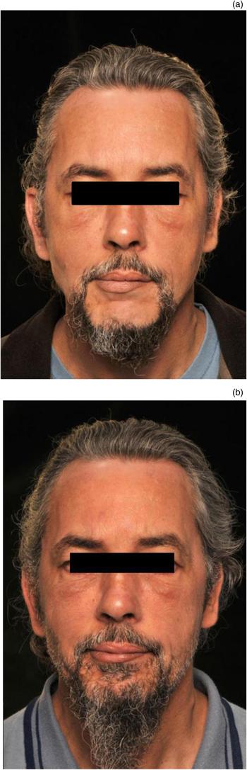
Ongoing follow up continues at the facial palsy clinic and involves physiotherapy, now exceeding 28 months’ duration. At present, he has bilateral grade IV facial motor function. This has significantly improved his quality of life compared to the bilateral debilitating total paralysis (Figure 4).
(a & b) Clinical post-operative photographs showing the patient's recovery.

Discussion
Managing bilateral lower facial nerve palsies is extremely difficult. Various techniques have been described for facial reanimation, including hypoglossal–facial nerve anastomosis using interposition cable grafts. There is considerable discussion in the literature regarding the best type of hypoglossal–facial nerve anastomosis for maximal function post-operatively in cases with a unilateral deficit.Reference Mohamed, Omi, Honda, Suzuki and Ishikawa2, Reference Han, Suh, Kim, Cho and Moon3 Variability in outcomes relates to the underlying aetiology, the duration of palsy, co-morbidities, surgical skill and the technique used, and the nature of rehabilitation.
In direct end-to-end procedures, tongue movements are completely sacrificed to allow full thickness anastomosis of the nerves, with the aim of optimal facial function. However, this may not be the most effective method.Reference Han, Suh, Kim, Cho and Moon3 End-to-side anastomoses, both with and without an interpositional jump graft, are equally effective at improving facial movements, whilst retaining tongue function.Reference May, Sobol and Mester4, Reference Beutner, Luers and Grosheva5 In bilateral facial palsy, it is essential to preserve tongue function, as a bilaterally de-innervated tongue causes significant issues with mastication, speaking and swallowing. Therefore, an end-to-side anastomosis would be the only reasonable technique in this individual case.
• Bilateral facial nerve palsy is a rare but seriously disfiguring condition
• There is very little world literature on the management of these cases
• This paper reports a case managed with bilateral hypoglossal–facial nerve anastomosis, with good outcomes
An interpositional jump graft does introduce possible difficulties, as the presence of two anastomoses theoretically delays re-innervation quality and timescale.Reference Guntinas-Lichius, Streppel and Stennert6 There is also loss of sensation over the auricular region. If an adequate length of viable facial nerve is obtainable, with or without a cortical mastoidectomy, it seems prudent to use this for a single anastomosis. However, in many cases the precise level of the facial nerve injury is unknown, and therefore this may not be feasible. In our patient, neither the location of facial nerve injury nor the primary aetiology of the facial nerve palsy was clearly known at the time of repair. Therefore, an interpositional jump graft was utilised to bypass the facial nerve in the middle-ear cleft.
Cases of bilateral House–Brackmann grade VI facial palsies are extremely rare, and are described infrequently in the literature. In 1991, May et al. described a series with 3 bilateral facial palsies among 30 patients: 2 in children with brainstem tumours, and 1 in a patient with bilateral temporal bone fractures.Reference May, Sobol and Mester4 They used a similar jump interpositional graft and sequential procedure when adequate tongue function was assured on the first side. Whilst our patient did not regain full function, he has a much-improved quality of life. The surgery enabled transformation from a devastating bilateral grade VI palsy to a manageable grade IV weakness. Similar outcomes were described by May et al. in the patient with bilateral temporal bone fractures who subsequently underwent bilateral hypoglossal–facial nerve anastomoses.Reference May, Sobol and Mester4
Conclusion
We present a rare case of bilateral total facial paralysis in a patient with a complex history of bilateral chronic middle-ear disease. Treatment involved bilateral hypoglossal–facial nerve anastomoses when multidisciplinary conservative measures failed. This resulted in reasonable outcomes for the patient, significantly improving his quality of life. Such complex cases should be managed by a dedicated facial palsy team, with access to facial physiotherapy and rehabilitation services to support facial reanimation surgery. Our experience emphasises the need for early appropriate referral to a specialist centre when the patient has a complex history with progressive complications.
Competing interests
None declared