Case report
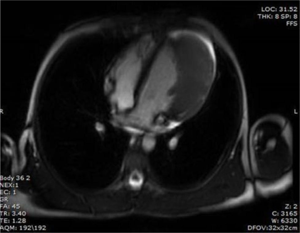
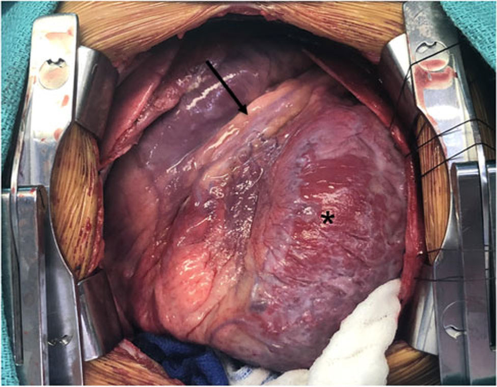
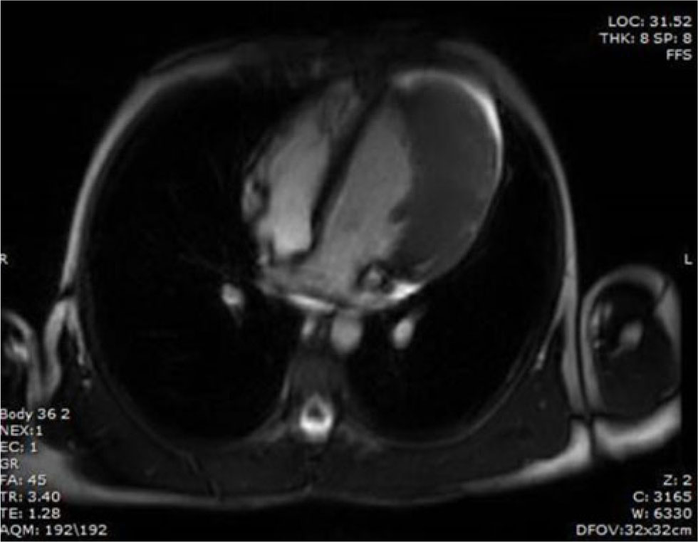
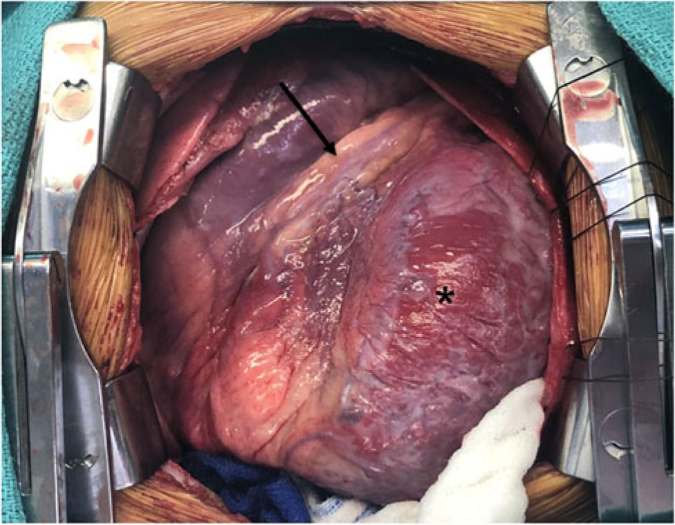
An 11-year-old male with no past medical history presented to a local emergency room with a chief complaint of cough. He had no intercurrent illness or complaints of chest pain, palpitations or exertional fatigue. There was no family history of sudden cardiac death, unexplained syncope or cardiomyopathy. A chest radiograph to rule out a pneumonia revealed cardiomegaly, for which he was referred to a cardiologist. Aside from the benign aforementioned clinical history, his examination was notable only for a slight palpable displacement of his apical point of maximum impact. His dermatologic examination revealed no rashes or café au lait spots. An electrocardiogram revealed sinus rhythm with inverted T waves in the lateral leads. An echocardiogram showed good biventricular function but a very thickened posterior–lateral wall of the left ventricle. A cardiac MRI with gadolinium revealed a 10.3-cm infiltrating, avidly enhancing mass of the left ventricle extending from the apex to the base and invading the anterolateral and inferolateral left ventricle myocardium (Fig 1). The right ventricle was normal. The remainder of the chest portion of the MRI demonstrated no lymphadenopathy. All inflammatory markers were normal. While in the hospital on telemetry, he had no ventricular ectopy. A decision was made that a definitive diagnosis necessitated tissue. Given the location and infiltrative nature of the mass, the biopsy was performed via a median sternotomy. Initial attempts while performing a core biopsy caused significant bleeding. Therefore, biopsies were done sharply with a knife, and the bleeding sites were sutured with adequate haemostasis. The tumour extended from the base to the apex of the heart to the left of the left anterior descending artery and posterior with a number of epicardial arteries and veins involved (Fig 2). The pathological specimen revealed ill-defined aggregates of closely packed thin-walled capillaries filled with blood cells. Given the infiltrative nature of the tumour and no discernible tissue planes, the tumour could not be resected. His post-operative course was unremarkable. Conservative medical management was initiated with propranolol and aspirin, and he had placement of an implantable loop recorder prior to discharge. A follow-up stress test 4-week post-surgery showed no concerning ST segment changes or ventricular ectopy. Monthly interrogations of his implantable loop recorder have not revealed any alarming ventricular arrhythmias. He has currently been followed for over 1 year and remains asymptomatic.
Horizontal long-axis image from a steady state free precision (SSFP) sequence demonstrating the infiltrative T2 hypointense mass involving the lateral wall and apex.
Operative picture of the patient’s heart. The patient’s head is towards the top of the picture. The asterisk depicts the tumour. The arrow points to the left anterior descending artery.
Discussion
Cardiac haemangiomas can occur at any age and constitute roughly 2.8% of cardiac tumours, with the left ventricle being a less likely chamber to present than the right-sided heart structures.Reference Li, Teng, Xu, Ma and Ni1 We believe that this is the largest reported left ventricle haemangioma presenting in a pre-adolescent patient. While the diagnosis and invasiveness of the tumour can be seen by CT or MRI, it is equally important to have an appreciation of the coronary flow pattern into these vascular tumours. Coronary CT angiogram demonstrated uniform dilatation of the left main, left anterior descending, and first diagonal coronary arteries feeding the large infiltrative tumour. The origin of haemangiomas is believed to represent some form of either a true neoplasm or hamartoma. While haemangiomas are relatively common on the skin of infants, they may also occur in internal organs but are rarely seen as an isolated cardiac tumour in children.Reference Liu, Hong, Zhang, Xu, Liu and Qiu2–Reference Delmo Walter, Javier, Sander, Hartmann, Ekkernkamp and Hetzer4 A review of 200 post-mortem adult cases of cardiac haemangiomas revealed 23.1% originated in the left ventricle, with the right atrium being the most common location.Reference Li, Teng, Xu, Ma and Ni1 Clinical manifestations of a primary vascular cardiac tumour vary from individuals being asymptomatic to some experiencing shortness of breath, chest pain, palpitations or exertional fatigue.Reference Li, Teng, Xu, Ma and Ni1 Our patient was asymptomatic at both rest and with exercise.
It is not uncommon for cardiac conditions such as a cardiomyopathy to be incidentally discovered in children and adolescents, while undergoing a chest radiograph for an unrelated finding or during a screening electrocardiogram where left ventricular hypertrophy or repolarisation abnormalities may be identified. While chest radiographs may be helpful to identify cardiomegaly, in the majority of cases where a cardiac haemangioma was subsequently identified, the heart size was initially reported as normal.Reference Li, Teng, Xu, Ma and Ni1, Reference Oueida, Lui, Al-Refae and Al-Omran5 The appreciation of cardiomegaly on the chest radiograph of this patient likely reflects the significant size of the mass (10.3 cm).
Similar to chest X-rays, electrocardiograms have mixed utility in patients with cardiac tumours. The finding of inverted T waves in the lateral leads may be seen with some cardiomyopathic processes with repolarisation changes or in other conditions such as arrhythmogenic right ventricular dysplasia. While our patient had inverted T waves in the lateral leads, it is not uncommon for electrocardiograms to be normal in patients with cardiac haemangiomas. One study found that electrocardiograms were normal in 36.5% of patients, with 40% showing non-specific T wave changes, 15.3% showing atrial–ventricular block and 9.4% showing diffusely low voltage.Reference Li, Teng, Xu, Ma and Ni1 One patient with cardiac haemangioma developed subsequent cardiac arrest, presumably secondary to ventricular fibrillation. Their only pertinent findings were isolated premature ventricular beats.Reference Miyake, Del Nido and Alexander6 There is clearly no pathognomonic electrical abnormality seen with vascular tumours, but rather a collection of non-specific abnormalities, which are likely dependent on lesion location.
Tumours that have strong gadolinium enhancement include angiosarcoma, pheochromocytoma or rhabdomyosarcoma. However, these were all unlikely given the age of the patient and the asymptomatic clinical presentation. Most cardiac haemangiomas show pedunculated lesions or polypoid lesions that can easily be resected.Reference Moniotte, Geva, Perez-Atayde, Fulton, Pigula and Powell7, Reference Esmaeilzadeh, Jalalian, Maleki, Givtaj, Mozaffari and Parsaee8 This particular mass, however, was quite unique and constituted a size comparable to the entirety of the left ventricle and was completely infiltrative within the muscle thus affording no curable plane for resection.
While resection is preferable in patients with symptoms and cardiac haemangiomas, the nature of this particular tumour made resection prohibitive. Aside from atrial–ventricular block and ventricular arrhythmias, sudden cardiac death has been reported in a healthy 15-year-old adolescent whose autopsy showed a large cardiac haemangioma at the apex.Reference Zerbo, Argo, Maresi, Liotta and Procaccianti9 However, other case reports have offered a conservative observational approach with no adverse sequelae.Reference Gribaa, Slim, Neffati and Boughzela10 Currently, this patient is being managed with beta-blockers, aspirin, and has an implantable loop recorder to monitor for any ventricular arrhythmias. Cardiac haemangiomas are benign tumours and as such lack the ability to metastasise so if they do grow it tends to be at an appreciably slower rate. Yet, given the rarity of this lesion and especially this size, the natural history remains unpredictable in that they can involute, stop growing or proliferate indefinitely. Heart failure does not seem to be a common clinical presentation either early or late for patients with an isolated haemangioma. The presence of a haemopericardium or non-sustained ventricular arrhythmias in the presence of a non-resectable tumour should prompt consideration for heart transplantation.
In summary, this case represents a rare presentation of an already rare paediatric cardiac tumour. The size of this tumour is believed to be the largest cardiac haemangioma seen in a pre-teen patient. This case shows the value, albeit non-specific, in non-invasive testing such as radiographs and electrocardiograms in identifying cardiomegaly and left ventricular hypertrophy. The size of this tumour and its infiltrative nature prohibited a resection. In the absence of symptoms, a conservative approach with careful attention to any ventricular arrhythmias or development of a pericardial effusion should be balanced against heart transplantation.
Acknowledgments
None.
Financial Support
This research received no specific grant from any funding agency, commercial or not-for-profit sectors.
Conflict of Interest
None.
Ethical Standards
Signed informed consent from the patient’s mother was obtained and documented.