Introduction
Although an aberrant origin of the subclavian artery from the aorta is common, an aberrant subclavian artery arising from the pulmonary artery is an extremely rare anomaly of the aortic arch and can result in various manifestations such as neurological symptoms, maldevelopment or underdevelopment of the upper extremity and cardiac stress. Reference Bae, Kang and Choo1 It can be present as an isolated lesion or additional finding in the setting of complex CHD like interrupted aortic arch. However, some cases remain asymptomatic and are only detected incidentally at an advanced age. Reference Malakan Rad and Pouraliakbar2 If not treated, it can result in subclavian steal syndrome or pulmonary steal syndrome. Reference Miller, Campbell, Barker and Hill3
Malformations of the aortic arch arteries can be explained by the double aortic arch hypothesis proposed by Edwards and are often associated with CHD. Reference Edwards4 Here, the differentiation of the fourth and sixth aortic arches, as well as the partial regression of the sixth aortic arch, seems crucial.
A left isolated subclavian artery from the pulmonary artery is commonly associated with a right aortic arch and occurs in only 0.8% of cases with right aortic arch. It was first described in 1908 by Ghon et al. Reference Ghon5 Due to the regression of the left aortic arch, the left subclavian artery arises from the pulmonary artery via the patent ductus arteriosus. It is particularly associated with tetralogy of Fallot, coarctation of the aorta, and left pulmonary stenosis. Reference Sen, Mohanty, Kulkarni and Rao6
Right isolated subclavian artery from the pulmonary artery is caused by the regression of the fourth aortic arch, specifically due to the loss of connection between the right aortic arch, right common carotid artery, and right subclavian artery. In these cases, the right ductus remains patent, connecting the right subclavian artery to the pulmonary artery. It is even more rare and four times less common as the isolated right subclavian artery. Reference Bae, Kang and Choo1
We report three cases with different isolated subclavian arteries. To explain the exact pathology, the following schematic diagrams of our patients are provided (Figure 1).
Schematic explanation of the patient’s anatomy. First case (left): right subclavian artery (RSA) branching of the right pulmonary artery. Second case (middle): brachiocephalic trunk with left common carotid artery (LCCA) and left subclavian artery (LSA) originates from the pulmonary artery. Third case (right): interrupted aortic arch type C with hypoplastic ascending aorta: right common carotid artery (RCCA) originates from ascending aorta, RSA originates from right pulmonary artery. LCCA, LSA, and descending aorta originate from pulmonary artery through patent ductus arteriosus.

1st Case: K.M
A 1-year-old female patient with haemodynamically relevant patent ductus arteriosus, small ventricular septal defect, and atrial septal defect type II was presented for interventional patent ductus arteriosus closure via cardiac catheterisation. During the procedure, several aortopulmonary collaterals were identified, and coils were implanted in the three largest collaterals. The following angiographic imaging showed a complex collateral convolute branching from the major arterial vessels, which communicated with the right subclavian artery, arising from the pulmonary artery (Figure 2). As a result, the right subclavian artery was perfused in a retrograde fashion through this collateral network, explaining the low blood pressure measured in the patient’s right arm. A significant left-to-right shunt was present from the collateral network into the pulmonary artery, leading to volume overload of the left ventricle. Transthoracic echocardiography showed turbulent shunt flow with a left-to-right shunt opening into the right pulmonary artery at the bifurcation.
Catheterisation of the right common carotid artery (RCCA) and angiographic visualisation of retrograde filling of right subclavian artery (RSA), pulmonary artery (PA) and vascular convolute. The white arrow marks the stenotic junction of RSA and right PA.

Cardiovascular surgery was performed to reimplant the right subclavian artery. Right subclavian artery was divided from the pulmonary artery, and the stump was ligated and oversewn. The right subclavian artery was reimplanted into the ascending aorta near the origin of the right common carotid artery.
Postoperative transthoracic echocardiography showed laminar flow within the pulmonary arteries and aortic arch. The right subclavian artery anastomosis to the aortic arch demonstrated near-laminar flow. Additionally, the right subclavian artery and right common carotid artery showed laminar flow and there was no blood pressure differences between the two arms afterwards.
2nd Case: B.A
A 33-year-old male patient with trisomy 21 and tetralogy of Fallot, without cyanotic spells, and a right aortic arch, presented for planning of corrective surgery. There was a reduced blood pressure in the left arm, and transthoracic echocardiography revealed a false origin of the brachiocephalic trunk from the pulmonary artery with stenosis before it enters the pulmonary artery. Additionally, there was a retrograde venous flow pattern over the false origin of the brachiocephalic trunk.
The preoperative CT image with 3D reconstruction showed a right aortic arch with the origins of the right common carotid artery and the right subclavian artery (Figure 3). It also confirmed that the left brachiocephalic trunk, along with left common carotid artery and left subclavian artery, originated from the pulmonary artery. This was visualised more precisely during the angiographic cardiac catheterisation.
3D-reconstructed CT-angiography with right subclavian artery (RSA) and right common carotid artery (RCCA) arising from the right aortic arch (RAAo). Brachiocephalic trunk with left common carotid artery (LCCA) and left subclavian artery (LSA) originates from the pulmonary artery (PA).

Subsequently, surgical correction of tetralogy of Fallot was performed, including transtricuspid ventricular septal defect closure with a Gore-Tex patch, patchplasty of the RVOT with bovine pericardium and preservation of the pulmonary valve. The left brachiocephalic trunk was then corrected by separating it from the pulmonary artery trunk and extending it with an 8 mm PET tube interposition. Finally, the interposition graft was implanted in the ascending aorta.
Postoperative transthoracic echocardiography showed laminar flow out of the left ventricle and right aortic arch. Sufficient anterograde flow was visualised in the anastomosed brachiocephalic trunk. At discharge, there was no pressure difference between the left and right arms.
3rd Case: O.H
A premature male infant, 35 + 4 weeks’ gestation, with prenatally diagnosed DiGeorge syndrome, presented with an interrupted aortic arch type C, hypoplastic ascending aorta and malformations of the upper thoracic vessels, diagnosed on the first day of life using transthoracic echocardiography.
Right common carotid artery originated from the ascending aorta and right subclavian artery originated from the right pulmonary artery. Left common carotid artery and left subclavian artery originated from patent ductus arteriosus. Additionally, the aortic valve was bicuspid and hypoplastic (valve diameter 3 mm (z -7)), and there was an atrial septal defect and a ventricular septal defect. Oxygen levels and blood pressure measurements were normal in all extremities. To prevent the patent ductus arteriosus from obliterating, intravenous Prostaglandin was started.
Due to the infants’ overall condition, the final corrective surgery was planned for a later date. Initially, palliation with bilateral pulmonary artery banding and ductal stenting was performed.
Two months later, reconstructive heart surgery in the form of a Yasui procedure was performed. Using circulatory arrest with selective head perfusion, aortic arch reconstruction was performed with bovine pericardium and the right subclavian artery was used as a free graft. The left carotid artery was reattached to the aortic arch. The patent ductus arteriosus was ligated, ductal tissue was removed and a Damus-Kaye-Stansel anastomosis was created. A Gore-Tex tunnel patch was implanted, connecting the left ventricle to the pulmonary valve via the ventricular septal defect. Finally, a 12 mm Contegra conduit was implanted between the right ventricle and the pulmonary artery. The atrial septal defect was closed directly. A secondary thoracic closure was performed five days later.
There was no evidence of insufficient arm perfusion or anastomotic insufficiency during the postoperative course. Unfortunately, the patient later died in another hospital due to his concomitant illnesses related to the underlying genetic disease.
Discussion
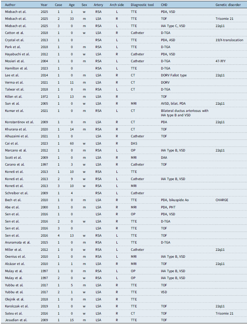
So far, there is only sparse information regarding this uncommon vascular variation. Based on our extensive literature research with PubMed database, the existing case reports (n = 44) have been compiled to better classify the above cases. Detailed information is listed in Table 1.
Case reports listed with clinical characteristics

CHD = congenital heart disease; RSA = right subclavian artery; LSA = left subclavian artery; L = left-sided aortic arch; R = right-sided aortic arch.
Patient age at the time of diagnosis ranged from 0 to 60 years, with 18 patients diagnosed immediately after birth (first month). Nineteen patients were male and 13 were female; consistent with existing data showing a higher prevalence in males. In our series, we had 2 males and 1 female patient.
Fifty-five per cent of patients had a left aberrant subclavian artery, all except one patient had a right aortic arch and 12 of whom had tetralogy of Fallot. The remaining 45% had a right aberrant subclavian artery, 7 of whom had dextro-transposition of the great arteries. We identified two patients with a right aberrant subclavian artery and one patient with left aberrant subclavian artery.
The majority of patients (86%) had associated CHD, which facilitated early diagnosis of aberrant subclavian arteries. Most common were dextro-transposition of the great arteries and tetralogy of Fallot, with six additional cases of interrupted aortic arch (all of whom had right subclavian artery) and two cases of double outlet right ventricle. Whereas our first patient presented with a simple patent ductus arteriosus only, the other patients showed complex anatomies including tetralogy of Fallot and interrupted aortic arch.
Eight patients were diagnosed with 22q11 deletion, two with trisomy 21. Other genetic disorders were CHARGE-syndrome, 19/X-translocation, and 47-XYY syndrome (Jacobs syndrome). One of our patients with right aberrant subclavian artery was diagnosed with 22q11 deletion, the patient with left aberrant subclavian artery and right aortic arch showed trisomy 21.
The most common symptom leading to clinical presentation was cyanosis in complex CHD, followed by failure to thrive, dizziness, and respiratory distress, according to the underlying pathophysiology of the cardiac defects. One patient (60 years old) remained asymptomatic until developing subclavian steal syndrome. Reference Luetmer and Miller7 Two patients showed reverse cyanosis postnatally due to transposition of the great arteries. Blood pressure differences were measured in 16 patients. In two of our patients diagnosed beyond the neonatal age, a pressure difference between the left and right was one of the clinical features indicative of a subclavian artery abnormality, the third case was diagnosed during neonatal workup.
In general, different imaging modalities were made to confirm the diagnoses; aberrant subclavian arteries were detected mostly by transthoracic echocardiography, while others were identified equally by using CT and catheterisation. We could identify all our cases by careful transthoracic echocardiography, and additional imaging modalities were performed for haemodynamic assessment (i.e. cardiac catheters) or surgical planning (i.e. CT).
Conclusion
Aberrant subclavian arteries are a very rare vascular anomaly, but the understanding of the embryology and anatomy is essential for the understanding of complex congenital heart variations. Clinical symptoms with different arm blood pressure may be indicative, and careful echocardiography may be adequate for diagnosis. Comprehensive imaging of the aortic arch and its branches may however be crucial in the preoperative evaluation of patients with CHD to enable timely surgical intervention. Cardiac catheterisation and MRI-CT serve as an excellent imaging modality for this assessment. Early re-anastomosis—if surgically feasible—is recommended to prevent long-term complications such as subclavian steal, pulmonary steal, or underdevelopment of the left upper limb.
Competing interests
None for this paper.