Introduction
Dogs and cats worldwide play an important social role as company for people, contributing to the physical and emotional development of children and improving the quality of life of their owners, particularly of seniors (Robertson et al., Reference Robertson, Irwin, Lymbery and Thompson2000). These animals, despite their social benefits, are also considered a risk to human health, as they can transmit infectious agents such as parasites, bacteria, fungi and viruses (Robertson & Thompson, Reference Robertson and Thompson2002).
Ancylostoma sp., among other intestinal helminthes carried by dogs and cats, demands great attention because of its zoonotic potential (Robertson & Thompson, Reference Robertson and Thompson2002), mainly soil contamination by faeces of infected animals. More than 1 billion people in the world are estimated to be infected by soil-transmitted helminthes (Crompton, Reference Crompton1999).
Nematode-antagonistic fungi can be classified into predatory, opportunistic (egg parasites and female plant-parasitic nematodes), endoparasitic and toxic metabolite producing fungi (Mota et al., Reference Mota, Campos and Araújo2003). Predatory fungi are the most studied nematode-antagonistic organisms because of their capacity of effectively reducing the nematode population in laboratory and field conditions (Larsen, Reference Larsen1999; Mota et al., Reference Mota, Campos and Araújo2003). Among predatory species, Arthrobotrys, Duddingtonia and Monacrosporium have stood out as potential agents in the biological control of parasitic nematodes in domestic animals (Larsen et al., Reference Larsen, Nansen, Wolstrup, Granvold, Henriksen and Zorn1995; Araújo et al., Reference Araújo, Gomes and Guimarães1998, Reference Araújo, Stephano and Sampaio1999, Reference Araújo, Sampaio, Vasconcellos and Campos2000; Alves et al., Reference Alves, Araújo, Guimarães, Assis, Sarti and Campos2003; Araújo & Ribeiro, Reference Araújo and Ribeiro2003; Dimander et al., Reference Dimander, Höglund, Uggla, Spörndly and Waller2003). A fungal isolate is considered as a potential biological controller of parasitic nematodes if it remains viable after passing though the gastrointestinal tract of animals, colonizes the faecal mass and captures hatched parasite larvae before they migrate to the soil (Araújo & Ribeiro, Reference Araújo and Ribeiro2003).
The objective of the present study was to compare the in vitro predatory activity of isolates of the nematophyagous fungi Duddingtonia flagrans (AC001), Monacrosporium thaumasium (NF34a), M. appendiculatum (CGI), M. sinense (SF53), Arthrobotrys conoides (I-40), A. cladodes (CG719) and A. robusta (I-31) on infective Ancylostoma sp. larvae parasitizing dogs. The capacity to remain viable after passing through the gastrointestinal tract of dogs while maintaining the predatory activity against infective Ancylostoma spp. larvae was also examined.
Material and methods
In vitro test
Brazilian isolates of Duddingtonia flagrans (AC001), Monacrosporium thaumasium (NF34a), M. appendiculatum (CGI), M. sinense (SF53), Arthrobotrys conoides (I-40), A. cladodes (CG719) and A. robusta (I-31) were stored in test tubes containing 2% cornmeal-agar, in the dark, at 4°C.
Panagrellus sp. free-living nematodes were kept in Petri dishes containing a medium of moistened and mashed oat flakes. Nematodes were recovered from the culture medium by soaking small amounts of the oats in distilled water in a Baermann apparatus and collecting the nematodes in haemolysis tubes after 6 h of decantation.
Culture discs, 4 mm in diameter, extracted from each isolate were transferred to 8.5-cm diameter Petri dishes containing 2% water-agar (2% WA) and stored in the dark at 25°C to induce conidia production. To each of these plates was added 1 ml of suspension containing 1000 Panagrellus sp., every 3 days for 21 days, until complete growth of fungal mycelium and complete nematode predation. After this period, 5 ml of distilled water was added to each Petri dish. Conidia and fragments of fungal mycelium were removed with a paintbrush and stored at 4°C.
Infective Ancylostoma sp. larvae (L3) were obtained from faeces of naturally infected dogs by vermiculite coproculture at 28°C, 80% humidity, for 15 days, and later using a Baermann funnel and water at 42–45°C with a decantation time of 12 h.
Treated groups, for each fungal species, consisting of 1000 conidia and 1000 L3 and a control group with 1000 L3 (without fungi) were formed in order to study the fungal predatory activity on Ancylostoma sp. L3.
Groups consisting of fungi and L3 were plated on 8.5-cm diameter Petri dishes, containing 2% WA culture medium previously marked with agar perforators, forming 4-mm diameter fields on the surface, incubated at 25°C in the dark. Plates were examined daily by light optical microscope ( × 10 objective lens) for 10 days. Ten fields per dish were randomly chosen and the number of non-predated L3 was counted to obtain the mean number of L3 per field.
On day 10, L3 larvae were recovered using a Baermann funnel (water at 42–45°C; 12-h decantation time) and counted to obtain the mean L3 number recovered from the control group and mean number of non-predated L3 from treated groups.
Data were examined by analysis of variance (test F), Tukey's test and linear regression, at 5% probability level. The average percentage of L3 reduction in the treated groups was calculated by the following formula: reduction (%) = [(mean L3 recovered from control group − mean L3 recovered from treated group)/mean L3 recovered from control group] × 100.
Passage test
The effectiveness of fungal isolates D. flagrans (AC001), M. thaumasium (NF34a) and A. robusta (I-31) against Ancylostoma sp. L3, after having passed through the gastrointestinal tract of dogs, was tested.
Fungal mycelium formation was induced by transferring 4-mm culture discs (2% WA) to 250 ml Erlenmeyer flasks, containing 150 ml of GPY (glucose–peptone–yeast extract) in liquid medium. Flasks were agitated at 120 rpm, in the dark, at 26°C for 10 days. After this period, mycelia were removed, filtered and weighed on an analytical scale.
Twenty-four adult dogs, no specific breed, average weight 14 kg (ranging from 11 to 17 kg), were treated with anthelmintics (pyrantel pamoate, febantel and praziquantel) 15 days before receiving fungal mycelia.
Animals were randomly separated into four groups of six dogs: three groups were treated with fungi and there was a non-treated control group. They received water and commercial dog food (18% crude protein, 6% ethereal extract, 6.5% crude fibre and 12% moisture) ad libitum. The treated groups received an oral dose of 1 g kg− 1 body weight of fungal mycelium, mixed with the same proportion of commercial canned dog food (8% crude protein, 4% ethereal extract, 1.5% crude fibre and 80% moisture). The control group received orally 1 g kg− 1 body weight of commercial canned dog food without fungal mycelium.
Faecal samples were collected at 8, 12, 24, 48, 72 and 96 h after treatment with the fungal mycelium and the control without fungi. Samples from each group were homogenized, then 2 g were placed in 5-cm Petri dishes containing 2% WA and incubated at 25°C in the dark with 1000 Ancylostoma sp. L3. Five replicates of each treatment and the control group were used for each established time. Plates were examined daily for conidia and conidiophores typical of the analysed isolates, according to the classification proposed by Van Oorschot (Reference Van Oorschot1985) and Liu & Zhang (Reference Liu and Zhang1994). On day 15, non-predated L3 larvae were recovered from the Petri dishes using the Baermann technique and the average number of non-predated L3 per dish was recorded.
Data were examined by analysis of variance and regression analysis. Means were compared using Tukey's test, 5% probability level.
Results and discussion
The tested isolates – D. flagrans (isolate AC001), M. thaumasium (isolate NF34a), M. appendiculatum (isolate CGI), M. sinense (isolate SF53), A. conoides (isolate I-40), A. cladodes (isolate CG719) and A. robusta (isolate I-31)– showed in vitro predatory capacity on Ancylostoma sp. L3. Trap formation and L3 predation by the fungal isolates were confirmed in the first assessment, after 24 h of interaction. This result agrees with the findings of Araújo et al. (Reference Araújo, Assis, Campos and Mota2006a) with Cooperia sp. and Oesophagostomum sp. in cattle, as well as Maciel et al. (Reference Maciel, Araújo and Cecon2006) with Ancylostoma sp. in dogs. Traps are formed in response to the presence of nematodes and may occur as a consequence of limiting nutritional conditions and/or water shortage (Scholler & Rubner, Reference Scholler and Rubner1994; Gronvold et al., Reference Gronvold, Nansen, Henriksen, Larsen, Wolstrup, Breciani, Rawate and Fribert1996). Nematode mobility acts as a stimulus for the induction of trap formation (Nansen et al., Reference Nansen, Foldager, Hansen, Henriksen and Jorgensen1988). The presence of Ancylostoma sp. L3 2% water-agar plates was essential for trap formation by fungal isolates, since the culture medium is nutritionally poor. Nematophagous fungi were not detected in control plates.
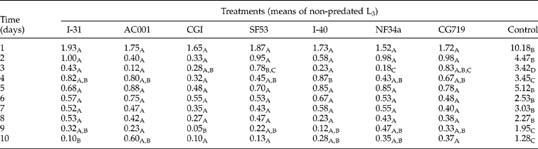
Means of non-predated L3 per field (4 mm diameter) of control plates were higher than means of non-predated L3 of plates treated with fungi in all periods (P < 0.05). There was a significant difference (P < 0.05) among the treatments for means of days 3, 4, 9 and 10. No differences were found (P>0.05) among means of non-predated L3 during the other days (table 1).
Daily means of non-predated infective Ancylostoma sp. (L3) larvae per 4-mm diameter field, on 2% water-agar medium, for 10 days, for treatments with fungal isolates Duddingtonia flagrans (AC001), Monacrosporium thaumasium (NF34a), M. appendiculatum (CGI), M. sinense (SF53), Arthrobotrys conoides (I-40), A. clododes (CG719), A. robusta (I-31) and control group (without fungi).
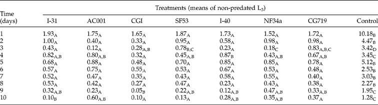
Means followed by at least one common capital letter in the row are not significantly different by Tukey's test at a 5% probability level.
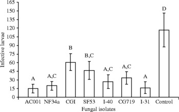
Predation was confirmed by the mean numbers of L3 recovered using the Baermann method at the end of the experiment (fig. 1). After 10 days, the mean numbers of L3 recovered from control plates were larger than the mean numbers of L3 recovered from groups treated with fungal isolates (P < 0.05). No difference was found among means of L3 recovered from treated groups for the isolates AC001, NF43a, I-40, CG719 and I-31 (P < 0.05). Comparison of treated groups with the control showed that the isolates AC001, I-31 and NF34a had the lowest L3 recovery, with L3 reduction of 87.02, 86.02 and 82.74%, respectively. These isolates were followed by I-40 with 76.89% reduction, CG719 with 71.33%, SF53 with 60.49%, and finally CGI with 47.93% reduction.
Means and standard deviation (bars) of infective non-predated Ancylostoma sp. larvae recovered from 2% water-agar plates by the Baermann method on the tenth day of treatment with the following fungal isolates: Duddingtonia flagrans (AC001), Monacrosporium thaumasium (NF34a), M. appendiculatum (CGI), M. sinense (SF53), Arthrobotrys conoides (I-40), A. clododes (CG719), A. robusta (I-31) and a control group (without fungi). Means followed by at least one common capital letter in the row are not significantly different by the Tukey's test at a 5% probability level.

The predatory fungal species used in this study have been reported as effective against parasitic nematodes in bovines (Araújo et al., Reference Araújo, Santos, Ferraz and Maia1993, Reference Araújo, Gomes and Guimarães1998; Alves et al., Reference Alves, Araújo, Guimarães, Assis, Sarti and Campos2003), small ruminants (Mota et al., Reference Mota, Bevilaqua and Araújo2000; Chandrawathani et al., Reference Chandrawathani, Jamnah, Waller, Larsen, Gillespie and Zahari2003; Araújo et al., Reference Araújo, Freitas, Vieira and Campos2006b) and equines (Castro et al., Reference Castro, Oliveira, Anjos, Ornela, Bittencourt, Araújo, Sampaio and Rodriges2003). Maciel et al. (Reference Maciel, Araújo and Cecon2006) evaluated in in vitro tests the predatory capacity of A. robusta (isolate I-31), D flagrans. (isolate CG768) and M. thaumasium (isolate NF34a) on Ancylostoma sp. L3. The authors found the highest predatory capacity for the isolate I-31, followed by CG768 and NF34a, with reductions of 97.75%, 89.89% and 88.76% respectively. In the present work, the fungal species D. flagrans (isolate AC001) presented the largest percentage of reduction, followed by A. robusta (isolate I-31) and M. thaumasium (isolate NF34a).
Linear regression coefficients calculated by using the means of L3 per 4-mm diameter field of treated and control groups were − 0.65 (control), − 0.068 (D. flagrans (AC001)), − 0.078 (M. thaumasium (NF34a)), − 0.096 (M. appendiculatum (CGI)), − 0.137 (M. sinense (SF53)), − 0.105 (A. conoides (I-40)), − 0.122 (A. cladodes (CG719)) and − 0.132 (A. robusta (I-31)). The negative values indicate a descending tendency of regression curves for the treatments, as there was a decrease in the means of non-predated L3 per 4-mm diameter field during the 10 days. This decrease was mainly caused by the capture of L3 by fungal traps. The decrease in the means of L3 per 4-mm diameter field in the control group during the 10 days of observation was caused by larval migration to the periphery of the plate, with higher humidity level. This migration was also reported by Araújo et al. (Reference Araújo, Assis, Campos and Mota2006a) in in vitro tests carried out in Petri dishes.
Although the predatory capacity against L3 of isolates AC001, I-31 and NF34a was not significantly different (P < 0.05) from I-40 and CG719, in vitro results showed they had higher percentages of reduction and were therefore used in the test of viability after passing through the gastrointestinal tract of dogs.
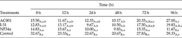
The mean number of Ancylostoma sp. L3 recovered from plates incubated with 2 g of faeces from animals of the treated groups (with the isolates AC001, I-31 and NF34a), and from animals of the control group (without fungi) are shown in table 2. The isolates AC001, I-31 and NF34a remained viable after passing through the gastrointestinal tract of dogs and were able to predate on Ancylostoma sp. L3.
Means numbers of Ancylostoma sp. infective larvae recovered from plates prepared with dog faeces sampled at 8, 12, 18, 24, 48, 72 and 96 h after feeding the dogs with 1 g mycelium/kg of live weight of the fungal isolates Duddingtonia flagrans (AC001), Arthrobotrys robusta (I-31) and Monacrosporium thaumasium (NF34a), and control (without fungi).
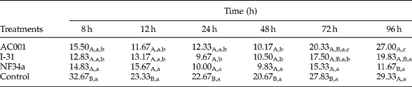
Means followed by at least one common capital letter, in the columns, are not significantly different by the Tukey's test at a 5% probability level. Means followed by at least one common small letter, in the rows, are not significantly different by the Tukey's test at a 5% probability level.
Conidia were identified only on day 14 in plates with faeces collected 12 h after fungal administration, for all tested isolates. It was not possible to identify conidia on the other days and collection times. Only mycelia were observed. Presence of nematophagous fungi was not detected in the control group.
Mean number of L3 recovered from the control group was larger than the mean number of L3 recovered from the treated groups (P < 0.05) until 48 h, except at 72 and 96 h, in which only the isolate NF34a showed mean number of L3 significantly smaller than the control group (P < 0.05). This suggests that this fungal isolate is eliminated in the faeces for a period of at least 96 h after ingestion of mycelia, whereas AC001 and I-31 were eliminated up to 48 h. It has been shown that the isolate NF34a remained viable after passing through the gastrointestinal tract of dogs up to 96 h.
Isolates AC001 and NF34a presented the lowest mean numbers of recovered L3 48 h after fungal administration, indicating a higher predatory activity. The isolate I-31 showed higher predatory activity 24 h after the ingestion of fungal mycelia. A higher larval predation within the same treatment is directly related with the period of higher fungal elimination through faeces. This could be influenced by the type and amount of food supplied during the experiment, as well as the tested fungal doses (Araújo & Ribeiro, Reference Araújo and Ribeiro2003). In this work, dogs received the same type and amount of food and fungal mycelium was administrated according to body weight.
Duddingtonia flagrans (AC001), A. robusta (I-31) and M. thaumasium (NF34a) showed predatory capacity on dog Ancylostoma sp. (L3) after passing through the gastrointestinal tract, remaining viable. They were also effective against L3 (P < 0.05) for the first 48 h of collection. After this period, only NF34a remained effective in samples collected 72 and 96 h after fungal administration (P < 0.05). At the end of the experiment (96 h), the reduction in the mean number of recovered L3 in the group treated with the isolate NF34a was 60.21%, whereas the groups treated with AC001 and I-31 had reduction percentages of 7.94 and 32.39%, respectively.
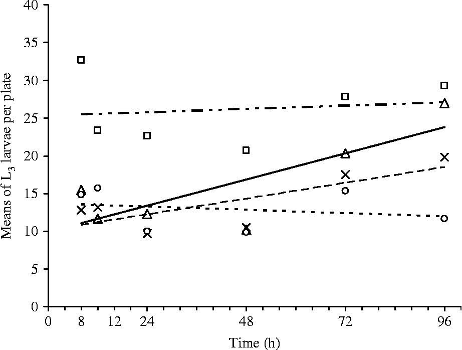
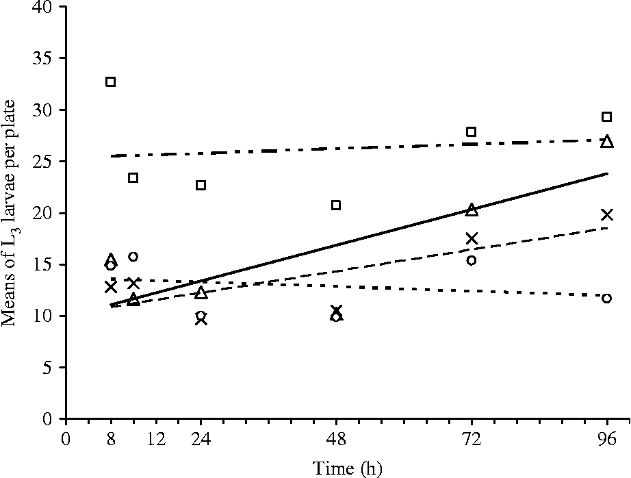
Regression curves (fig. 2) show that the isolate M. thaumasium (NF34a) was the only one with a negative linear correlation coefficient (r = − 0.017), whereas D. flagrans (AC001) and A. robusta (I-31) had positive values (r = 0.145 and r = 0.087, respectively). A negative coefficient indicates the existence of an inverse correlation between variables, showing that there was a reduction in the number of recovered larvae over time. A positive coefficient indicates that there was an increase in the number of recovered larvae over time, indicating the decrease in effectiveness.
Linear regression curves of Ancylostoma sp. infective larvae (L3), recovered from 2% water-agar plates (5 cm in diameter) incubated at 25°C for 15 days with the isolates Duddingtonia flagrans (AC001, △, —; y = 9.8941+0.1448*h; R 2 = 0.6327), Arthrobotrys robusta (I-31, × , – –; y = 10.1407+0.0871h; R 2 = 0.5960), Monacrosporium thaumasium (NF34a, ○, - - -; y = 13.6431+0.0174h; R 2 = 0.0515) and control (without fungi, □, – - - –y = 25.280+0.0185h; R 2 = 0.0203) as a function of time (8–96 h).
This is the first report on the viability of nematophagous fungi after passing through the gastrointestinal tract of dogs while still remaining effective against Ancylostoma sp. L3. Since the isolates remained viable and effective in the control of Ancylostoma sp. after passing through the gastrointestinal tract of dogs, their administration to animals contaminated by Ancylostoma sp. can decrease environmental contamination, acting directly on larvae present in the soil.
All tested fungal isolates showed in vitro action against Ancylostoma sp. L3, with a larger predatory activity of the nematophagous fungi D. flagrans (isolate AC001), A. robusta (isolate I-31) and M. thaumasium (isolate NF34a), which stayed viable after passing through the gastrointestinal tract of dogs. These fungal species are therefore promising for future use in the biological control of Ancylostoma sp. in dogs.
Acknowledgements
The authors would like to thank CNPq for financial support.