Case report
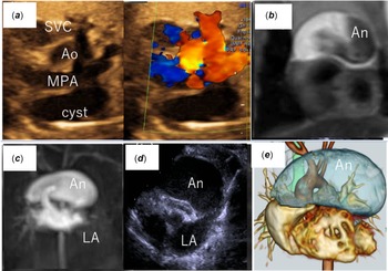
Fetal echocardiography at 29 weeks of gestation revealed a large cystic mass (29 × 14 mm) on the left side of the main pulmonary artery (Fig 1a). There was no growth of the mass during the fetal period. At birth (39 weeks’ gestation, 2964 g, male), the newborn was asymptomatic and clinical examination was unremarkable. Chest radiography revealed cardiomegaly with a more prominent left heart border. Electrocardiogram showed sinus rhythm with a normal axis. On day 2, transthoracic echocardiography showed a cyst size of 39 × 26 mm, and the cyst was considered a pericardial type. There were no other intracardiac abnormalities, and cardiac function was normal. MRI on day 8 revealed a cyst size of 46 × 26 × 25 mm, suggestive of enlargement. However, no thrombus formation was identified. The patient was asymptomatic and kept on observation. At day 20, respiratory distress and feeding difficulties appeared. Dynamic contrast-enhanced MRI showed further cyst enlargement to 50 × 34 × 27 mm (Fig 1b) and connection to the left atrial appendage in the arterial phase (Fig 1c). Colour Doppler echocardiography also revealed blood flow originating from the left atrium (Fig 1d). The cyst was diagnosed as a left atrial appendage aneurysm. On cardiac CT, the left atrial appendage aneurysm extended anterosuperiorly to the heart and compressed the pulmonary artery, with mild-moderate pulmonary stenosis (Fig 1e).
( a ) Three-vessel view on fetal echocardiography at 34 weeks’ gestation revealed a large cystic mass on the left side of the main pulmonary artery. ( b ) MRI confirmed post-natal enlargement of the aneurysm. ( c ) Contrast-enhanced MRI revealed that the aneurysm was connected to the left atrial appendage. ( d ) Transthoracic echocardiography view showing a large aneurysm arising from the left atrial appendage. ( e ) Left atrial appendage aneurysm (LAAA) on CT images. The LAAA extended anterosuperiorly to the heart, excluding the pulmonary artery. SVC = superior vena cava; AO = aorta; MPA = main pulmonary artery; An = aneurysm; LA = left atrium.

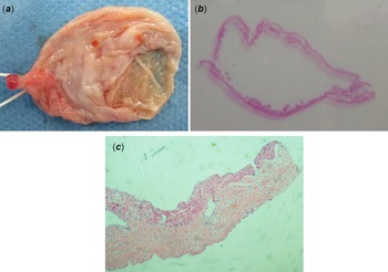
Blood tests showed normal D-dimer levels (0.7 μg/mL) but elevated brain natriuretic peptide (265 pg/mL). The patient was recommended for surgery because of the rapid cyst enlargement and symptoms of cardiac failure. Surgery was performed on day 29 via a median sternotomy under cardiopulmonary bypass. Intraoperatively, there was an intact pericardium, while the huge aneurysm compressed the main pulmonary artery. The aneurysm was larger than the left ventricle. No thrombus was detected within the aneurysm and the aneurysm was completely resected (65 × 35 × 30 mm) (Fig 2a). His pre-operative symptoms, including respiratory distress and feeding difficulties, improved. Histopathologically, the three-layered structure of the left atrial appendage aneurysm (similar to that of the left atrial appendage) was retained, but the atrial wall had a thin myocardial layer accompanied by myxoid degeneration (Fig 2b, c). There was no evidence of specific inflammation or malignancy. No post-operative complications were observed. Post-operatively, his symptoms improved completely. Follow-up echocardiography at 12 months after surgery showed no evidence of recurrence.
( a ) Surgical removal of the left atrial appendage aneurysm (LAAA). The connection to the left atrium was ligated (size = 65×35×30 mm). ( b, c ) Haematoxylin and eosin-stained section of the aneurysm. Histopathology of the atrial wall showed a thin myocardial layer, accompanied by myxoid degeneration. There were no areas of tissue loss, and similar changes were seen throughout the circumference (c, ×400 magnification).

Discussion
Congenital left atrial appendage aneurysm is rare, with approximately 50 congenital left atrial appendage aneurysm cases (including several cases diagnosed during the fetal period) previously reported. Although asymptomatic patients are sporadically diagnosed during adulthood, Reference Aryal, Hakim and Ghimire1 there are no reports of rapid left atrial appendage aneurysm enlargement and symptomatic features in the neonatal period, or of cardiac failure symptoms associated with left atrial appendage aneurysm expansion. Previous surgical cases in neonates have reported left atrial appendage aneurysm sizes of approximately 30 mm Reference Fountain-Dommer, Wiles and Shuler2–Reference Strecker, Koch and Schild4 , while the aneurysm in the present case was nearly twice as large. With rapid left atrial appendage aneurysm enlargement, the circulatory blood volume decreased and the main pulmonary artery was compressed. Thus, the heart failure symptoms (i.e., respiratory distress and feeding difficulties) were likely a result of the left atrial appendage aneurysm enlargement, which is supported by the improvement in heart failure symptoms after left atrial appendage aneurysm resection.
Transthoracic echocardiography is considered an adequate and primary method for left atrial appendage aneurysm detection. However, in our case, the mass was found by fetal echocardiography and was misidentified as a cyst. By contrast, MRI is important for assessing giant left atrial appendage aneurysms, overall aneurysm spread (i.e., by accurate size measurement), the presence of thrombi, and in ruling out a differential diagnosis. Reference Oz, Oguz, Karcaaltincaba, Yilmaz and Haliloglu5 In the present case, dynamic contrast-enhanced MRI showed enhancement in the arterial phase and continuity with the left atrium, which contributed to the definitive diagnosis. Interestingly, stasis maps provided by 4D flow Cardiovascular MRI were reported to provide objective information in therapeutic decision-making. Reference Pradella6
Congenital left atrial appendage aneurysms can be intrapericardial (associated with an intact pericardium) or extrapericardial (associated with pericardial defects). The intrapericardial type is thought to be caused by congenital abnormalities in the left atrial wall and/or appendage. Reference Cho, Park, Lee, Choo and Sung7 The congenital form arises from dysplasia of the musculi pectinati and related atrial muscle bands. Acquired forms often occur in association with conditions leading to elevated left atrial pressure, including organic mitral valve diseases. In the present case, pathological examination revealed no epicardial defect, an intrapericardial type left atrial appendage aneurysm, and marked thinning in part of the atrial muscle, accompanied by myxoid degeneration. There was no evidence of inflammation, abnormal muscle bundles, or degeneration. Increased blood flow after birth may have contributed to thinning of the left atrial appendage. Further enlargement of the left auricular mass may have also caused aneurysm wall thinning, creating a vicious cycle that facilitated further aneurysm enlargement.
In rare cases, mitral regurgitation or cardiac tamponade in the neonatal period due to left ventricular compression can occur. Reference Mansour, Aldousany, Arce, Subramanian and Ashraf8 Thus, careful follow-up with consideration for early surgery is required. In the present case, surgery was chosen because of the rapid aneurysm enlargement and symptoms of heart failure. During the operative period (including anaesthesia), we had concerns over potential circulation failure following left atrial appendage aneurysm excision. However, his vital signs were stable and cardiac failure symptoms resolved post-operatively. Resection through a median sternotomy during cardiopulmonary bypass is a safe method. Reference Sankhyan9
Conclusion
Congenital left atrial appendage aneurysm, which can be observed during the fetal period, may rapidly enlarge after birth and requires follow-up, even if asymptomatic. MRI may be useful for accurate left atrial appendage aneurysm size assessment. Because the only effective treatment is surgery, the timing of surgery must be comprehensively determined.
Acknowledgements
We thank Edanz (https://jp.edanz.com/ac) for editing a draft of this manuscript.
Author contributions
N.Y., M.H., and H.M. participated in clinical data collection. H.M. critically reviewed the manuscript and supervised the study. N.Y. wrote the manuscript. All authors read and approved the final manuscript.
Financial support
This research received no specific grant from any funding agency, commercial, or not-for-profit sectors.
Conflicts of interest
None.
Ethical standards
This study was exempted from ethical approval procedures because it was a case report of a single patient. Written informed consent was obtained from the parents for publication.