Introduction
Since the extraction of bryostatin from the bryozoans Bugula neritina in the late 1960s by George Pettit and the discovery of its anti-cancer activity (Pettit et al., Reference Pettit, Cragg, Herald, Schmidt and Lohavanijaya1982), researchers have been interested in screening the biological activities of extracts from marine organisms by various extraction techniques, solvents and experimental protocols. Consequently, the chemistry of marine natural products has grown enormously. More than 20,000 natural products have been isolated and identified from marine organisms worldwide (Hu et al., Reference Hu, Chen, Hu, Yu, Zhu, Lin, Chen and Yuan2015).
Most recent studies have focused on the benthic organisms according to their ability to produce bioactive metabolites in response to various ecological stresses such as competition for space and food, maintenance of unfouled surface and predator deterrence (Abdul Jaffar Ali & Tamilselvi, Reference Abdul Jaffar Ali and Tamilselvi2016).
Among benthic organisms, invertebrates are particularly interesting because they constitute a rich source of novel and bioactive secondary metabolites (Zhang et al., Reference Zhang, Yi and Tang2006). Echinoderms have especially attracted the attention of scientists. According to Blunt et al. (Reference Blunt, Copp, Munro, Northcotec and Prinsep2003), 6% of the natural products involved in preclinical and clinical trials were extracted from echinoderms.
Among echinoderms, the sea cucumbers are interesting in terms of ecological, socio-economic and biological perspectives. They are an important component of the marine ecosystem and are considered as resources for coastal livelihoods (Wolkenhauer et al., Reference Wolkenhauer, Uthicke, Burridge, Skewes and Pitcher2010). They are recognized as a human food source, particularly in some parts of Asia, and they have been harvested for more than 20 years (Purcell et al., Reference Purcell, Samyn and Conand2012).
Furthermore, sea cucumbers have been examined as a rich source of natural active compounds with biological and pharmacological activities (Bordbar et al., Reference Bordbar, Anwar and Saari2011; Dhinakaran & Lipton, Reference Dhinakaran and Lipton2014). Several studies reported that chemicals extracted from sea cucumber species were shown to have high antioxidant potential (Althunibat et al., Reference Althunibat, Hashim, Taher, Daud, Ikeda and Zali2009, Reference Althunibat, Ridzwan, Taher, Daud, Jauhari Arief Ichwan and Qaralleh2013), with antibacterial (Anbukkarasu et al., Reference Anbukkarasu, Subramanian, Elayarperumal and Masilamani2014), antifungal (Santafé et al., Reference Santafé, Guzmán and Torres2014) and anti-cancer properties (Wargasetia & Widodo, Reference Wargasetia and Widodo2017).
There are about 1250 species of sea cucumber in the world (Chao et al., Reference Chao, Chen and Alexander1995) and 47 species have been found in the Mediterranean Sea (Ramón et al., Reference Ramón, Jordi and Massutí2010). Among them, the present study focuses on the sea cucumber Holothuria forskali. As far as we know, no research article has yet been published covering the analytical screening and biological activities of the sea cucumber H. forskali from the Tunisian coasts.
This study is an attempt to compile data of analytical investigations of the bioactive components and biological activities of H. forskali, in particular, the antibacterial effect of molecules extracted with ethyl acetate as a solvent.
Materials and methods
Samples collection
A total of 76 specimens of sea cucumbers H. forskali Delle Chiaje, 1823 were collected by scuba diving at about 5 m depth from a rocky shore population in Haouaria (north-eastern Tunisia) in April 2017. Sea cucumbers were transferred to the laboratory in a filtered seawater tank equipped with a bubbler and maintained under ambient temperature. Specimens were measured underwater and subsequently removed and weighed immediately. Their length and weight were respectively 21.5 ± 2.6 cm and 324.5 ± 4.6 g fresh weight. Samples were cleaned of sand and left-over debris with distilled water three times (about 200 ml each time) during about five seconds. Sea cucumbers were dissected and tissues of digestive tract, gonad, muscle, body wall and respiratory tree were removed. For each organ, samples were pooled, lyophilized and then kept frozen at −80°C until extraction.
Extract preparation
Extractions using ethyl acetate (Fisher Chemical) were carried out as described by Althunibat et al. (Reference Althunibat, Ridzwan, Taher, Daud, Jauhari Arief Ichwan and Qaralleh2013). This solvent was chosen based on its chemical and biological characteristics. Besides its medium polarity which can allow the extraction of several polar and non-polar biological compounds, ethyl-acetate is a suitable extraction solvent for both GC-MS and LC-MS analytical techniques. In addition, ethyl acetate is a class 3 solvent with low toxic potential on biological matrices (FDA, 2012). Therefore, it has been widely used as an extraction solvent for numerous antimicrobial studies (Yeo et al., Reference Yeo, Chia, Lee, Sow and Yap2014).
The freeze-dried samples of each organ were pooled, homogenized using a blender and suspended with stirring in the dark in the solvent individually during one night at 4°C (1 g of sample into 100 ml of solvent). The mixture was filtered by passage through a Whatman filter paper No. 42 (125 mm) and the filtrate was centrifuged (15 min; 30,000 g; 4°C) and the residue was removed. The solvent was evaporated using a rotary evaporator (Buchi Rota-vapor R-200, Switzerland). The powdered extracts of each sample were weighed and stored at −20°C. All extractions were performed in triplicate.
Antibacterial assay
The antibacterial activities of H. forskali ethyl acetate extracts were tested by the diffusion method using Muller–Hinton agar as described by Kiani et al. (Reference Kiani, Heidari, Rassa, Kadkhodazadeh and Heidari2014). Dimethyl sulphoxide, DMSO (Sigma Aldrich, UK) was used to dissolve the crude ethyl acetate extracts obtained from different organs of H. forskali.
Low (0.4 mg ml−1) and high (2 mg ml−1) concentrations of all extracts were chosen based on previous studies examining the antibacterial activities of crude sea cucumbers' ethyl acetate extracts (Mokhlesi et al., Reference Mokhlesi, Saeidnia, Gohari, Shahverdi, Nasrolahi, Farahani, Khoshnood and Eśhaghi2012; Adibpour et al., Reference Adibpour, Nasr, Nematpour, Shakouri and Ameri2014; Pringgenies et al., Reference Pringgenies, Ridlo and Sembiring2017; Torres, Reference Torres2017). The two selected concentrations of all extracts were assessed against Bacillus subtilis ATCC 6633 and Bacillus cereus ATCC 11778 as gram-positive bacteria, and Escherichia coli ATCC 8739 and Pseudomonas aeruginosa as gram-negative bacteria. Agar surface of each plate was seeded by sterile cotton swab with the reference bacterial strains. After inoculation, wells of 6 mm diameter were dug using sterile cork borers and then filled with 100 μl of dissolved extracts. After 30 min, the plates were incubated at 37°C for 24 h. The antibacterial activity was determined by measuring the diameter of the inhibition zone (mm). Ampicillin (30 μg disc−1) and erythromycin (30 μg disc−1) were included as positive controls while sterile DMSO (100%) served as negative control.
Gas chromatography-mass spectrometry (GC-MS) analysis
For the analytical screening, the GC-MS technique is a powerful tool and is highly commended for analysis of small molecules, especially non-polar components, volatile essential oils and fatty acids (Gu et al., Reference Gu, Lin, Pease, Chetwyn and Yehl2015).
In the present study, GC-MS analyses were carried out in a GC Agilent model 7890B coupled with a 240 Ion trap mass spectrometer (Agilent Technologies, USA). The GC column was HP-5 (5%-Phenyl)-methylpolysiloxane: 30 mm × 0.25 mm × 0.25 μm. The GC conditions were as follows: The injector was operated at 250°C and ion-source temperature was 280°C. The oven temperature was programmed from 110°C (isothermal for 2 min) with an increase of 10 to 200°C min−1, then 5 to 280°C min−1, ending with a 9 min isothermal at 280°C. Helium was used as the carrier gas at constant flow rate 1 ml min−1. Interpretation on mass spectrum of GC-MS was using the database of National Institute: Standard and Technology (NIST mass spectral search 2014, Version 2.2 with 242,466 compounds). The name, retention time, peak area, molecular weight, molecular formula and structure of the components of the sea cucumber extracts were ascertained.
Liquid chromatography-mass spectrometry (LC-MS) analysis
A wide range of metabolites in fluids and tissues can currently be measured using LC-MS which is among the techniques of choice for screening of multiple substances in biological samples. Indeed, LC-MS is characterized by its high specificity and sensitivity and its short chromatographic run times (Grebe & Singh, Reference Grebe and Singh2011).
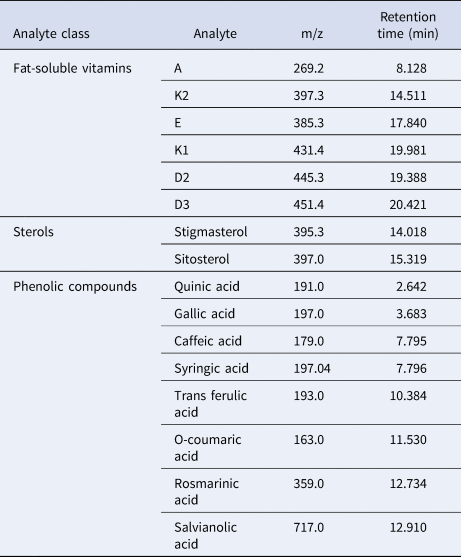
Molecules belonging to fat-soluble vitamins, phytosterols and phenolic compounds were quantitatively analysed in ethyl acetate extract with separate methods validated in-house. Analysis was conducted on a Shimadzu LCMS-2030 (Shimadzu Corp., Japan) equipped with an electrospray ionization source with negative ionization mode according to in-house validated methods. Chromatographic separation was done on an Inertsil 3 μm C18 column (GL Science Corp., Japan) maintained at 40°C with different mobile phases at a flow rate of 0.5 ml min−1. For vitamins, the mobile phase was composed of two mixtures of (A) 5 mmol ammonium formiate+0.1% formic acid in H2O, v/v and (B) 0.1% of formic acid in methanol, with a linear gradient elution: 0–15 min, 70–100% B; 15–45 min, 100% B. For phytosterols, separation was completed with a linear gradient elution, the mobile phase was composed of a mixture of 1% aqueous acetonitrile solution (A) and methanol (B) as follows: from 0 to 30 min, 90% B, from 30–35 min of 90–100% B and finally, 10 min of 100% B. For phenolic compounds, the mobile phase was composed of a linear gradient elution by two mixtures: (A) 0.1% aqueous formic acid and (B) 0.1% formic acid in methanol: 0–40 min of 20–100% A and 40–60 min 100% B. Acetonitrile, methanol, formic acid and water were obtained from Fisher Scientific (Optima™ LC/MS Grade). Instrumental MS detection conditions were the same for both compound groups. Detection was made in negative ion mode with a capillary voltage of −3.5 V, a mobilizing gas flow of 1.5 l min−1, a dry gas flow rate of 14 l min−1, a dissolving line temperature of 250°C, a block source temperature of 400°C and a voltage detector of 1.2 V. Scanned ions for different analytes are summarized in Table 1.
Retention times and characteristic ions (m/z) of the target analytes for the LC-MS analysis

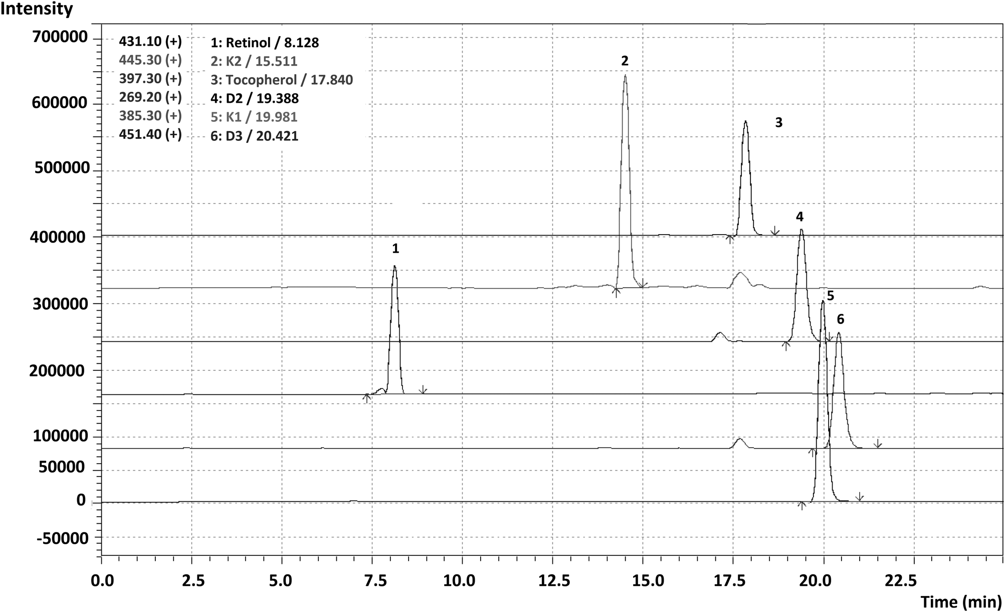
The identification of scanned molecules was done by comparison with retention times and MS spectra obtained with pure standard solutions of relative compounds (Figure 1) and quantification was achieved with external calibration curves.
Chromatographic profile of the standard solution of fat-soluble vitamins. 1: Retinol / 8.128, K2: 14.511, Tocopherol: 17.840, D2: 19.388, K1: 19.981, D3: 20.421.

Results
Antibacterial activity
Antibacterial properties of ethyl acetate extracts of the sea cucumber organs were tested against four bacterial strains: E. coli, B. cereus, B. subtilis and P. aeruginosa. The ethyl acetate extracts revealed moderate antibacterial activity (Table 2). The best effect of the ethyl acetate extracts of all organs except those of the gonad, at 2 mg ml−1 concentration, was found against E. coli strains. For this bacterium, the diameter of the inhibition zone ranged from 11.2 ± 1.2 mm to 18.2 ± 1.9 mm. At 0.4 mg ml−1, the ethyl acetate extract of the digestive tract and gonad revealed an inhibition zone for B. subtilis, however, for the body wall and the muscle extracts antibacterial activity was recorded only against E. coli. At the concentration of 2 mg ml−1, the body wall extract showed an inhibition of the growth of all bacterial strains. Digestive tract extract showed a high activity against E. coli that reached the positive control to some extent. The muscle and the digestive tract extracts revealed an antibacterial activity against three strains; however, the gonad extract was active against only two strains. The gonad extract exhibited highest activity against B. cereus and B. subtilis while the respiratory tree extract showed an effect only against E. coli.
Antibacterial activities expressed as the diameter of the plated inhibition zones (mm) of ethyl acetate extract of Holothuria forskali organs against bacterial pathogens

BW, Body wall; MU, Muscle; RT, Respiratory tree; DT, Digestive tract; GO, Gonad.
C1: 0.4 mg ml−1, C2: 2 mg ml−1, Ctrl+: Positive control erythromycin (10 μg disc−1) and Ctrl−: Negative control d.d.H2O.
Qualitative and quantitative analysis of biomolecules
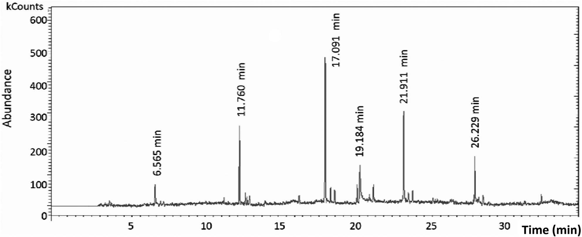
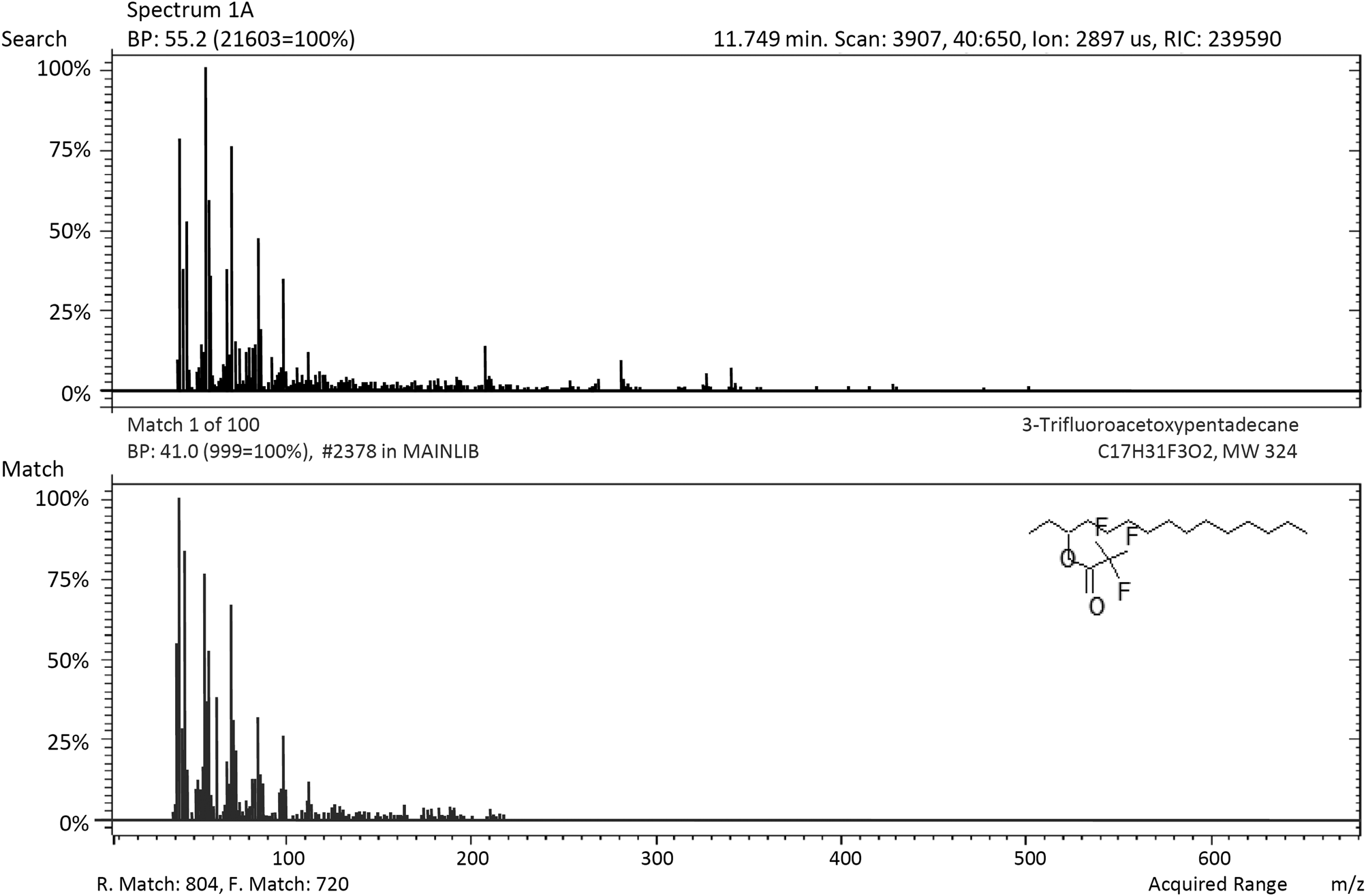
GC-MS and LC-MS analyses were performed on extracts of the body wall which showed an inhibitory effect against all tested bacterial strains. GC-MS/MS scanning showed a higher diversity of extracted biomolecules and allowed a good identification of analysed compounds. A total of 25 major compounds were identified. Among these compounds, six are the most concentrated: Morphinan-4,5-epoxy-3,6-diol, 6amino (C26H27N5O6); 3-trifluoroacetoxypentadecane (C17H31F3O2); Stigmasta-5,22-dien-3-ol, acetate (C31H50O2); 1-Phthalanol,1,3,3-trimethyl 1-phenyl (C11H14O2); Ergosta-5,22-dien-3-ol, acetate (C30H48O2) and Octadecenoic acid. Figures 2 and 3 show some examples of the obtained GC-MS chromatogram and mass spectrometry, for the body wall extract of H. forskali.
GC-MS chromatogram of the ethyl acetate extract of Holothuria forskali body wall.

Example of mass spectra of identified compounds in the ethyl acetate extract of the sea cucumber Holothuria forskali body wall.

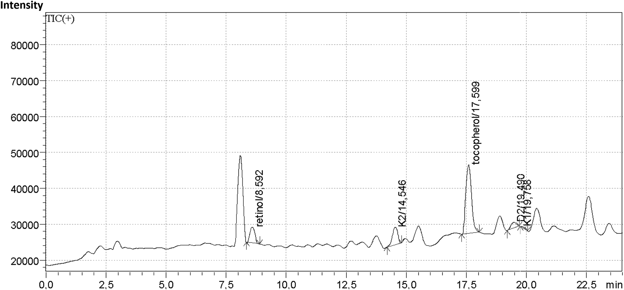
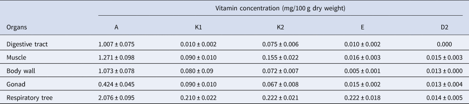
Concerning the LC-MS analysis, among six studied fat-soluble vitamins, five were detected in analysed extract. Figure 4 shows the chromatographic profile of ethyl acetate extract. Quantitative analysis revealed that vitamin A was by far the most abundant vitamin followed by K2 and D2. The higher relative concentrations were recorded in respiratory tree extract for vitamin A and vitamin K2 respectively, equal to 2.076 ± 0.095 mg/100 g dry weight and 0.222 ± 0.021 mg/100 g dry weight. The detailed concentrations of fat-soluble vitamins in different organs are summarized in Table 3.
LC-MS chromatogram showing the peaks of five fat-soluble vitamins identified in the ethyl acetate extract of Holothuria forskali body wall.

Concentrations of the fat-soluble vitamins identified in the ethyl acetate extract of Holothuria forskali organs

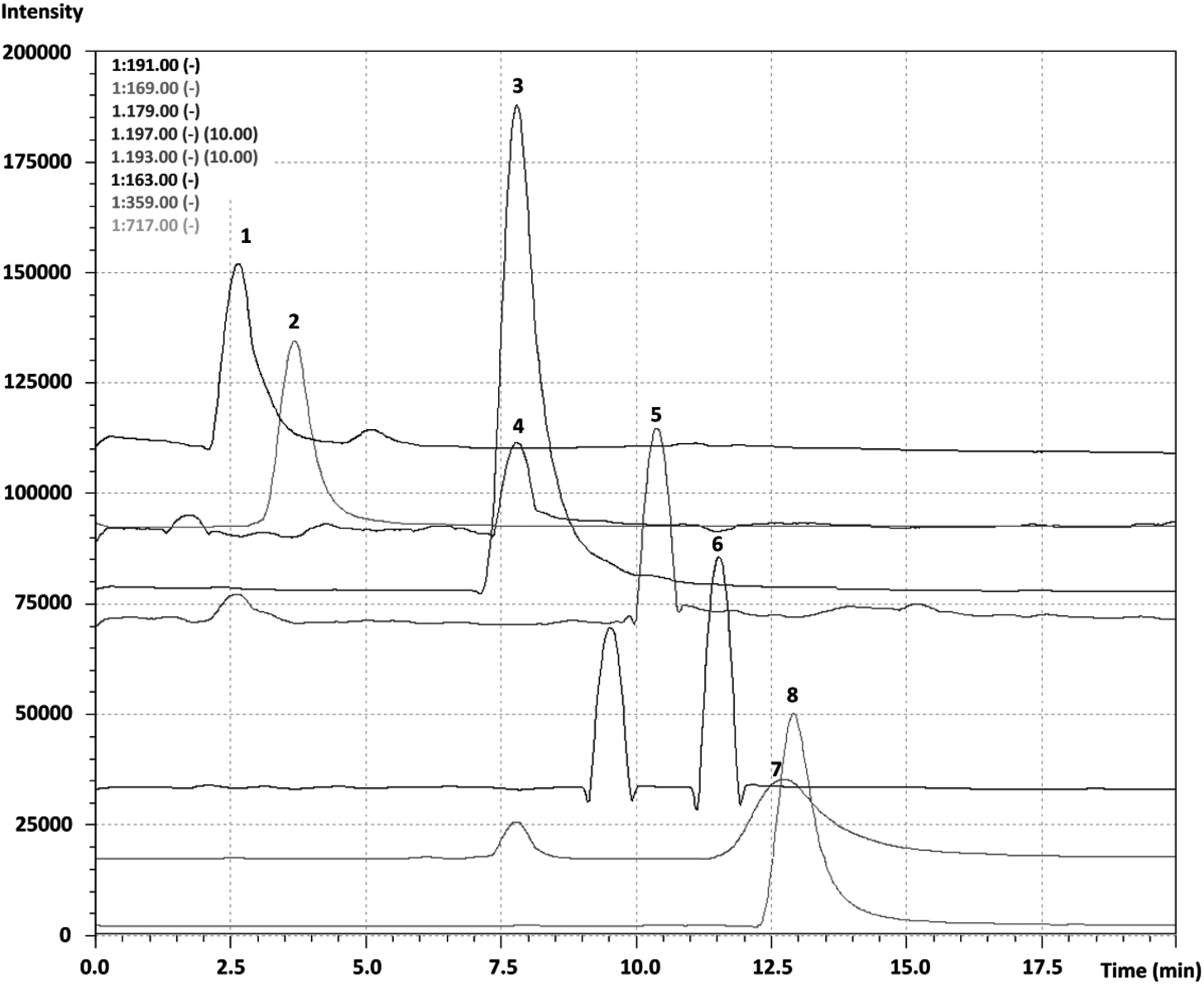
The LC-MS analysis of the ethyl acetate extract has also confirmed the presence of phenolic compounds, quinic acid, gallic acid, caffeic acid, syringic acid, trans ferulic acid, o-coumaric acid, rosmarinic acid and salvianolic acid (Figure 5).
LC-MS chromatograms of phenolic compound standard solution. 1: Quinic acid, 2: Gallic acid, 3: Caffeic acid, 4: Syringic acid, 5: Trans ferulic acid, 6: O-coumaric acid, 7: Rosmarinic acid, 8: Salvianolic acid.

The quinic acid concentration far exceeded those of other compounds (Table 4). The highest values of quinic acid content (0.476 ± 0.044 μg ml−1 and 0.471 ± 0.055 μg ml−1) were obtained from muscle and digestive tract extracts respectively.
Concentrations (μg ml−1) of phenolic compound and phytosterols identified in the ethyl acetate extract of Holothuria forskali body wall by LC-MS

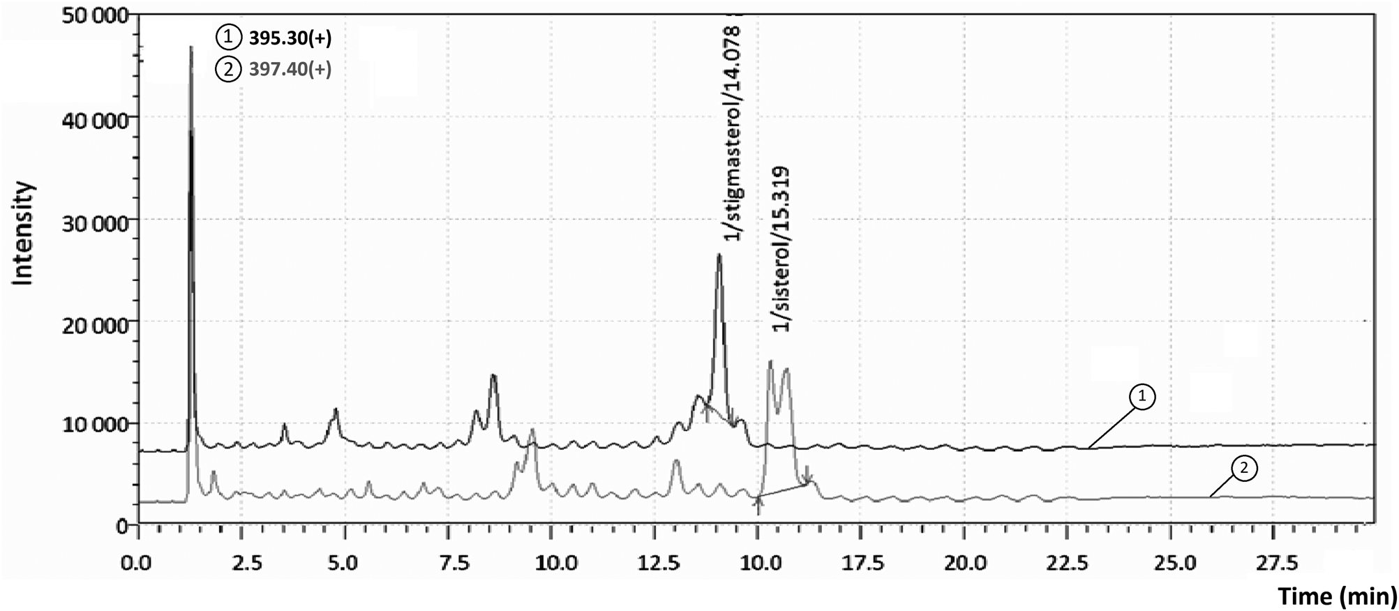
Analysis of phytosterols in H. forskali ethyl acetate extract revealed the presence of stigmasterol and sitosterol as shown in Figure 6 and Table 4. The stigmasterol content of digestive tract and respiratory tree extracts are higher than those of the other organs whereas the high value of sitosterol content was recorded in the body wall.
Stigmasterol LC-MS chromatograms of ethyl acetate extract of Holothuria forskali body wall.

Discussion
The ethyl acetate extracts from different organs of the sea cucumber H. forskali were screened for antibacterial activity against Gram-positive and Gram-negative bacteria. The results showed that the examined extracts exhibited antibacterial activity which differs depending on organs.
In the literature, the antibacterial activity of sea cucumber extracts varies according to species, solvent and doses. Similarly to the present study, Mohammadizadeh et al. (Reference Mohammadizadeh, Ehsanpor, Afkhami, Mokhlesi, Khazaali and Montazeri2013) and Sarhadizadeh et al. (Reference Sarhadizadeh, Afkhami and Ehsanpour2014) have recorded antibacterial activity of the ethyl acetate extracts of H. scabra and Stichopus hermanni respectively. Omran & Allam (Reference Omran and Allam2013) have demonstrated antimicrobial activity of sea cucumber H. polii. Recently, Ghadiri et al. (Reference Ghadiri, Kazemi, Heidari and Rassa2018) noted an antibacterial activity of ethanol, methanol and acetone extracts from H. parva. These authors have used as extract solvents phosphate-buffered saline, ethanol and acetonitrile and they reported an antimicrobial activity against infective streptococci. Abraham et al. (Reference Abraham, Nagarajan and Shanmugam2002) and Adibpour et al. (Reference Adibpour, Nasr, Nematpour, Shakouri and Ameri2014), however, reported that alcohol extracts of several sea cucumber species (e.g. Actinopyga echinites, Actinopyga miliaris, H. atra, H. scabra and H. leucospilota) were not effective against Bacillus sp. whereas E. coli and P. aeruginosa were sensitive to the tested extracts.
It is well known that the benthic marine environment can effectively be described as a soup of microorganisms (Benkendorff et al., Reference Benkendorff, Davis and Bremner2001). In this context, it was established that coastal marine sediment may be particularly considered as a reservoir for several pathogenic and antimicrobial-resistant bacterial strains (Vignaroli et al., Reference Vignaroli, Luna, Rinaldi, Di Cesare, Danovaro and Biavasco2012; Yang et al., Reference Yang, Wang, Shu, Liu, Geng, Hu and Feng2013). Therefore sedentary bottom dweller and deposit-feeding organisms occurring in nearshore and estuarian ecosystems such as sea cucumbers are constantly exposed to high concentrations of harmful pathogens (Haug et al., Reference Haug, Kjuul, Styrvold, Sandsdalen, Olsen and Stensvåg2002). To survive and to protect themselves against microbial infections and surface fouling microorganisms, numerous benthic marine invertebrates such as sea cucumbers possess an efficient chemical defence mechanism involving the production of secondary metabolites with antimicrobial activities (Benkendorff et al., Reference Benkendorff, Davis and Bremner2001; Haug et al., Reference Haug, Kjuul, Styrvold, Sandsdalen, Olsen and Stensvåg2002; Anbukkarasu et al., Reference Anbukkarasu, Subramanian, Elayarperumal and Masilamani2014; Ghadiri et al., Reference Ghadiri, Kazemi, Heidari and Rassa2018). In this framework, the antimicrobial properties of sea cucumber extracts have been ascribed to the presence of glycosides, commonly in the form of triterpene glycosides (saponin) (Chludil et al., Reference Chludil, Muniain, Seldes and Maier2002; Mokhlesi et al., Reference Mokhlesi, Saeidnia, Ahmad, Gohari Shahverdi Ahmad, Mollazadeh-Moghaddam and Eśhaghi2011). Indeed, these secondary metabolites have been reported to possess a wide spectrum of pharmacological effects including antifungal, antibacterial and antiviral activities (Caulier et al., Reference Caulier, Dyck, Gerbaux, Eeckhaut and Flammang2011). Holothurian triterpenoid glycosides consisting of an aglycone based on holostane-3b-ol and a carbohydrate chain present a very high diversity depending on species and body component (Caulier et al., Reference Caulier, Dyck, Gerbaux, Eeckhaut and Flammang2011, Reference Caulier, Mezali, Soualili, Decroo, Demeyer, Eeckhaut, Gerbaux and Flammang2016). Furthermore, it is well established that saponins present different properties in relation to their ecological roles (Caulier et al., Reference Caulier, Mezali, Soualili, Decroo, Demeyer, Eeckhaut, Gerbaux and Flammang2016). Such molecular diversity could be a selective advantage for these organisms. It also may act as a true chemical signature for holothuriid species (Caulier et al., Reference Caulier, Dyck, Gerbaux, Eeckhaut and Flammang2011, Reference Caulier, Mezali, Soualili, Decroo, Demeyer, Eeckhaut, Gerbaux and Flammang2016).
In the current study, GC-MS analysis was performed in order to identify the bioactive compounds of the H. forskali extracts which are most likely implicated in antibacterial activity. Results revealed a mixture of several compounds; among them 25 are major and six of these are the most concentrated. According to the literature, these compounds possess interesting biological activities. Referring to Subavathy & Thilaga (Reference Subavathy and Thilaga2016), 3-trifluoroacetoxypentadecane has an antibacterial activity, while Stigmasta-5,22-dien-3-ol, acetate (which is a steroid) has antimicrobial, anti-inflammatory, anti-arthritic, anti-asthma, diuretic, hepatoprotective and antioxidant activities (Selvi & Murugesh, Reference Selvi and Murugesh2017). In vitro experiments conducted by Byju et al. (Reference Byju, Anuradha, Vasundhara, Nair and Kumar2014) have demonstrated that ergosta-5,22-dien-3-ol, acetate extracted from the soft coral Subergorgia reticulata may exert an anti-carcinogenic activity. The sixth most concentrated compound was 1-Phthalanol,1,3,3-trimethyl 1-phenyl (C11H14O2), which is classified among phthalates. It is known that phthalates are esters of phthalic acid used as additives to plastics in order to make the material softer and more flexible. They are one of the most frequently detected persistent organic pollutants in the marine environment (Gao & Wen, Reference Gao and Wen2016). The identification of phthalate in the H. forskali extract reflects the presence of this pollutant in the sampling site. Indeed, sea cucumbers are considered to be non-selective feeders that ingest large quantities of sediment to extract organic matter (Uthicke & Karez, Reference Uthicke and Karez1999; Slater et al., Reference Slater, Jeffs and Sewell2011). This is why the recorded concentration was relatively high. Our results are in agreement with the literature mentioning the ingestion of microplastic by some sea cucumber species: Thyonella gemmate, Cucumaria frondosa, H. floridana, H. grisea and H. atra (Graham & Thompson, Reference Graham and Thompson2009; Sharifi et al., Reference Sharifi, Yegdaneh, Sajjadi and Shushizadeh2017).
Furthermore, the identified molecules in the extracts of sea cucumber H. forskali body wall have been reported in other sea cucumber species but in different amounts (Van Dyck et al., Reference Van Dyck, Gerbaux and Flammang2009; Dhinakaran & Lipton, Reference Dhinakaran and Lipton2014). The chemical composition depends on many parameters such as species, specimen size, harvesting period and environmental conditions (Ahmed et al., Reference Ahmed, Poot-Salazar, Mohammad Ali and Bat2018; González-Wangüemert et al., Reference González-Wangüemert, Roggatz, Rodrigues, Barreira, Moreira da Silva and Custódio2018).
In the present study, the LC-MS analysis has revealed some other compounds in the H. forskali body wall including vitamins, phytosterols and polyphenols. The quantitative analysis revealed that vitamin A was by far the most abundant fat-soluble vitamin followed by K2 and D2. According to the literature, the impressive profile of valuable nutrients such as vitamins in sea cucumbers is well recognized (Bordbar et al., Reference Bordbar, Anwar and Saari2011). Stigmasterol and β-Sitosterol which are two phytosterols were also detected in the body wall of H. forskali. Our findings corroborate with those of Drazen et al. (Reference Drazen, Phleger, Guest and Nichols2008) which showed that holothurians are characterized by high amounts of phytosterols (mainly C28 and C29-sterol). In addition, quinic acid, gallic acid, caffeoylquinic acid and salvianolic acid have been detected in the ethyl acetate extract of H. forskali. These compounds are phenolic acids that can be extracted from plants. Such findings revealed tight trophic coupling to phytodetritus, as suggested by Drazen et al. (Reference Drazen, Phleger, Guest and Nichols2008). It is known that quinic acid has antioxidant, anti-inflammatory, hepato-protective and several other medicinal properties (Pero & Lund, Reference Pero and Lund2009). According to Miyamae et al. (Reference Miyamae, Kurisu, Han, Isoda and Shigemori2011), caffeoylquinic acid has numerous bioactivities such as antioxidant, antibacterial, anti-carcinogenic, antihistaminic, and other biological effects. Gallic acid is very important antioxidant with confirmed antihyperlipidemic action and cardioprotective activity (Zanwar et al., Reference Zanwar, Badole, Shende, Hegde, Bodhankar, Watson, Preedy and Zibadi2014). Salvianolic acid is known for its cardioprotective and chemopreventative properties (Ho & Hong, Reference Ho and Hong2011).
Based on the present findings, we suggest developing H. forskali as a valuable natural marine resource with potential functional applications in various nutraceutical, cosmetic and pharmaceutical industrial products.
The performed analytical methods only allowed us to identify the known compounds but an effort will be made to explore the chemical arsenal of sea cucumbers using other techniques such as Nuclear Magnetic Resonance (NMR).
As a conclusion, this study is a preliminary attempt to evaluate the biological activities of the organs of H. forskali. Findings revealed that this holothurian species possesses antibacterial activity. Although this activity was moderated and depended on the extracting organ, it could be concluded that H. forskali can be explored as a potential source of high-value bioactive compounds. Indeed, it is known that microbial communities vary according to organs or tissues (Enomoto et al., Reference Enomoto, Nakagawa and Sawabe2012; Zhang et al., Reference Zhang, Qing, Shilin, Da, Jianmin, Libin, Ye, Lina and Hongsheng2019). In other words, it would seem that organs that are the most exposed to potential pathogens show a higher antibacterial activity. This activity is most likely related to secondary metabolites synthesized by the host and/or its symbiotic bacteria. The findings will guide future efforts to optimize extraction, separation and characterization of other interesting bioactive molecules.