Coarctation of the aorta is a relatively common congenital heart disease, representing 5–10% of all congenital heart lesions. The condition has been reparable since the mid-forties Reference Crafoord1 but late complications such as arterial hypertension and left ventricular hypertrophy still pose a significant risk to the patients after intervention. Reference Guntheroth2–Reference Choudhary, Canniffe, Jackson, Tanous, Walsh and Celermajer9 Coarctation of the aorta is also associated with bicuspid aortic valve in 60–85% of the cases, which often leads to the development of aortic valve disease. Reference Teo, Cannell, Babu-Narayan, Hughes and Mohiaddin10,Reference Ward11
Ascending aortic dilation is known to occur in patients with coarctation of the aorta, Reference Luijendijk, Franken and Vriend12,Reference von Kodolitsch, Aydin and Koschyk13 especially in the presence of bicuspid aortic valve, Reference Rinnström, Engström and Johansson14 and this is an important complication to consider in clinical practice since the incidence of life-threatening complications such as aortic dissection and rupture appears directly related to the diameter of the aorta. Reference Covella, Milan and Totaro15,Reference Elefteriades16 Coarctation repair in patients with bicuspid aortic valve may provide some protection, Reference Thanassoulis, Yip and Filion17 but for patients with repaired coarctation of the aorta, evaluation of aortic diameter remains an important aspect in the periodic follow-up. Reference Baumgartner, Bonhoeffer and De Groot18 Furthermore, arterial hypertension is common in patients with previously repaired coarctation of the aorta and is known to be independently associated with dilation of the aorta. Reference Ljungberg, Johansson and Engström19
There are currently limited data regarding the prevalence of ascending aortic dilation, and factors associated with ascending aortic dilation in patients with repaired coarctation of the aorta. This study, therefore, aimed to identify factors associated with ascending aortic dilation in adults with repaired coarctation of the aorta, as well as to investigate its prevalence.
Methods
SWEDCON
This register study is based on data in SWEDCON, the Swedish National Register on Congenital Heart Disease (www.ucr.uu.se/swedcon/). Since 1998, the register has covered all seven health care regions in Sweden, although registration started earlier in some centres. Longitudinal data are collected by each centre and contain information on diagnoses, interventions, demographics, functional class, symptoms, quality of life (EQ-5D), social variables, electrocardiograms, exercise tests, self-reported level of physical exercise, medications, pacemakers/implantable cardioverter defibrillators, and echocardiographs performed by dedicated examiners. All data collected until 6 October 2017 were searched.
Patients
SWEDCON was used to identify all patients with repaired coarctation of the aorta and echocardiographic data on aortic dimensions, who were at least 18 years of age at the time of echocardiography, excluding those with complex pathologies.
In the SWEDCON register, 263 adult (≥ 18 years) patients with repaired coarctation of the aorta, echocardiographic data, and no complex pathologies were identified. Only patients with sufficient data to allow for comparison with reference values were included, reducing the population to 178 patients. In a final step, 13 patients were excluded due to their echocardiographic examinations and closest clinical visits being more than 3 years apart. This yielded a final study population of 165 patients.
Definition of aortic dilation
The latest available data on aortic dimensions were extracted from echocardiographic examinations reported in the SWEDCON register. The diameters of the sinuses of Valsalva (aortic root) and the ascending aorta were indexed to body surface area calculated using the DuBois method Reference Du Bois and Du Bois20 and compared to previously published reference data grouped by sex and age. Reference Mirea, Maffessanti and Gripari21 If either indexed diameter was > 2 standard deviations over reference mean, the patient was considered to have aortic dilation.
Definition of aortic valve disease
Cases of aortic valve disease were identified by register diagnosis and/or intervention. The variable, therefore, includes patients with aortic stenosis, and/or aortic insufficiency, as well as patients with aortic valve prostheses, and those having been treated with other aortic valve surgeries. Bicuspid aortic valve without functional complications was not considered an aortic valve disease by this definition.
Blood pressure
Data on blood pressure were acquired from the closest clinical visit within 3 years from the echocardiographic examination. Blood pressure was included in the analyses as systolic or diastolic arm blood pressure, respectively, as well as arm–leg blood pressure gradient, and as a nominal variable describing whether or not the patient was hypertensive (≥ 140/90 mmHg) during the visit.
Statistics
All calculations were performed using SPSS 23–25 (IBM, Armonk, NY, United States of America). Differences in means and ratios between groups were tested with Student’s t-test or chi2-test as appropriate. Variables associated with aortic dilation were identified using univariable logistic regression analysis.
The null hypothesis was rejected for p-values < 0.05.
Results
General findings
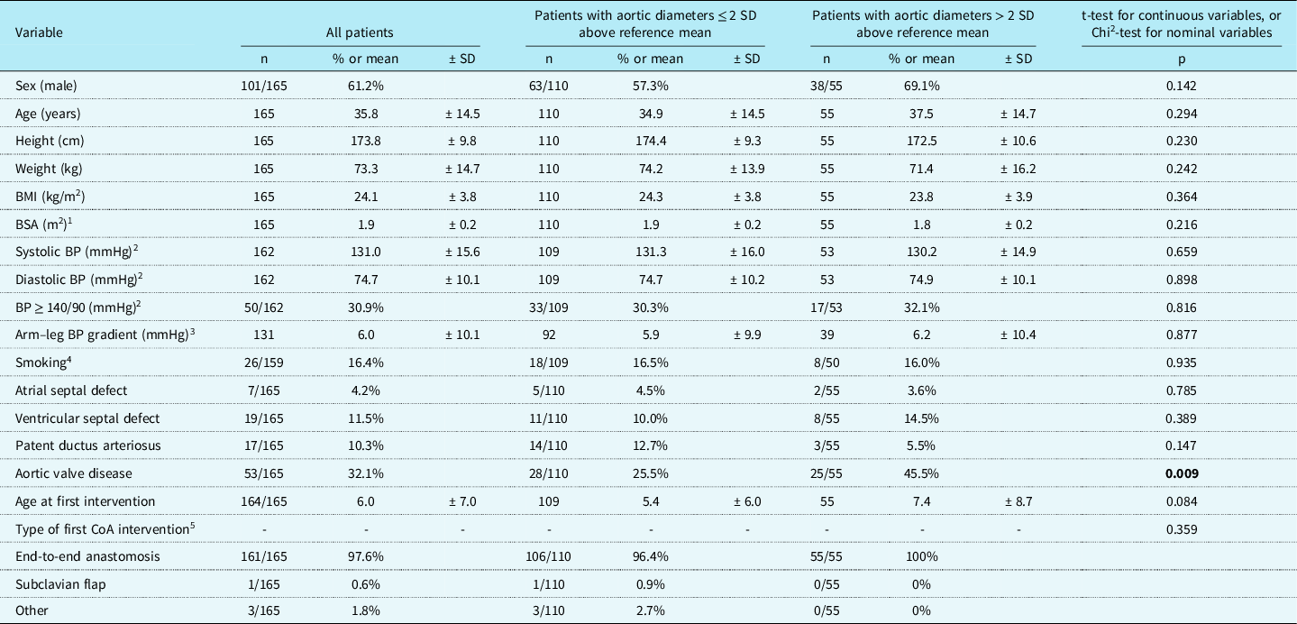
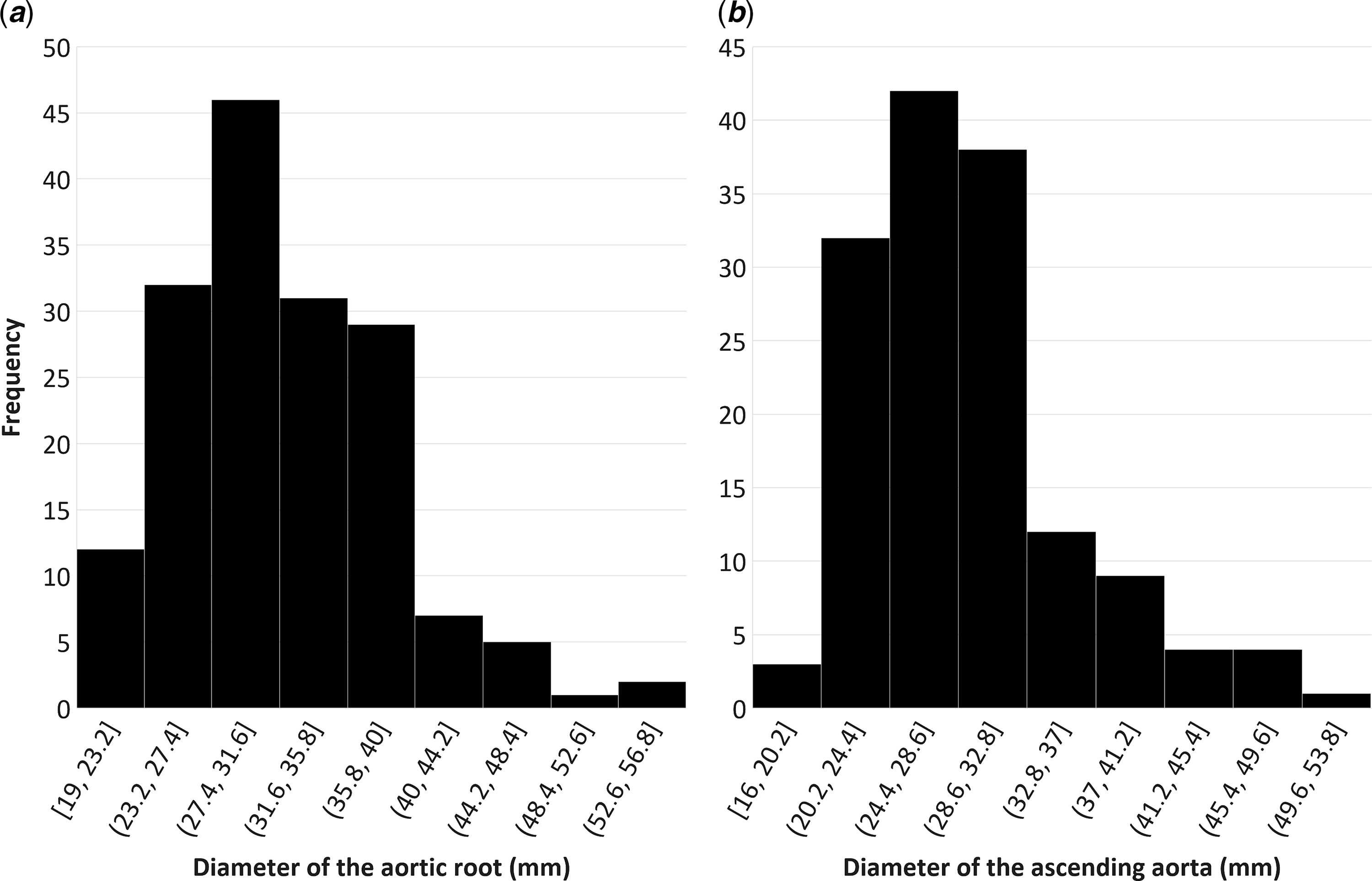
Within the population of 165 adult patients with repaired coarctation of the aorta, 101 (61.2%) were male, and the mean age was 35.8 ± 14.5 years. The majority of the interventions (97.6%) were end-to-end anastomoses. Aortic dilation, defined as aortic diameters > 2 standard deviations above reference mean, was found in 55 (33.3%) of the 165 included patients (Table 1). The average diameter of the aortic root was 31.8 ± 6.7 mm in the entire study population, 29.1 ± 4.8 mm for patients without aortic dilation, and 37.3 ± 6.5 mm for patients with aortic dilation. The average diameter of the ascending aorta was 29.3 ± 6.4 mm in the entire study population, 27.4 ± 4.2 mm for patients without aortic dilation, and 35.5 ± 8.0 mm for patients with aortic dilation. Figure 1 shows the distribution of aortic dimensions in the study population. Aortic valve disease, defined as register diagnosis and/or intervention, was found in 32.1% of the patients.
Patient characteristics

1 BSA denotes body surface area
2 BP denotes blood pressure. Systolic and diastolic blood pressures were measured in the arm
3 BP gradient denotes the systolic arm–leg blood pressure gradient as a continuous variable, with negative values defined as no gradient
4 Smoking is defined as either previous or current regular smoking habits
5 CoA denotes coarctation of the aorta
Diameter of ( a. ) the aortic root (n = 165, mean = 31.8 mm ± 6.7 mm), and ( b. ) the ascending aorta (n = 145, mean = 29.3 mm ± 6.4 mm).

Factors associated with aortic dilation
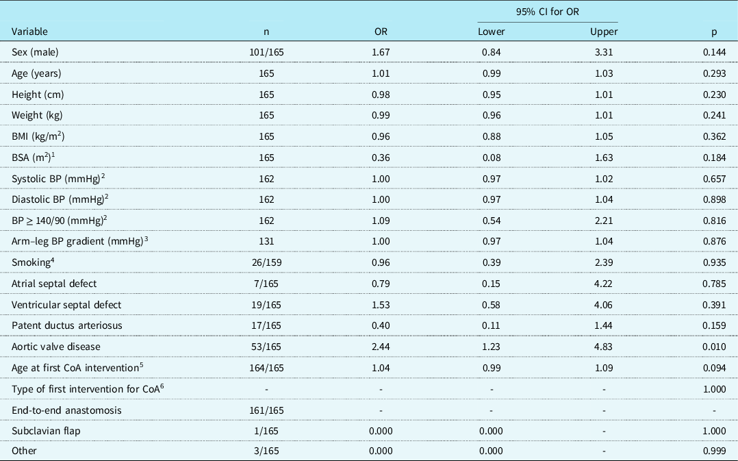
In univariable logistic regression, only aortic valve disease was associated with aortic dilation (p = 0.010, OR 2.44 [1.23, 4.83]) (Table 2).
Univariate logistic regressions with aortic dilation (aortic diameters > 2 SD above reference mean) as a dependent variable

1 BSA denotes body surface area
2 BP denotes blood pressure. Systolic and diastolic blood pressures were measured in the arm
3 BP gradient denotes the systolic arm–leg blood pressure gradient as a continuous variable, with negative values defined as no gradient
4 Smoking is defined as either previous or current regular smoking habits
5 CoA denotes coarctation of the aorta
6 End-to-end anastomosis is used as the reference category for the type of first CoA intervention
Discussion
The findings show that ascending aortic dilation is common in patients with previously repaired coarctation of the aorta. Only the presence of manifest aortic valve disease was associated with ascending aortic dilation, whereas sex, age, blood pressure, diagnosed hypertension, and systolic arm-leg blood pressure gradient were not. Patients with manifest aortic valve disease should, therefore, also be closely monitored for ascending aortic dilation.
Aortic valve disease
The high prevalence of aortic valve disease in the studied population is likely due to the high prevalence of bicuspid aortic valve, previously shown to be present in approximately 60–85% of patients with coarctation of the aorta, Reference Teo, Cannell, Babu-Narayan, Hughes and Mohiaddin10,Reference Rinnström, Engström and Johansson14,Reference Jurcut, Daraban and Lorber22 as opposed to 1–2% of the general population. Reference Braverman, Guven, Beardslee, Makan, Kates and Moon23 Importantly, dilation of the ascending aorta can also occur after aortic valve replacement.
It is worth noting that 30 of the 55 patients with ascending aortic dilation (54.5%) did not have aortic valve disease. Coarctation of the aorta and aortic dilation may represent two separate signs of a more fundamental underlying aortic pathology independent of valvular morphology. However, since bicuspid aortic valve without diagnosed functional complications was not included in the definition of aortic valve disease used in this study, there might still have been an overrepresentation of bicuspid aortic valve among the 30 patients with aortic dilation and no known aortic valve disease.
Of the 53 patients with manifest aortic valve disease, 28 (52.1%) had normal aortic diameters. These patients may still be at risk of developing ascending aortic dilation in the future, but longitudinal data are needed to further investigate this.
Blood pressure
It is worth noting that blood pressure was not associated with aortic dilation in this study, especially since hypertension is common in patients with repaired coarctation of the aorta. Reference Canniffe, Ou, Walsh, Bonnet and Celermajer8,Reference Rinnström, Dellborg and Thilén24,Reference Rinnström, Dellborg and Thilén25 Hypertension has previously been shown to be a major factor in aortic dissection, Reference Hiratzka, Bakris and Beckman26,Reference Pape, Tsai and Isselbacher27 but while hypertension is commonly regarded as a predisposing condition for the development of thoracic aortic aneurysms, the link between hypertension and ascending aortic dilation is still somewhat controversial. Reference Covella, Milan and Totaro15,Reference Simone and Chinali28
Age
Increasing age is known to be associated with increasing diameter of the ascending aorta, but, somewhat surprisingly, this association was not found in the study population. The reason for this is unclear. It might be that the ascending aorta in patients with coarctation of the aorta has other properties and that remodelling over time, therefore, differs from the general population. The study population is also relatively young, which may obscure the effect of age on the aortic dimensions.
General aspects
In the cohort, many patients had dilated aortas in relation to published reference values, but none reached dimensions requiring intervention. This may indicate that patients are referred for surgery in a proper way and/or that the ascending aorta in coarctation of the aorta is wider than in the general population without being prone to further dilation in such a young cohort.
Limitations
This study is crosssectional and provides no data on the development of aortic dilation over time. No reliable data were available regarding the prevalence of bicuspid aortic valve in this cohort. Bicuspid aortic valve is known to be common in coarctation of the aorta, Reference Rinnström, Engström and Johansson14 but this study cannot show whether or not a tricuspid aortic valve in coarctation of the aorta protects from aortic valve dilation. This study is based on register data and thus limited to the patients and parameters available within the register. The echocardiographic data have been obtained by many different investigators, and thus the possibility of inter-examiner variability cannot be excluded. Also, the patients included were those with available data regarding aortic dimensions in SWEDCON, which may introduce a selection bias.
Conclusions
In conclusion, aortic dilation is common in this study population of adult patients post-repair of coarctation of the aorta and is strongly associated with manifest aortic valve disease. No association was found between aortic dilation and age or blood pressure, suggesting that, in this relatively young cohort, aortic dilation in adults with repaired coarctation of the aorta does not primarily result from hypertension.
Acknowledgements
None.
Financial support
This work was supported by the Swedish Heart and Lung Foundation, the Heart Foundation of Northern Sweden, Umeå University, and the County Council of Västerbotten.
Conflicts of interest
None.
Ethical standards
The study was approved by the Regional Ethical Review Board in Umeå, Sweden (Dnr 08-218 M and 2012-445-32 M).